Chapter 1 Extradural Benign Tumors
VERTEBRAL HEMANGIOMA
EPIDEMIOLOGY
DISTRIBUTION
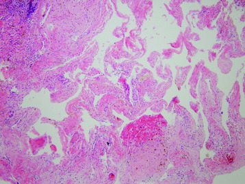
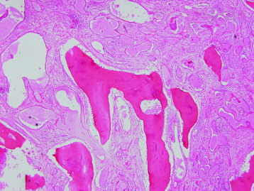
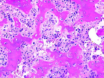
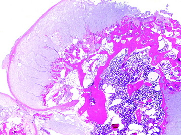
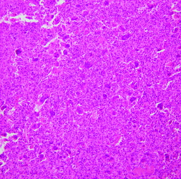
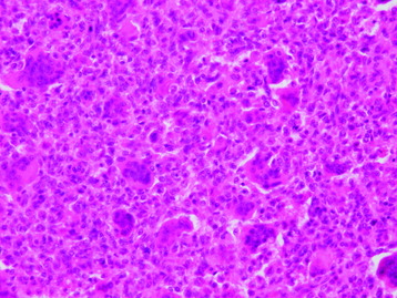
HISTOLOGY
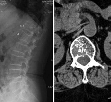
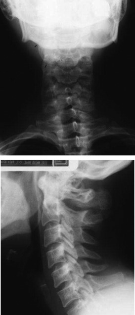
RADIOLOGY
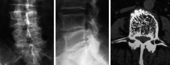
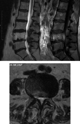
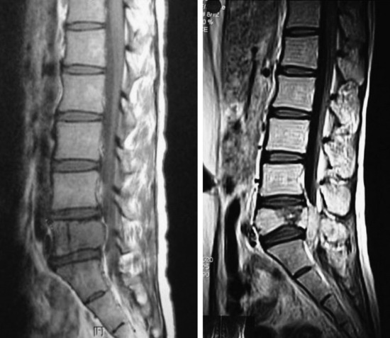
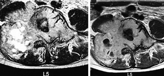
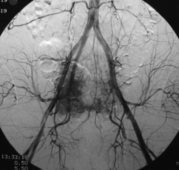
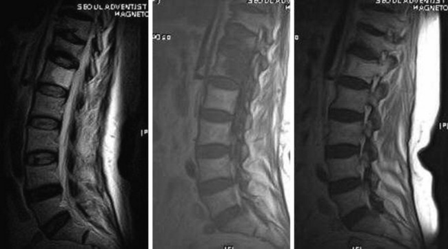
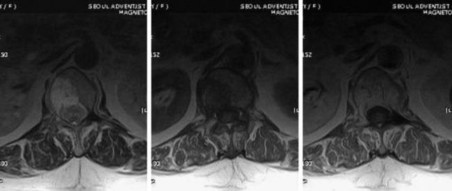
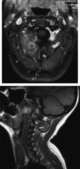
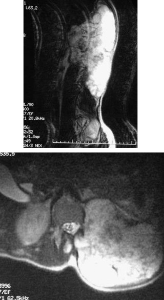
MR is extremely sensitive in detecting spinal hemangiomas. On both T1- and T2-weighted images, these lesions tend to have increased signal intensity, reflecting the adipose tissue rather than the hemorrhagic component. The hyperintense stroma surrounds foci of very low signal intensity, which represent the thickened vertical trabeculae (Fig. 1-5).3 In cases of hemangiomas that are rich in vascular element, the signal change is high on T2-weighted images (T2WI), low on T1-weighted images (T1WI), and shows homogenous enhancement with contrast (Figs. 1-6 and 1-7).
OSTEOID OSTEOMA/OSTEOBLASTOMA
DISTRIBUTION
Osteoid osteomas typically affect long bones, whereas osteoblastomas often involve the spine.
HISTOLOGY/GRADING
OSTEOCHONDROMA
DISTRIBUTION
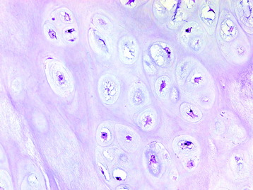
HISTOLOGY/GRADING
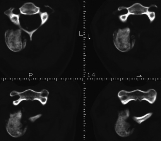
RADIOLOGY
X-Ray and CT
GIANT CELL TUMOR
EPIDEMIOLOGY
DISTRIBUTION
HISTOLOGY
RADIOLOGY
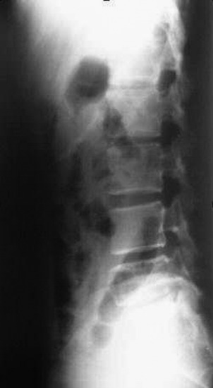
Fig. 1-19 On lateral view of lumbar spine, osteolytic expansile lesion is seen in the vertebral body.