Eventration: Diaphragmatic contour is eccentric, with upward bulging of affected portion of diaphragm
– Usually anteromedial aspect of right hemidiaphragm
Paralysis: Asymmetric elevation of involved hemidiaphragm with basal atelectasis
• US
Sonography superior to fluoroscopy for diaphragmatic abnormalities
Diaphragmatic paralysis: No motion or paradoxical motion during inspiration
Eventration seen best during inspiration
• Chest fluoroscopy
Paralysis: Movement of affected hemidiaphragm is paradoxical (upward during inspiration or sniff test)
– Mediastinum shifts away from paralyzed side during inspiration
– Much less sensitive for bilateral paralysis
Eventration: Paradoxical motion may or may not be present depending on size of eventration
• CT
Liver or spleen “mushrooms” through site of eventration
PATHOLOGY
• Eventration is a congenital abnormality due to incomplete muscularization of central membranous diaphragm in utero
• Paralysis can occur due to abnormalities of brain, spinal cord, neuromuscular junction, phrenic nerve, or muscle
CLINICAL ISSUES
• No treatment for asymptomatic patients
• Surgical plication of diaphragm for symptomatic patients
(Left) Coronal CECT demonstrates a focal “hump” or “bulge” in the right hemidiaphragm, characteristic of eventration. Eventration typically occurs in the anteromedial aspect of the right hemidiaphragm.
(Right) Sagittal CECT shows focal eventration or thinning of the central portion of the right hemidiaphragm. Compare this with the normal thickness of the more anterior portion of the diaphragm . Note the “mushrooming” of the liver at the site of eventration.
(Left) Coronal CECT demonstrates marked asymmetric elevation of the left hemidiaphragm.
(Right) Axial CECT in the same patient demonstrates a soft tissue mass in the expected location of the phrenic nerve (later found to be lymphoma). Diaphragmatic paralysis can occur due to abnormalities in the brain, spinal cord, neuromuscular junction, phrenic nerve, or muscle.
Coronal CECT demonstrates a wide “bulge” in the right hemidiaphragm, containing liver, representing a diaphragmatic eventration.
Coronal CECT in a patient with eventration shows part of the liver bulging into the lower thorax. The diaphragm is eccentric, with the liver occupying the bulge created by the eventrated segment.


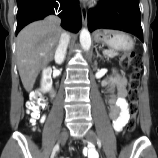
 in the right hemidiaphragm, characteristic of eventration. Eventration typically occurs in the anteromedial aspect of the right hemidiaphragm.
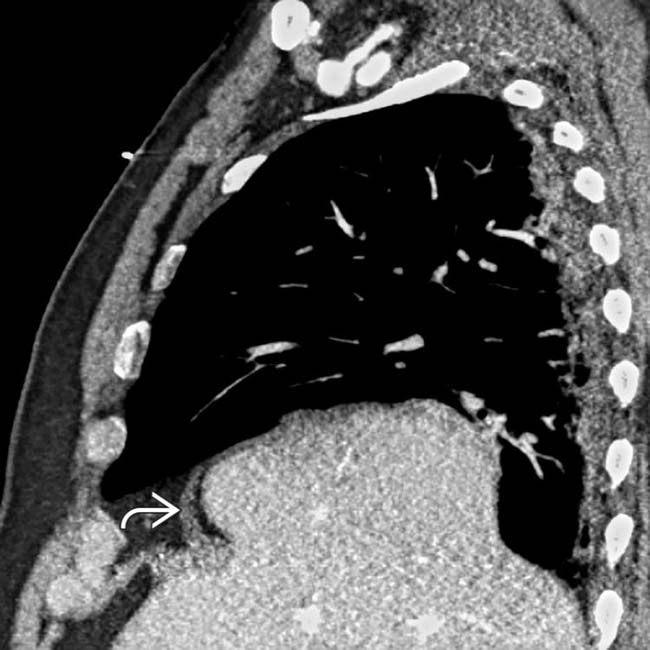
 . Note the “mushrooming” of the liver at the site of eventration.
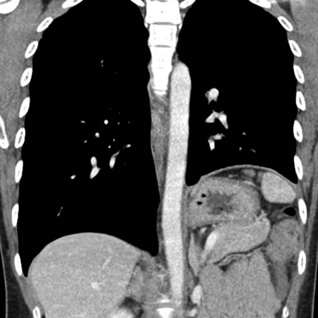
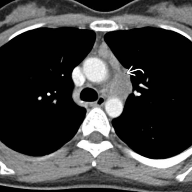
 in the expected location of the phrenic nerve (later found to be lymphoma). Diaphragmatic paralysis can occur due to abnormalities in the brain, spinal cord, neuromuscular junction, phrenic nerve, or muscle.
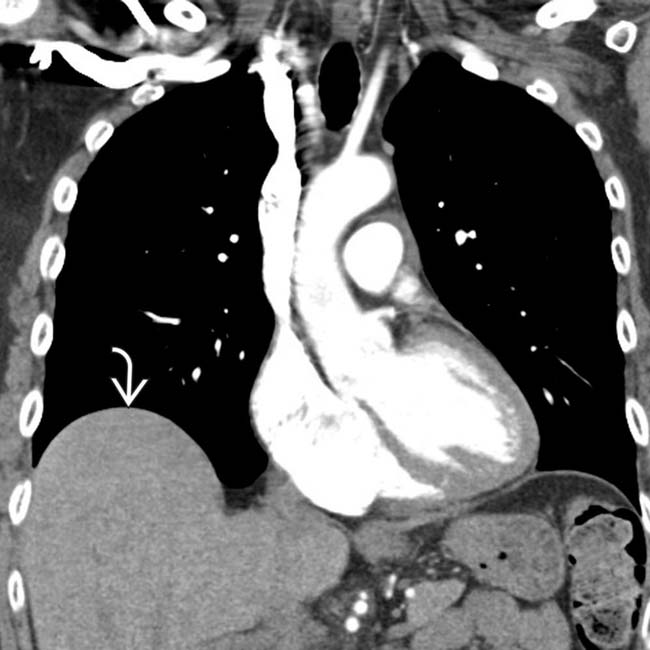
 in the right hemidiaphragm, containing liver, representing a diaphragmatic eventration.
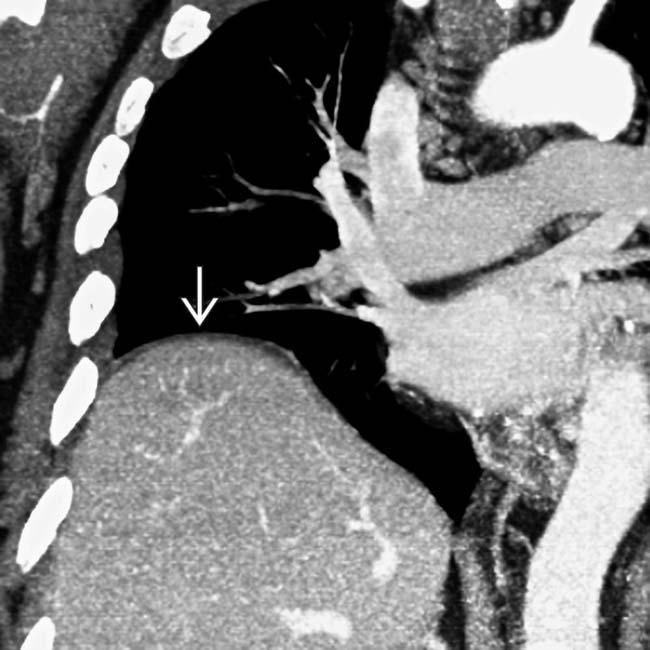
 bulging into the lower thorax. The diaphragm is eccentric, with the liver occupying the bulge created by the eventrated segment.


