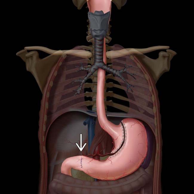
[level-membership-for-radiology-category] Transthoracic esophagectomy: Usually performed through right intercostal approach (Ivor Lewis procedure)

• Stomach is ideal conduit, as it has reliable blood supply and can reach high into thorax or neck for anastomosis


• Postoperative complications
 Essentially all patients have some degree of dysphagia, early satiety, and reflux following esophagectomy
Essentially all patients have some degree of dysphagia, early satiety, and reflux following esophagectomy


 is done to facilitate gastric emptying.
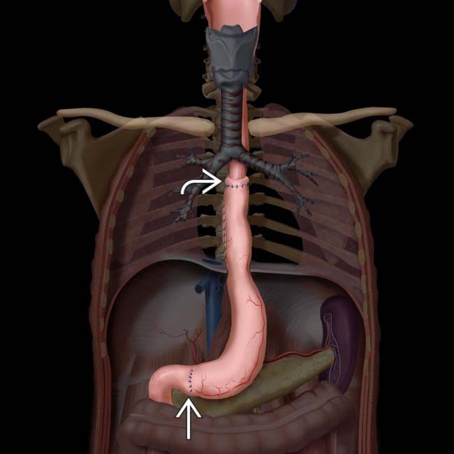
 and the pyloroplasty
  .
.
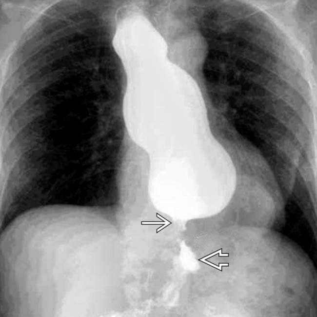
 anastomosed to the cervical esophagus. Note the position of the gastric staple line
  along the right side of the conduit.
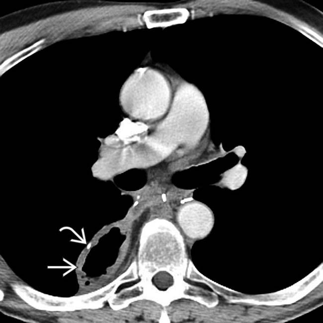
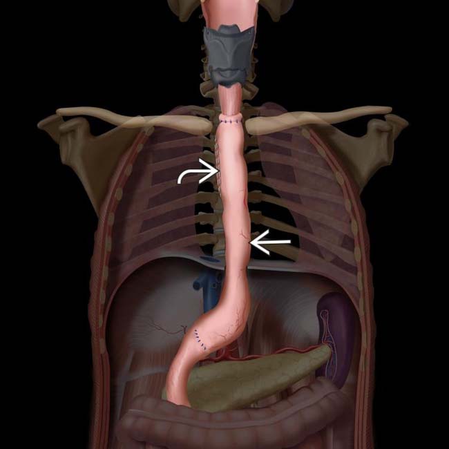
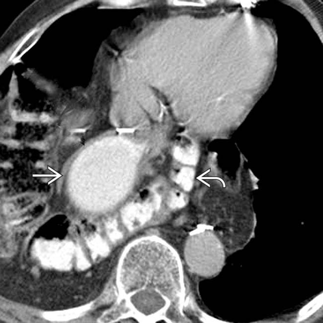
 in the paravertebral location. Note the position of the gastric staple line
  . The conduit is not filled with retained fluid, and there is no evidence of lung injury from reflux.
IMAGING
Surgical Procedures


• Transthoracic esophagectomy
 Usually performed through right intercostal approach (Ivor Lewis procedure)
 Many variations exist
Usually performed through right intercostal approach (Ivor Lewis procedure)
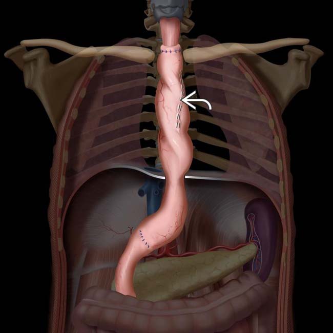
– Generally begins with laparotomy for mobilization of stomach, which is then used to create gastric tube/conduit that will replace resected esophagus


– Pyloroplasty or pyloromyotomy is performed to facilitate gastric emptying and to minimize gastroesophageal reflux


 Many variations exist
– e.g., left thoracotomy approach, transhiatal open approach (without thoracotomy), minimally invasive procedures (performed through ports in thorax and abdomen without open incision into either)


Complications
• Perioperative complications
 Injury to recurrent laryngeal or vagus nerve (5-10%)
 Injury to recurrent laryngeal or vagus nerve (5-10%)


• Postoperative complications
 Essentially all patients have some degree of dysphagia, early satiety, and reflux following esophagectomy
 Anastomotic leak (10-16%)
 Delayed e mptying of conduit
Essentially all patients have some degree of dysphagia, early satiety, and reflux following esophagectomy


 Anastomotic leak (10-16%)
– Occurs more commonly with neck anastomoses, but thoracic anastomotic leaks cause more serious complications


 Delayed e mptying of conduit


– Mechanical obstruction


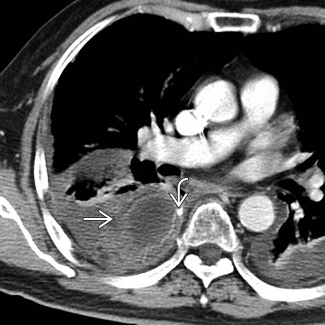
 and evidence of severe aspiration pneumonia and pleural effusion. The conduit is probably twisted, as evidenced by the position of the gastric staple line
  .
.
 , as well as severe lung disease and pleural effusions.
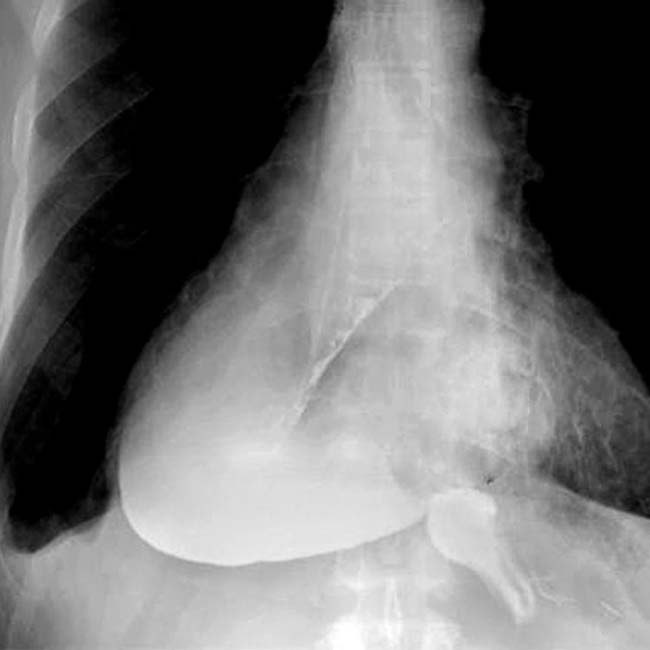
 above the diaphragm
  . The conduit is dilated with an air-fluid level, indicating partial obstruction. The conduit was pulled down into the abdomen at revision laparoscopy with resolution of symptoms.
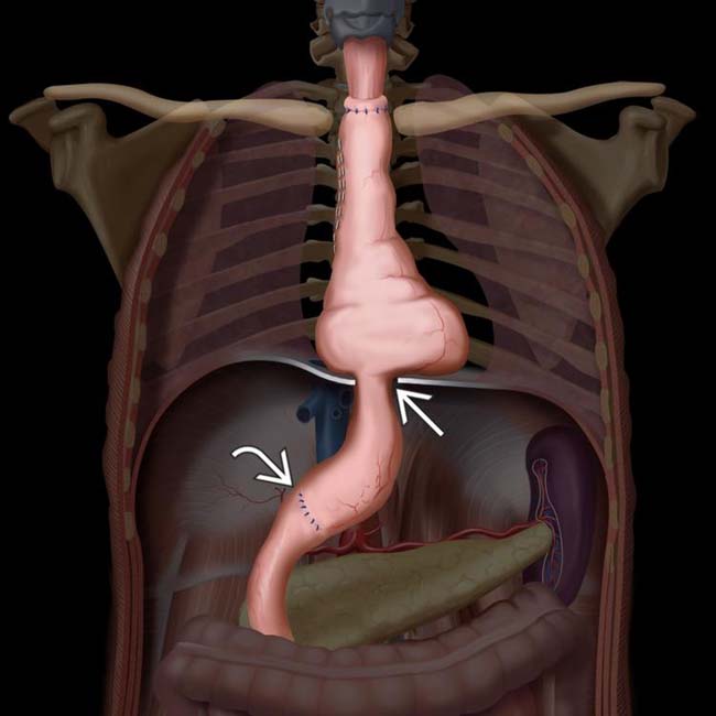
 , not the pylorus, which has been widened by pyloroplasty
  .
.
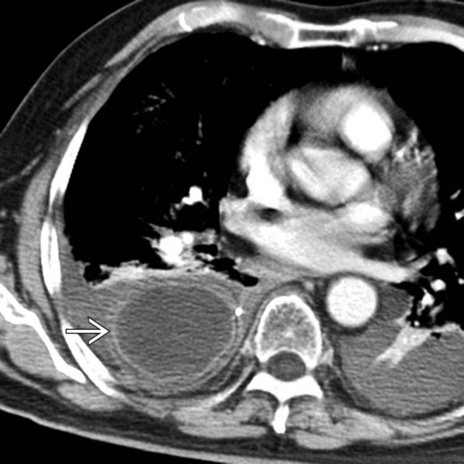
 , which is proximal to the collapsed gastric antrum
  in this patient with mechanical obstruction of the conduit at the diaphragmatic hiatus (the most common site).
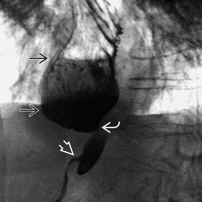
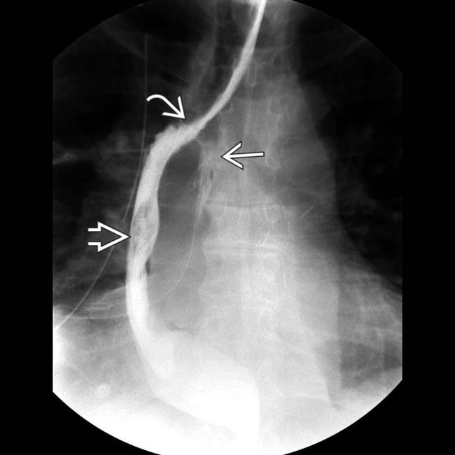
 . The stomach is narrowed as it traverses the diaphragm
  , but the pyloroplasty
  is neither the site nor the cause of the delayed emptying.
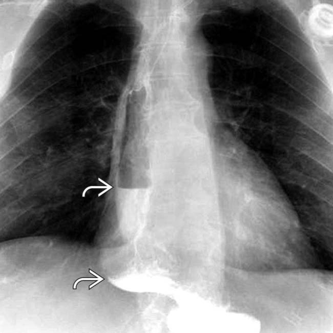
 on upright film. Vagal nerve injury is the most common etiology.
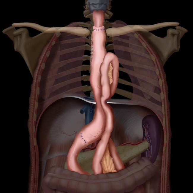
 and omental fat posterior to the gastric conduit
  .
.
 , which has rotated to the left anterolateral position.
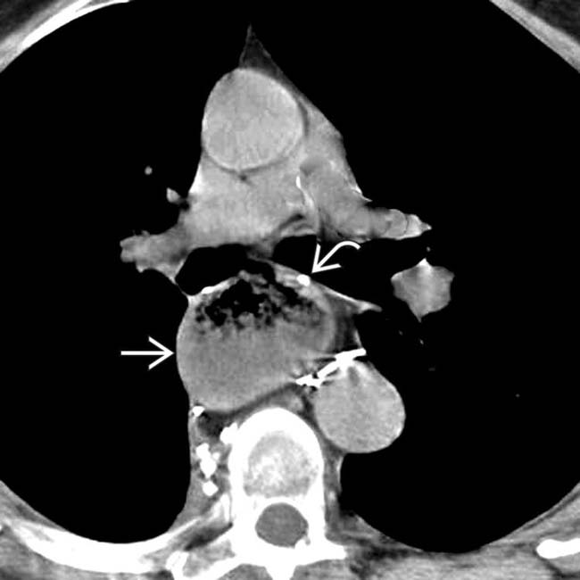
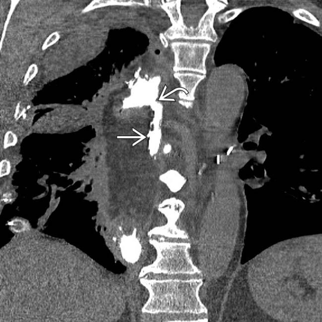
 , suggesting impaired emptying. The position of the gastric staple line
  indicates rotation or volvulus of the conduit, as it is expected to be at the 9-o’ clock position
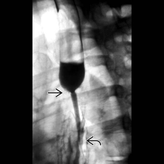
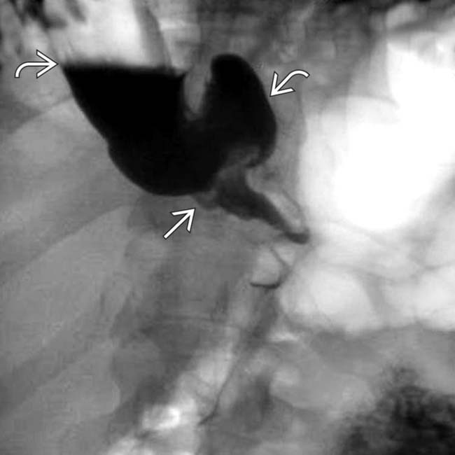
 at the esophagogastric anastomosis with delayed emptying of the esophagus, evident as an air-fluid level. Note the smooth surface of the esophagus and the rugal fold pattern of the gastric conduit
  .
.
 between the esophagus and the gastric conduit
  . There is an anastomotic leak
  into the mediastinum.
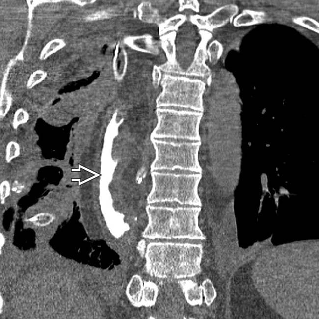
 .
.
 from the esophagogastric anastomosis
  into the mediastinum.
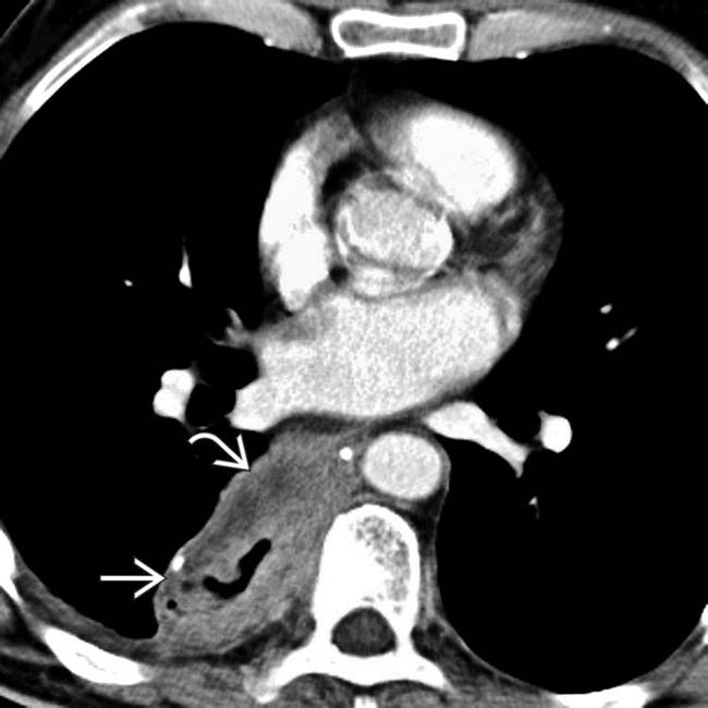
 . There is a large soft tissue mass
  abutting the conduit and extending into the mediastinum in this patient with a recurrent tumor.
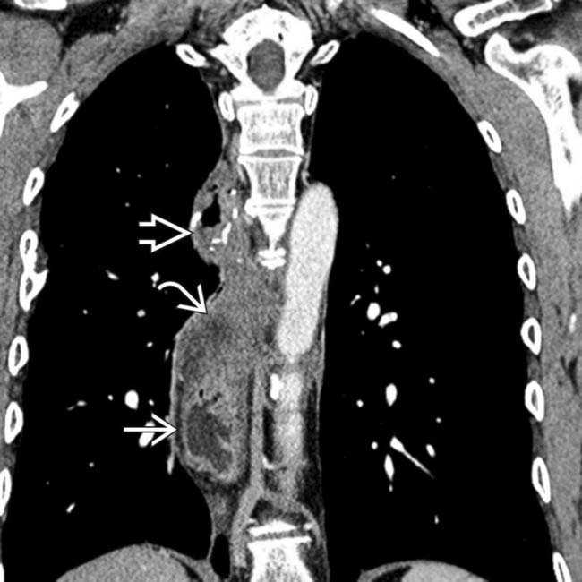
 and extensive mediastinal mass effect
  , representing recurrent esophageal tumor. A portion of the anastomotic staple line
  is seen.
[/level-membership-for-radiology-category][not-level-membership-for-radiology-category] Transthoracic esophagectomy: Usually performed through right intercostal approach (Ivor Lewis procedure)
• Stomach is ideal conduit, as it has reliable blood supply and can reach high into thorax or neck for anastomosis
• Postoperative complications
Essentially all patients have some degree of dysphagia, early satiety, and reflux following esophagectomy
Essentially all patients have some degree of dysphagia, early satiety, and reflux following esophagectomy is done to facilitate gastric emptying. anastomosed to the cervical esophagus. Note the position of the gastric staple line along the right side of the conduit. in the paravertebral location. Note the position of the gastric staple line . The conduit is not filled with retained fluid, and there is no evidence of lung injury from reflux.IMAGING
Surgical Procedures
• Transthoracic esophagectomy
Usually performed through right intercostal approach (Ivor Lewis procedure)
Many variations exist
Usually performed through right intercostal approach (Ivor Lewis procedure)
– Generally begins with laparotomy for mobilization of stomach, which is then used to create gastric tube/conduit that will replace resected esophagus
– Pyloroplasty or pyloromyotomy is performed to facilitate gastric emptying and to minimize gastroesophageal reflux
Many variations exist
– e.g., left thoracotomy approach, transhiatal open approach (without thoracotomy), minimally invasive procedures (performed through ports in thorax and abdomen without open incision into either)
Complications
• Perioperative complications
Injury to recurrent laryngeal or vagus nerve (5-10%)
Injury to recurrent laryngeal or vagus nerve (5-10%)
Buy Membership for Radiology Category to continue reading. Learn more here
[/not-level-membership-for-radiology-category]
