Esophageal Scleroderma

Published on 13/07/2015 by admin

Filed under Radiology

Last modified 22/04/2025

Print this page

rate 1 star rate 2 star rate 3 star rate 4 star rate 5 star
Your rating: none, Average: 0 (0 votes)

This article have been viewed 4573 times

 GI tract, lungs, heart, kidneys, and nervous system

• GI: 3rd most common manifestation after skin changes and Raynaud phenomenon

image Seen in up to 90% of patients
image Most common sites

– Esophagus > duodenum > anorectal > small bowel > colon
• Interstitial pulmonary fibrosis, often severe
• Scleroderma is classified into 2 types

image Diffuse scleroderma and CREST syndrome
• Diffuse scleroderma

image Cutaneous and visceral involvement 

– Often severe
• CREST syndrome: Minimal cutaneous and late visceral

image C: Calcinosis of skin
image R: Raynaud phenomenon
image E: Esophageal dysmotility
image S: Sclerodactyly (involvement of fingers)
image T: Telangiectasia
• Esophagography

image Atony or aperistalsis: Lower 2/3 (smooth muscle)
image Mild to moderate dilatation of esophagus
image Patulous lower esophageal sphincter (early)
image Ulcers, fusiform peptic stricture (later)
image Gastroesophageal reflux (70% of patients)
image 40% develop Barrett esophagus

TOP DIFFERENTIAL DIAGNOSES

• Esophageal achalasia
• Reflux esophagitis (with stricture)
• Esophageal carcinoma
• Iatrogenic
image
(Left) Upright film from an esophagram in a 29-year-old woman with dysphagia and shortness of breath shows a dilated, atonic esophagus image with a distal esophageal stricture image
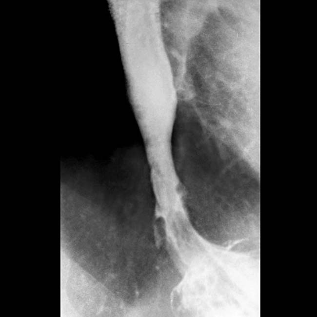
. Esophageal peristalsis was completely absent.

image
(Right) Chest CT in the same patient shows interstitial fibrosis and a massive dilated esophagus image
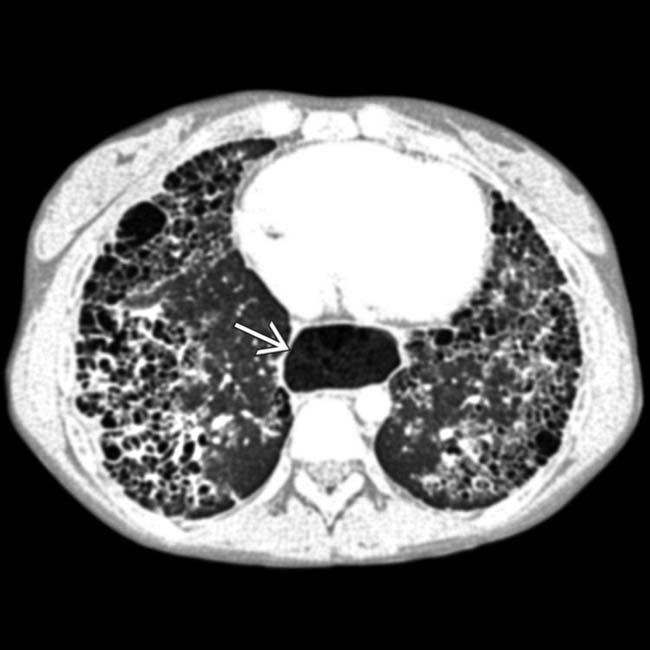
, all findings due to scleroderma.
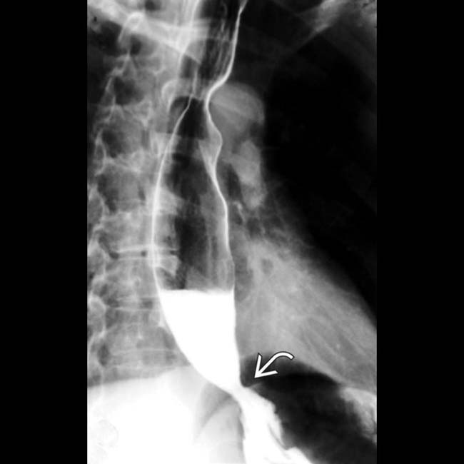
image
(Left) Film from an esophagram in a young woman with dysphagia shows a dilated esophagus with a persistent air-fluid level, indicating delayed emptying. There is stricture of the distal esophagus image.

image
(Right) Subsequent film in the same patient shows a dilated duodenum with functional narrowing of its 3rd portion. The duodenum is often dilated and atonic in patients with scleroderma.

TERMINOLOGY

Synonyms

• Progressive systemic sclerosis

Definitions

• Multisystem disorder of small vessels and connective tissue (collagen vascular disease) of unknown etiology

IMAGING

General Features

• Best diagnostic clue

image Dilated atonic esophagus with distal stricture (late findings)
• Other general features

image Multisystemic disorder with immunologic and inflammatory changes
image Characterized by atrophy, fibrosis, and sclerosis of skin, vessels, and organs
image Involves skin, synovium, and parenchyma of multiple organs

– Gastrointestinal tract, lungs, heart, kidneys, and nervous system
image Gastrointestinal (GI) tract scleroderma

– 3rd most common manifestation after skin changes and Raynaud phenomenon
– Seen in up to 90% of patients
– Most common sites: Esophagus > duodenum > anus/rectum > small bowel > colon
– Most frequent cause of chronic intestinal pseudo-obstruction
image Scleroderma is classified into 2 types

– Diffuse scleroderma
– CREST syndrome (more benign course)
image Diffuse scleroderma: Cutaneous and visceral involvement

– Interstitial pulmonary fibrosis, often severe
– Organ failure more likely
– Associated with antitopoisomerase 1 antibody (anti-Scl 70)
image CREST syndrome: Minimal cutaneous and late visceral involvement

– C: Calcinosis of skin
– R: Raynaud phenomenon
– E: Esophageal dysmotility
– S: Sclerodactyly (involvement of fingers)
– T: Telangiectasia
– Associated with anticentromere antibodies

Radiographic Findings

• Fluoroscopic-guided esophagography

image Normal peristalsis above aortic arch (striated muscle)
image Atony or aperistalsis: Lower 2/3 (smooth muscle)
image Mild to moderate dilatation of esophagus
image Patulous lower esophageal sphincter (LES)

– Early finding of scleroderma
image Erosions, superficial ulcers, fusiform peptic stricture

– Due to reflux esophagitis
image Gastroesophageal reflux (70% cases)

– 40% develop Barrett esophagus
image Hiatal hernia
• Upper GI series

image Stomach: Gastric dilation and delayed emptying
• Small bowel follow-through

image Duodenal dilation

– Often mimics SMA syndrome
– Abrupt transition to nondilated duodenum after crossing over spine in midline
image Pathognomonic: Hidebound sign

– Dilated jejunum with crowded, thin, circular folds
– Seen in > 60% cases of scleroderma-related pseudo-obstruction
– Due to muscle atrophy and its uneven replacement by collagen in longitudinal fibers + intense fibrosis of submucosa
image Wide-mouthed sacculations (true diverticula) on antimesenteric border
image Prolonged transit time with barium retention in duodenum and small bowel up to 24 hours
image ± pneumatosis intestinalis ± pneumoperitoneum

– Can be due to steroid medication plus dilated bowel lumen
image ± transient, nonobstructive intussusceptions
• Barium enema

image Sacculations on antimesenteric border of colon
image Marked dilatation (simulates Hirschsprung disease)
image Chronic phase: Complete loss of haustrations

– Simulates cathartic colon or chronic ulcerative colitis
image Stercoral ulceration (from retained fecal material)

DIFFERENTIAL DIAGNOSIS

Esophageal Achalasia

• Grossly dilated esophagus with smooth, beak-like tapering at lower end
• Scleroderma shows moderate dilatation of esophagus with fusiform stricture

Reflux Esophagitis (With Stricture)

• Longer tapered distal stricture
• Less luminal dilation
• Distinguished from scleroderma by normal peristalsis

Esophageal Carcinoma

• Abrupt proximal borders of strictured segment (“rat tail” appearance)
• Mucosal irregularity, shouldering, mass effect

Iatrogenic

• e.g., fundoplication and vagotomy

image Tight wrap narrows esophageal lumen
image Vagotomy, scarring decrease peristalsis

PATHOLOGY

General Features

• Etiology

image Unknown; autoimmune condition with genetic predisposition
image May be initiated by environmental antigens like silica and L-tryptophan
image Immunologic mechanism (delayed hypersensitivity)

– Increased production of cytokines (TNF-α or IL-1) → collagen production
– Vascular damage and activation of fibroblasts
• Genetics

image Localized: Anticentromere antibodies associated with HLA-DR1, HLA-DR4, and HLA-DR5
image Diffuse: Antitopoisomerase 1 antibodies (anti-Scl 70) associated with HLA-DR5
• Associated abnormalities

image Systemic lupus, polymyositis, or dermatomyositis

Gross Pathologic & Surgical Features

• Rubber-hose inflexibility: Lower 2/3 of esophagus
• Thin and ulcerated mucosa
• Dilated gas and fluid-containing small bowel loops with sacculations

Microscopic Features

• Perivascular lymphocytic infiltrates
• Early capillary and arteriolar injury
• Atrophy and fragmentation of smooth muscle → collagen deposition and fibrosis

CLINICAL ISSUES

Presentation

• Most common signs/symptoms

image Esophagus

– Dysphagia, regurgitation
– Epigastric fullness and burning pain
image Small bowel

– Bloating, abdominal pain
– Weight loss, diarrhea, anemia
image Colon

– Chronic constipation
• Lab data

image Increased erythrocyte sedimentation rate (ESR)
image Iron, B12, and folic acid deficiency anemias
image Increased antinuclear antibodies (ANA)
image CREST syndrome: Anticentromere antibodies
image Diffuse scleroderma

– Antitopoisomerase 1 antibody

Demographics

• Age

image Onset usually by 30-50 years
• Gender

image M:F = 1:3
• Ethnicity

image African American > Caucasian
• Epidemiology

image Incidence: ∼ 20 cases per million adults in USA
image Prevalence: 200 per million adults

Natural History & Prognosis

• Complications

image Barrett esophagus → adenocarcinoma
image Bowel pseudo-obstruction
• Prognosis

image Limited disease with ANA bodies: Good prognosis
image Diffuse disease: Poor with involvement of kidneys, heart, and lungs rather than GI tract

Treatment

• Small, frequent meals; elevation of head of bed
• Avoid tea and coffee
• Cimetidine, ranitidine, omeprazole
• Metoclopramide, laxatives
• Patients with severe malabsorption

image Parenteral hyperalimentation

DIAGNOSTIC CHECKLIST

Consider

• Rule out other pathologies that cause distal esophageal stricture ± dysmotility
• Check for family history of collagen vascular diseases

Image Interpretation Pearls

• Mild to moderate dilatation of esophagus with distal fusiform stricture + decreased or absent peristalsis

image
(Left) Film from an esophagram in a 50-year-old man shows absence of primary peristalsis, with repetitive and deep tertiary contractions image
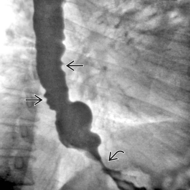
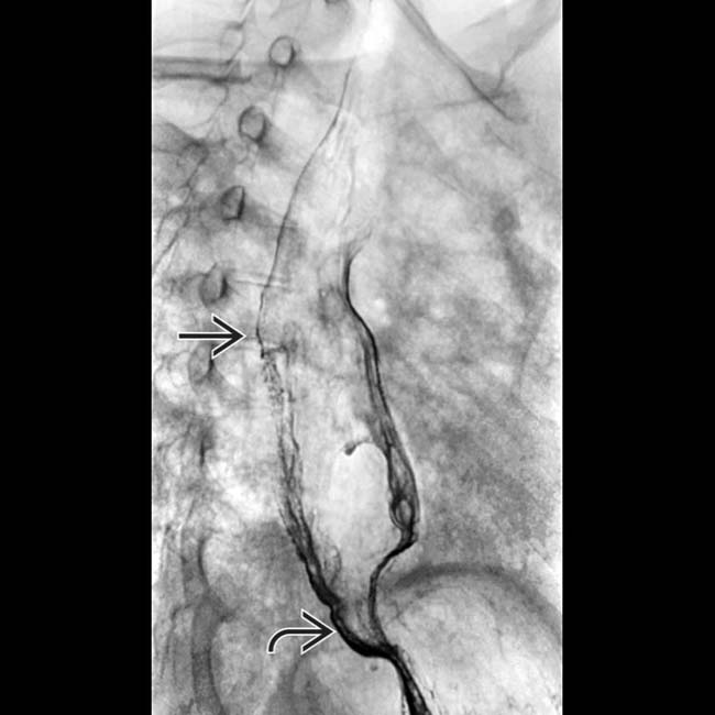
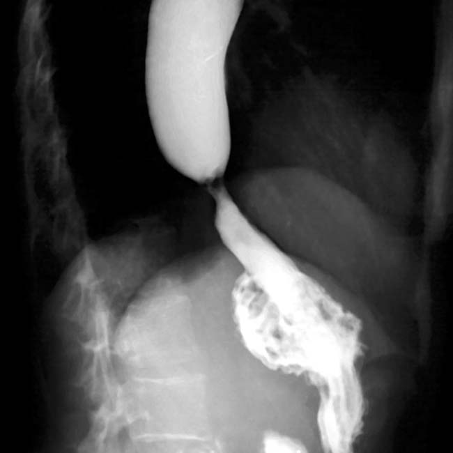
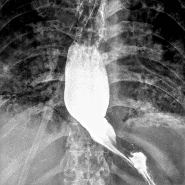
. The esophageal lumen is mildly dilated, and there is a stricture at the GE junction image.
image
(Right) Film obtained 2 hours after the esophagram in the same patient shows markedly delayed passage of the barium through the small bowel. The folds within the jejunum are thin and closely spaced image
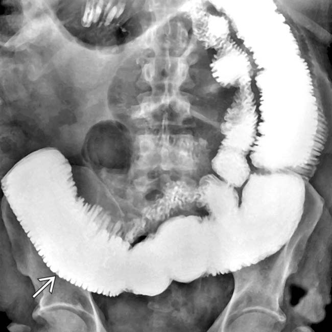
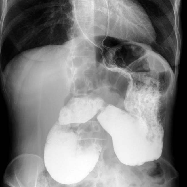
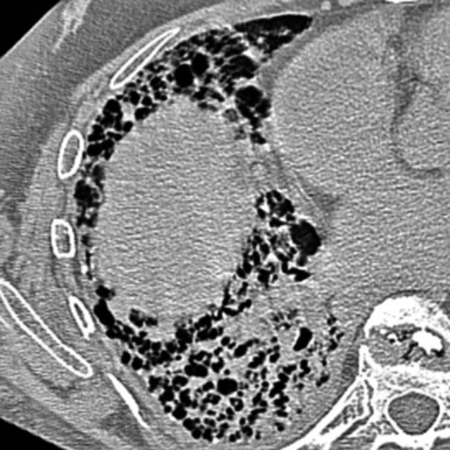
, and the lumen is dilated with minimal peristalsis in this classic “hidebound” bowel.
image
(Left) A single film from a barium esophagram in a middle-aged woman with scleroderma shows a dilated, atonic esophagus with a tight stricture image 
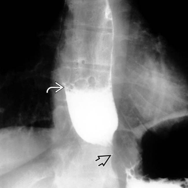
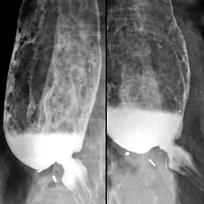
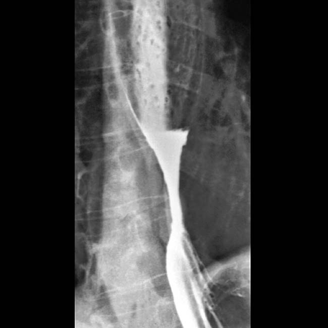
at the GE junction. The esophagus was slow to empty, even in the upright position, with a fluid-barium level seen image.

image
(Right) A subsequent film in the same patient shows a markedly dilated 2nd part of the duodenum image
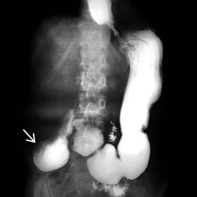
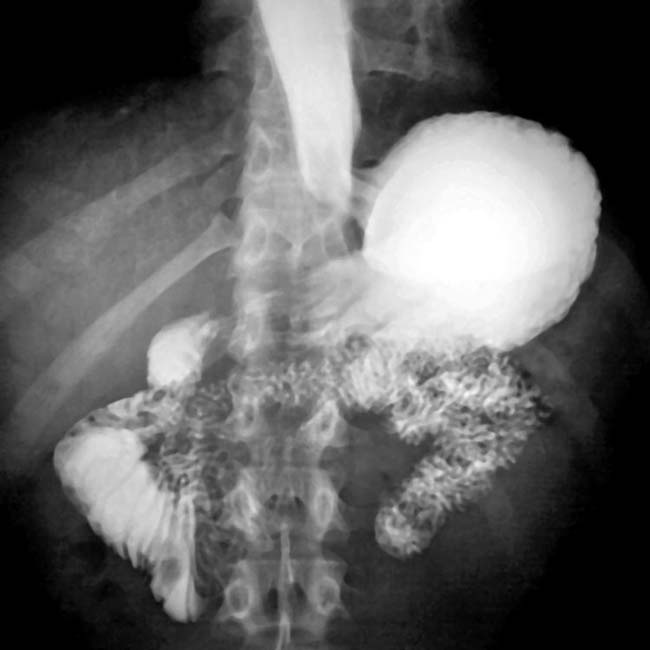
, with abrupt narrowing as it crosses the spine. The duodenum is the most frequently affected part of the GI tract beyond the esophagus.
image
(Left) Film from an upper GI series shows reflux into the esophagus through a patulous GE junction image
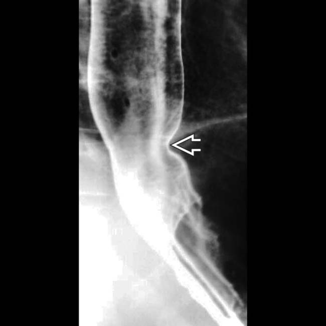
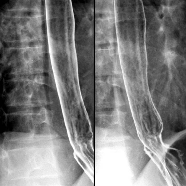
. The esophagus had no primary peristalsis. These are relatively early signs of scleroderma, and no stricture or ulcerations have yet developed.
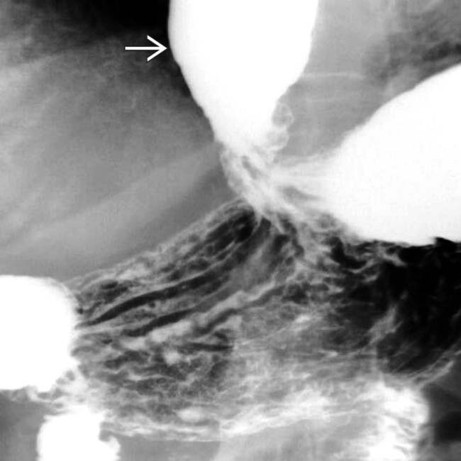
image
(Right) Supine film from an upper GI series in the same patient shows reflux of barium into the dilated esophagus image.
image
Single-contrast upper GI shows a dilated, atonic esophagus with patulous LES. The dilated duodenum abruptly narrows as it crosses the spine and is also due to scleroderma.

image
Air-contrast esophagrams show a markedly dilated esophagus and distal stricture due to scleroderma.
image
Single-contrast esophagram shows a short, smooth stricture with a dilated atonic esophagus.
image
Air-contrast esophagram shows a dilated, atonic esophagus with an air-contrast level and a long, smooth distal stricture in this patient with a small hiatal hernia with “short esophagus.”
image
Single-contrast esophagram shows a hiatal hernia, shortening of the esophagus, and a long. smooth distal stricture.
image
Two views from an air-contrast esophagram show an atonic, but not dilated esophagus at the patulous GE junction.
image
Axial NECT shows “honeycomb” lung in this patient with interstitial fibrosis due to scleroderma.
image
Upper GI shows a dilated, atonic esophagus with distal stricture. Note the underlying lung disease.

SELECTED REFERENCES

1. Simeón-Aznar, CP, et al. Systemic sclerosis sine scleroderma and limited cutaneous systemic sclerosis: similarities and differences. Clin Exp Rheumatol. 2014. [ePub].

2. Savarino, E, et al. Gastroesophageal reflux and pulmonary fibrosis in scleroderma: a study using pH-impedance monitoring. Am J Respir Crit Care Med. 2009; 179(5):408–413.

3. Hinchcliff, M, et al. Systemic sclerosis/scleroderma: a treatable multisystem disease. Am Fam Physician. 2008; 78(8):961–968.

4. Vonk, MC, et al. Oesophageal dilatation on high-resolution computed tomography scan of the lungs as a sign of scleroderma. Ann Rheum Dis. 2008; 67(9):1317–1321.

5. Sifrim, D, et al. Non-achalasic motor disorders of the oesophagus. Best Pract Res Clin Gastroenterol. 2007; 21(4):575–593.

6. Ntoumazios, SK, et al. Esophageal involvement in scleroderma: gastroesophageal reflux, the common problem. Semin Arthritis Rheum. 2006; 36(3):173–181.

7. Mayes, MD. Scleroderma epidemiology. Rheum Dis Clin North Am. 2003; 29(2):239–254.

8. Goldblatt, F, et al. Antibody-mediated gastrointestinal dysmotility in scleroderma. Gastroenterology. 2002; 123(4):1144–1150.

9. Coggins, CA, et al. Wide-mouthed sacculations in the esophagus: a radiographic finding in scleroderma. AJR Am J Roentgenol. 2001; 176(4):953–954.

10. Duchini, A, et al. Gastrointestinal hemorrhage in patients with systemic sclerosis and CREST syndrome. Am J Gastroenterol. 1998; 93(9):1453–1456.

11. Weston, S, et al. Clinical and upper gastrointestinal motility features in systemic sclerosis and related disorders. Am J Gastroenterol. 1998; 93(7):1085–1089.

12. Lock, G, et al. Gastrointestinal manifestations of progressive systemic sclerosis. Am J Gastroenterol. 1997; 92(5):763–771.

13. Young, MA, et al. Gastrointestinal manifestations of scleroderma. Rheum Dis Clin North Am. 1996; 22(4):797–823.

14. Kahan, A, et al. Gastrointestinal involvement in systemic sclerosis. Clin Dermatol. 1994; 12(2):259–265.

15. Ott, DJ. Esophageal motility disorders. Semin Roentgenol. 1994; 29(4):321–331.

Sjogren, RW. Gastrointestinal motility disorders in scleroderma. Arthritis Rheum. 1994; 37(9):1265–1282.

Levine, MS, et al. Update on esophageal radiology. AJR Am J Roentgenol. 1990; 155(5):933–941.

Marshall, JB, et al. Gastrointestinal manifestations of mixed connective tissue disease. Gastroenterology. 1990; 98(5 Pt 1):1232–1238.