Escherichia coli

Published on 25/03/2015 by admin

Filed under Pediatrics

Last modified 22/04/2025

Print this page

rate 1 star rate 2 star rate 3 star rate 4 star rate 5 star
Your rating: none, Average: 2 (1 votes)

This article have been viewed 1631 times

Chapter 192 Escherichia coli

Escherichia coli are important causes of enteric infections as well as urinary tract infections (Chapter 532), sepsis and meningitis in the newborn (Chapter 103), and bacteremia and sepsis in immunocompromised patients (Chapter 171) and in patients with intravascular devices (Chapter 172).

E. coli species are members of the Enterobacteriaceae family. They are facultatively anaerobic, gram-negative bacilli that usually ferment lactose. Most fecal E. coli do not cause diarrhea. Six major groups of diarrheagenic E. coli have been characterized on the basis of clinical, biochemical, and molecular-genetic criteria: enterotoxigenic E. coli (ETEC); enteroinvasive E. coli (EIEC); enteropathogenic E. coli (EPEC); Shiga toxin–producing E. coli (STEC), also known as enterohemorrhagic E. coli (EHEC) or verotoxin producing E. coli (VTEC); enteroaggregative E. coli (EAEC or EggEC); and diffusely adherent E. coli (DAEC).

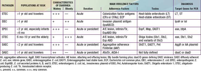
Because E. coli are normal fecal flora, pathogenicity is defined by demonstration of virulence characteristics and association of those traits with illness (Table 192-1). The mechanism by which E. coli produces diarrhea typically involves adherence of organisms to a glycoprotein or glycolipid receptor, followed by production of some noxious substance that injures or disturbs the function of intestinal cells. The genes for virulence properties and for antibiotic resistance are often carried on transferable plasmids, pathogenicity islands, or bacteriophages. In the developing world, the various diarrheagenic E. coli cause frequent infections in the first few years of life. They occur with increased frequency during the warm months in temperate climates and during rainy season months in tropical climates. Most diarrheagenic E. coli strains (except STEC) require a large inoculum of organisms to induce disease. Infection is most likely when food-handling or sewage-disposal practices are suboptimal. The diarrheagenic E. coli are also important in North America and Europe, although their epidemiology is less well defined in these areas than in the developing world. Recent data in North America suggest that the various diarrheagenic E. coli could be the etiology of as much as 30% of infectious diarrhea in children <5 yr of age.

Enterotoxigenic Escherichia Coli

ETEC account for a sizeable fraction of dehydrating infantile diarrhea in the developing world (10-30%) and of traveler’s diarrhea (20-60% of cases). ETEC are also responsible for 3-39% of overall diarrhea episodes in children in the developing world. The typical signs and symptoms include explosive watery, nonmucoid, nonbloody diarrhea, abdominal pain, nausea, vomiting, and little or no fever. The illness is usually self-limited and resolves in 3-5 days, but occasionally lasts >1 wk.

ETEC cause few or no structural alterations in the gut mucosa. Diarrhea is caused by colonization of the small intestine and subsequent elaboration of enterotoxins. ETEC strains secrete a heat-labile enterotoxin (LT) and/or a heat-stable enterotoxin (ST). LT, a large molecule consisting of five receptor-binding subunits and one enzymatically active subunit, is structurally, functionally, and immunologically related to cholera toxin produced by Vibrio cholerae. LT stimulates adenylate cyclase, resulting in increased cyclic adenosine monophosphate (cAMP). ST is a small molecule not related to LT or cholera toxin. ST stimulates guanylate cyclase, resulting in increased cyclic guanosine monophosphate (cGMP). The genes carrying these toxins are encoded on plasmids.

Colonization of the intestine requires fimbrial colonization factor antigens (CFs or CFAs), which promote adhesion to the intestinal epithelium. CFs are antigenic fimbriae that are currently targets for vaccine development. There are at least 25 CF types; these antigens are composed of coli surface (CS) antigens and can be expressed alone or in combination. Prevalent colonization factors include CFA/I, CS1-CS7, CS14, and CS17. However, CFs have not been detected on all ETEC strains. A large proportion of strains produce a type IV pilus called longus, which functions as a colonization factor and is found among several other gram-negative bacterial pathogens. ETEC strains also have the common pilus, produced by commensal and pathogenic E. coli strains. Among the nonfimbrial adhesions, TibA is a potent bacterial adhesin that mediates bacterial attachment and invasion of cells.

For many years, the O serogroup was used to distinguish pathogenic from commensal E. coli. Because the pathogenic E. coli are now defined and classified by using probes or primers for specific virulence genes, determining the O serogroup has become less important. Of the >180 E. coli serogroups, only a relatively small number typically are ETEC. The most common O groups are O6, O8, O128 and O153, and these serogroups only account for half of the ETEC strains based on some large retrospective studies.

Enteroinvasive Escherichia Coli

Clinically, EIEC infections present either with watery diarrhea or a dysentery syndrome with blood, mucus, and leukocytes in the stools, as well as fever, systemic toxicity, crampy abdominal pain, tenesmus, and urgency. The illness resembles bacillary dysentery, because EIEC share virulence genes with Shigella spp. EIEC are mostly described in outbreaks; however, endemic disease occurs in developing countries where these bacteria can be isolated. In some areas of the developing world as many as 5% of sporadic diarrhea episodes and 20% of bloody diarrhea cases are caused by EIEC strains.

EIEC cause colonic lesions with ulcerations, hemorrhage, mucosal and submucosal edema, and infiltration by polymorphonuclear leukocytes. EIEC strains behave like Shigella in their capacity to invade gut epithelium and produce a dysentery-like illness. The invasive process involves initial entry into cells, intracellular multiplication, intracellular and intercellular spread, and host-cell death. All bacterial genes necessary for entry into the host cell are clustered within a 30-kb region of a large virulence plasmid; these genes are closely related to those found on the invasion plasmid of Shigella spp. This region carries genes encoding the entry-mediating proteins, which code for proteins forming a type III secretion apparatus required for secreting the invasins (IpaA-D and IpgD). IpaB and IpaC have been identified as the primary effector proteins of epithelial cell invasion. The type III secretion apparatus is a system triggered by contact with host cells; bacteria use it to transport proteins into the host cell plasma membrane and inject toxins into the cytoplasm.

EIEC encompass a small number of serogroups (O28ac, O29, O112ac, O124, O136, O143, O144, O152, O159, O164, O167, and some untypable strains). These serogroups have lipopolysaccharide (LPS) antigens related to Shigella LPS, and, like shigellae, are nonmotile (they lack H or flagellar antigens) and are usually not lactose fermenting.

Enteropathogenic Escherichia Coli

EPEC are a major cause of acute and persistent diarrhea in children <2 yr of age in developing countries (20-30% of infant diarrhea). In developed countries, EPEC are responsible for occasional outbreaks in daycare centers and pediatric wards. Profuse watery, nonbloody diarrhea with mucus, vomiting, and low-grade fever are common symptoms. Persistent diarrhea (>14 days) can lead to malnutrition, a potentially serious outcome of EPEC infection in infants in the developing world. Studies have shown that breast-feeding is protective against diarrhea due to EPEC.

EPEC colonization causes blunting of villi, inflammatory changes, and sloughing of superficial mucosal cells; these lesions can be found from the duodenum through the colon. EPEC induce a characteristic attaching and effacing (A/E) histopathologic lesion, which is defined by the intimate attachment of bacteria to the epithelial surface and effacement of host cell microvilli. Factors responsible for the A/E lesion formation are encoded by the locus of enterocyte effacement (LEE), which is a pathogenicity island that contains the genes for a type III secretion system, the translocated intimin receptor (Tir) and intimin, and multiple effector proteins such as the E. coli–secreted proteins (EspA-B-D). Some strains adhere to the host’s intestinal epithelium in a pattern known as localized adherence (LA); this trait is mediated in part by the type IV bundle forming pilus (Bfp) encoded on a plasmid (the EAF plasmid). After initial contact, proteins are translocated through filamentous appendages forming a physical bridge between the bacteria and the host cell; bacterial effectors (EspB, EspD, Tir) are translocated through these conduits. Tir moves to the surface of host cells, where it is bound by a bacterial outer membrane protein intimin (encoded by the eae gene). Intimin-Tir binding triggers polymerization of actin and other cytoskeletal components at the site of attachment. The result of these cytoskeletal changes is intimate bacterial attachment to the host cell, enterocyte effacement, and pedestal formation.

Other LEE-encoded effectors include Map, EspF, EspG, EspH, and SepZ. Various other effector proteins are encoded outside the LEE and secreted by the type III secretion system (the non–LEE-encoded proteins or Nle). The contribution of these putative effectors (NleA/EspI, NleB, NleC, NleD, etc.) to virulence is still under investigation. There is variability in presence and expression of virulence genes among EPEC strains.

The eae (intimin) and bfpA (bundle-forming pilus) genes are use for identifying EPEC and for subdividing this group of bacteria into typical and atypical strains. E. coli strains that are eae+/bfpA+ are classified as typical EPEC; most of these strains belong to classic O : H serotypes. On the other hand, E. coli strains that are eae+/bfpA are classified as atypical EPEC. Typical EPEC have been considered for many years to be the leading cause of infantile diarrhea in developing countries and were considered rare in industrialized countries where atypical EPEC seemed to be a more important cause of diarrhea. However, current data suggest that atypical EPEC are more prevalent than typical EPEC in both developed and developing countries, even in persistent diarrhea cases.

The classic EPEC serogroups include strains of 12 O serogroups: O26, O55, O86, O111, O114, O119, O125, O126, O127, O128, O142, and O158. However, various E. coli strains defined as EPEC based on presence of the intimin gene, belong to nonclassic EPEC serogroups, especially the atypical strains.

Shiga Toxin–Producing Escherichia Coli

STEC have been shown to cause a wide spectrum of diseases. STEC infections may be asymptomatic. Patients who develop intestinal symptoms can have mild diarrhea or severe hemorrhagic colitis. The gastrointestinal illness is characterized by abdominal pain with diarrhea that is initially watery but within a few days can become blood-streaked or grossly bloody. Although this pattern resembles that of shigellosis or EIEC disease, it differs in that fever is an uncommon manifestation. Most persons infected with STEC recover from the infection without further complication. However, 5-10% of children with STEC hemorrhagic colitis go on within a few days to develop systemic complications such as hemolytic-uremic syndrome (HUS), characterized by acute kidney failure, thrombocytopenia, and microangiopathic hemolytic anemia (Chapter 512). Severe illness occurs most often among children from 6 months to 10 years of age. The elderly can also develop HUS or thrombotic thrombocytopenic purpura.

STEC are transmitted person to person (e.g., in families and day care centers) as well as by food and water; ingestion of a small number is sufficient to cause disease with some strains. Poorly cooked hamburger is a common cause of food-borne outbreaks, although many other foods (apple cider, lettuce, spinach, mayonnaise, salami, dry fermented sausage, and unpasteurized dairy products) have also been incriminated.

STEC affect the colon most severely. These organisms adhere to intestinal cells, and most strains that affect humans produce attaching-effacing lesions like those seen with EPEC. The attachment mechanism has genes (intimin, tir, EspA-D, etc.) very closely related to those of EPEC. However, in addition to enterocyte attachment, these bacteria produce toxins that kill cells. These toxins (Shiga toxins [Stx]) are the key virulence factors of STEC. In the past these toxins were also called verotoxins or Shiga-like toxins. There are two major Shiga toxin families, Stx1 and Stx2, with multiple subtypes. Some STEC produce only Stx1 and others produce only Stx2, but many STEC have genes for several toxins. Stx1 is essentially identical to Shiga toxin, the protein synthesis–inhibiting exotoxin of Shigella dysenteriae serotype 1, whereas Stx2 and variants of Stx2 are more distantly related to Shiga toxin.

These toxins are composed of a single A subunit noncovalently associated with a pentamer composed of identical B subunits. The B subunits bind to globotriaosylceramide (Gb3), a glycosphingolipid receptor on host cells. The A subunit is taken up by endocytosis. The toxin target is the 28S rRNA, which is depurinated by the toxin at a specific adenine residue, causing protein synthesis to cease and affected cells to die. These toxins are carried on lambdoid bacteriophages that are normally inactive when inserted into the bacterial chromosome; when the phages are induced to replicate (e.g., by the stress induced by many antibiotics), they cause lysis of the bacteria and release of large amounts of toxin. It is generally thought that the toxins enter the systemic circulation after translocation across the intestinal epithelium and damage vascular endothelial cells, resulting in activation of the coagulation cascade, formation of microthrombi, intravascular hemolysis, and ischemia.

Clinical outcome of STEC infection depends on both epithelial attachment and the toxin(s) produced by the infecting strain. The Stx2 family of toxins is associated with a higher risk of causing HUS. Strains that make only Stx1 often cause only watery diarrhea and are uncommonly associated with HUS.

The most common STEC serotypes are E. coli O157 : H7, E. coli O111 : NM, and E. coli O26 : H11, although several hundred other STEC serotypes have also been described.

Enteroaggregative Escherichia Coli

EAEC are associated with acute and persistent pediatric diarrhea in developing countries, most prominently in children <2 yr of age. EAEC are also etiologic agents in AIDS-associated chronic diarrhea and acute traveler’s diarrhea. Typical EAEC illness is manifested by watery, mucoid, secretory diarrhea with low-grade fever and little or no vomiting. The watery diarrhea can persist ≥14 days. In some studies many patients have grossly bloody stools. EAEC have been associated with growth retardation and malnutrition in infants in the developing world.

EAEC form a characteristic biofilm on the intestinal mucosa and induce shortening of the villi, hemorrhagic necrosis, and inflammatory responses. The proposed model of pathogenesis of EAEC involves three phases: adherence to the intestinal mucosa by way of the aggregative adherence fimbriae or related adhesins; enhanced production of mucus; and production of toxins and inflammation that results in damage of the mucosa and intestinal secretion. Diarrhea caused by EAEC is predominantly secretory. The intestinal inflammatory response (elevated fecal lactoferrin, interleukin [IL]-8 and IL-1β) may be related to growth impairment and malnutrition.

EAEC are recognized by adherence to HEp-2 cells in an aggregative, stacked-brick–like pattern, called aggregative adherence (AA). EAEC virulence factors include the aggregative adherence fimbriae (AAF-I and AAF-II) that confers the AA phenotype. Some strains produce toxins including the plasmid-encoded enterotoxin EAST1, homolog of the ETEC heat-stable toxin; an autotransporter toxin called Pet; and the chromosomally encoded enterotoxin ShET1. Other virulence factors include outer membrane and secreted proteins such as dispersin.

Strains of E. coli categorized as EAEC belong to multiple serogroups, including O3, O7, O15, O44, O77, O86, O126, and O127. EAEC is a heterogeneous group of E.coli. The original diagnostic criteria (HEp-2 cell adherence pattern) identified many strains that are probably not true pathogens; genetic criteria appear to more reliably identify true pathogens. A transcriptional activator called AggR controls expression of plasmid-borne and chromosomal virulence factors. Identification of AggR or members of the AggR regulator appears to reliably identify illness-associated pathogenic EAEC strains.

Diffusely Adherent Escherichia Coli

Although the status of DAEC as true pathogens has been in doubt, multiple studies in both developed and developing countries have associated these organisms with diarrhea, particularly in children after the first year or two of life. Discrepancies among epidemiologic studies may be explained by age-dependent susceptibility to diarrhea or by the use of inappropriate detection methods. Data suggest that these organisms also cause traveler’s diarrhea in adults. DAEC produces acute watery diarrhea that is usually not dysenteric but is often prolonged.

DAEC strains have been identified on the basis of their diffuse adherence pattern (DA) on cultured epithelial cells. Two putative adherence factors have been described for DAEC strains. One of the adherence factors is the surface fimbriae (designated F1845) that are responsible for the diffuse adherence phenotype in a prototype strain. These fimbriae are homologous with members of the Afa/Dr family of adhesins, which are identified by hybridization with a specific probe, daaC, common to operons encoding Afa/Dr adhesions. A second putative adhesin associated with the DA phenotype is an outer membrane protein, designated AIDA-I. The contribution of other putative effectors (icuA, fimH, afa, agg-3A, pap, astA, shET1) to virulence is still under investigation.

Bacteria expressing Afa/Dr adhesins interact with membrane-bound receptors, including the recognition of decay-accelerating factor. The structural and functional lesions induced by DAEC include loss of microvilli and decrease in the expression and enzyme activities of functional brush border–associated proteins. Afa/Dr DAEC isolates produce a secreted autotransporter toxin that induces marked fluid accumulation in the intestine.

Serogroups associated with DAEC strains are less well defined than are those of other diarrheagenic E. coli.

Diagnosis

The clinical features of illness are seldom distinctive enough to allow confident diagnosis, and routine laboratory studies are of very limited value. Diagnosis currently depends heavily on laboratory studies that are not readily available to practitioners. Practical, non–DNA-dependent, methods for routine diagnosis of diarrheagenic E. coli have been developed primarily for the STEC. Serotype O157 : H7 is suggested by isolation of an E. coli that fails to ferment sorbitol on MacConkey sorbitol medium; latex agglutination confirms that the organism contains O157 LPS. Other STEC can be detected in routine hospital laboratories using commercially available enzyme immunoassay or latex agglutination to detect Shiga toxins, although variable sensitivity of commercial immunoassays has limited their value.

The diagnosis of other diarrheagenic E. coli infection is typically made based on tissue culture assays (e.g., HEp-2-cells assay for EPEC, EAEC, DAEC) or identification of specific virulence factors of the bacteria by phenotype (e.g., toxins) or genotype. DNA probes for genes encoding the various virulence traits are the best diagnostic tests but are currently available only as a research tool. Multiplex, real-time, or conventional polymerase chain reaction (PCR) can be used for presumptive diagnosis of isolated E. coli colonies. The genes commonly used for diagnostic PCR are LT and ST for ETEC, IpaH or iaL for EIEC, eae and bfpA for EPEC, eae, Stx1 and Stx2 for STEC, AggR or the AA plasmid for EAEC, and daaC or daaD for DAEC. Suspected organisms can be forwarded to reference or research laboratories for definitive evaluation, although such effort is seldom necessary.

Other laboratory data are at best nonspecific indicators of etiology. Fecal leukocyte examination of the stool is often positive with EIEC or mildly elevated with other diarrheagenic E. coli. With EIEC and STEC there may be an elevated peripheral blood polymorphonuclear leukocyte count with a left shift. Fecal lactoferrin, IL-8, and IL-1β can be used as inflammatory markers. Electrolyte changes are nonspecific, reflecting only fluid loss.

Treatment

The cornerstone of management is appropriate fluid and electrolyte therapy. In general, this therapy should include oral replacement and maintenance with rehydration solutions such as those specified by the World Health Organization. Pedialyte and other readily available oral rehydration solutions are acceptable alternatives. After refeeding, continued supplementation with oral rehydration fluids is appropriate to prevent recurrence of dehydration. Early refeeding (within 6-8 hr of initiating rehydration) with breast milk or infant formula or solid foods should be encouraged. Prolonged withholding of feeding can lead to chronic diarrhea and malnutrition. If the child is malnourished, oral zinc should be given to speed recovery and decrease the risk of future diarrheal episodes.

Specific antimicrobial therapy of diarrheagenic E. coli is problematic because of the difficulty of making an accurate rapid diagnosis of these pathogens and the unpredictability of antibiotic susceptibilities. Treatment is complicated by the fact that these organisms are often multiply resistant to antibiotics due to their previous exposure to inappropriate antibiotic therapy. Multiple studies in developing countries have found diarrheagenic E. coli strains to be commonly resistant to antibiotics such as trimethoprim-sulfamethoxazole (TMP-SMX) and ampicillin (60-70%). There are no randomized controlled studies of antibiotics for the treatment of diarrheagenic E. coli diarrhea in children; most data come from case series or clinical trials in traveler’s diarrhea. ETEC respond to antimicrobial agents such as TMP-SMX when the E. coli strains are susceptible. ETEC cases from traveler’s diarrhea trials respond to ciprofloxacin, azithromycin, and rifaximin. However, other than for a child recently returning from travel in the developing world, empirical treatment of severe watery diarrhea with antibiotics is seldom appropriate.

EIEC infections may be treated before the availability of culture results because the clinician suspects shigellosis and has begun empirical therapy. If the organisms prove to be susceptible, TMP-SMX is an appropriate choice. Although treatment of EPEC infection with TMP-SMX intravenously or orally for 5 days may be effective in speeding resolution, the lack of a rapid diagnostic test makes treatment decisions difficult. Ciprofloxacin or rifaximin are useful for EAEC traveler’s diarrhea, but pediatric data are sparse. Specific therapy for DAEC has not been defined.

The STEC represent a particularly difficult therapeutic dilemma; many antibiotics can induce toxin production and phage-mediated bacterial lysis with toxin release. Antibiotics should not be given for STEC infection because they can increase the risk of hemolytic-uremic syndrome (Chapter 512).

Bibliography

Centers for Disease Control and Prevention. Detection of Enterobacteriaceae isolates carrying metalol-beta-lactamase—United States, 2010. MMWR Morb Mortal Wkly Rep. 2010;59:750-751.

Centers for Disease Control and Prevention. Guidance for control of infections with carbapenem-resistant or carbapenem-producing Enterobacteriaceae in acute facilities. MMWR Morb Mortal Wkly Rep. 2009;58:256-260.

Chen HD, Frankel G. Enteropathogenic Escherichia coli: unraveling pathogenesis. FEMS Micro Rev. 2005;29:83-98.

Cohen MB, Nataro JP, Bernstein DI, et al. Prevalence of diarrheagenic E. coli in acute childhood enteritis; a prospective controlled study. J Pediat. 2005;146:54-61.

Donnenberg MS, Whittam TS. Pathogenesis and evolution of virulence in enteropathogenic and enterohemorrhagic Escherichia coli. J Clin Invest. 2001;107:539-548.

Garmendia J, Frankel G, Crepin VF. Enteropathogenic and enterohemorrhagic Escherichia coli infections: translocation, translocation, translocation. Infect Immun. 2005;73:2573-2585.

Guion CE, Ochoa TJ, Walker CM, et al. Detection of diarrheagenic Escherichia coli by use of melting-curve analysis and real-time multiplex PCR. J Clin Microbiol. 2008;46:1752-1757.

Harrington SM, Dudley EG, Nataro JP. Pathogenesis of enteroaggregative Escherichia coli infection. FEMS Microbiol Lett. 2006;254:12-18.

Le Bouguenec C, Servin AL. Diffusely adherent E. coli strains expressing AFA/Dr adhesins (AFA/Dr DAEC): hitherto unrecognized pathogens. FEMS Microbiol Lett. 2006;256:185-194.

Nataro JP, Kaper JB. Diarrheagenic Escherichia coli. Clin Micro Rev. 1998;11:142-201.

Qadri F, Svennerholm AM, Faruque AS, Sack RB. Enterotoxigenic Escherichia coli in developing countries: epidemiology, microbiology, clinical features, treatment, and prevention. Clin Microbiol Rev. 2005;18:465-483.

Stenutz R, Weintraub A, Widmalm G. The structures of E. coli O-polysaccharide antigens. FEMS Microbiol Rev. 2006;30:382-403.