Epstein-Barr Virus

Published on 25/03/2015 by admin

Filed under Pediatrics

Last modified 22/04/2025

Print this page

rate 1 star rate 2 star rate 3 star rate 4 star rate 5 star
Your rating: none, Average: 0 (0 votes)

This article have been viewed 1948 times

Chapter 246 Epstein-Barr Virus

Infectious mononucleosis is the best-known clinical syndrome caused by Epstein-Barr virus (EBV). It is characterized by systemic somatic complaints consisting primarily of fatigue, malaise, fever, sore throat, and generalized lymphadenopathy. Originally described as glandular fever, it derives its name from the mononuclear lymphocytosis with atypical-appearing lymphocytes that accompany the illness. Other pathogens may cause a mononucleosis-like illness.

Epidemiology

The epidemiology of infectious mononucleosis is related to the epidemiology and age of acquisition of EBV infection. EBV infects >95% of the world’s population. It is transmitted via penetrative sexual intercourse and in oral secretions such as “deep kissing” and sharing water bottles. Among children, transmission may occur by exchange of saliva from child to child, such as occurs between children in out-of-home child care. Non-intimate contact, environmental sources, and fomites do not contribute to spread of EBV.

EBV is shed in oral secretions consistently for >6 mo after acute infection and then intermittently for life. As many as 20-30% of healthy EBV-infected persons excrete virus at any particular time. Immunosuppression permits reactivation of latent EBV; 60-90% of EBV-infected immunosuppressed patients shed the virus. EBV is also found in male and female genital secretions and, especially type 2, is spread through sexual contact.

Infection with EBV in developing countries and among socioeconomically disadvantaged populations of developed countries usually occurs during infancy and early childhood. In central Africa, almost all children are infected by 3 yr of age. Primary infection with EBV during childhood is usually inapparent or indistinguishable from other infections of childhood; the clinical syndrome of infectious mononucleosis is practically unknown in undeveloped regions of the world. Among more affluent populations in industrialized countries, infection during childhood is also common but occurs less frequently, presumably because of high standards of hygiene, with approximately 30% of infections during adolescence and young adulthood. Primary EBV infection in adolescents and adults manifests in >50% of cases as the classic triad fatigue, pharyngitis, and generalized lymphadenopathy, which constitute the major clinical manifestations of infectious mononucleosis. This syndrome may be seen at all ages but is rarely apparent in children <4 yr of age, when most EBV infections are asymptomatic, or in adults >40 yr of age, of whom most individuals have already been infected by EBV. The true incidence of the syndrome of infectious mononucleosis is unknown but is estimated to occur in 20-70/100,000 persons/year; in young adults, the incidence increases to about 1/1,000 persons/year. The prevalence of serologic evidence of past EBV infection increases with age; almost all adults in the USA are seropositive.

Pathogenesis

After acquisition in the oral cavity, EBV initially infects oral epithelial cells, possibly contributing to the symptoms of pharyngitis. After intracellular viral replication and cell lysis with release of new virions, virus spreads to contiguous structures such as the salivary glands, with eventual viremia and infection of B lymphocytes in the peripheral blood and the entire lymphoreticular system, including the liver and spleen. The atypical lymphocytes that are characteristic of infectious mononucleosis are CD8+ T lymphocytes, which exhibit both suppressor and cytotoxic functions that develop in response to the infected B lymphocytes. This relative as well as absolute increase in CD8+ lymphocytes results in a transient reversal of the normal 2 : 1 CD4+/CD8+ (helper/suppressor) T-lymphocyte ratio. Many of the clinical manifestations of infectious mononucleosis may result, at least in part, from cytokine release from the host immune response, which is effective in reducing the EBV load to <1 copy/105 circulating B lymphocytes, equivalent to <10 copies/µg of DNA from whole blood. The EBV load is more variable among immunocompromised persons and can be >4,000 copies/µg of DNA.

Epithelial cells of the uterine cervix may become infected by sexual transmission of the virus, although local symptoms have not been described after sexual transmission. EBV is consistently found intracellularly in smooth muscle cells of leiomyosarcomas of immunocompromised persons but not in leiomyosarcomas of immunocompetent persons.

EBV, like the other herpesviruses, establishes lifelong latent infection after the primary illness. The latent virus is carried in oropharyngeal epithelial cells and systemically in memory B lymphocytes as multiple episomes in the nucleus. The viral episomes replicate with cell division and are distributed to both daughter cells. Viral integration into the cell genome is not typical. Only a few viral proteins, including the EBV-determined nuclear antigens (EBNAs), are produced during latency. These proteins are important in maintaining the viral episome during the latent state. Progression to viral replication begins with production of EBV early antigens (EAs), proceeds to viral DNA replication, is followed by production of viral capsid antigen (VCA), and culminates in cell death and release of mature virions. Reactivation with viral replication occurs at a low rate in populations of latently infected cells and is responsible for intermittent viral shedding in oropharyngeal secretions of infected individuals. Reactivation is apparently asymptomatic and is not recognized to be accompanied by distinctive clinical symptoms.

Oncogenesis

EBV was the first human virus to be associated with malignancy. EBV infection may result in a spectrum of proliferative disorders ranging from self-limited, usually benign disease such as infectious mononucleosis to aggressive, nonmalignant proliferations such as the virus-associated hemophagocytic syndrome to lymphoid and epithelial cell malignancies. Benign EBV-associated proliferations include oral hairy leukoplakia, primarily in adults with AIDS, and lymphoid interstitial pneumonitis, primarily in children with AIDS. Malignant EBV-associated proliferations include nasopharyngeal carcinoma, Burkitt lymphoma, Hodgkin disease, lymphoproliferative disorders, and leiomyosarcoma in immunodeficient states, including AIDS. There is no firm evidence of development of EBV quasispecies that would contribute to the pathogenesis of EBV-positive malignancies.

Nasopharyngeal carcinoma occurs worldwide but is 10 times more common in persons in southern China, where it is the most common malignant tumor among adult men. It is also common among whites in North Africa and Inuit in North America. Patients usually present with cervical lymphadenopathy, eustachian tube blockage, and nasal obstruction with epistaxis. All malignant cells of undifferentiated nasopharyngeal carcinoma contain a high copy number of EBV episomes. Persons with undifferentiated and partially differentiated, nonkeratinizing nasopharyngeal carcinomas have elevated EBV antibody titers that are both diagnostic and prognostic. High levels of immunoglobulin A (IgA) antibody to EA and VCA may be detected in asymptomatic individuals and can be used to follow response to tumor therapy (Table 246-1). Cells of well-differentiated, keratinizing nasopharyngeal carcinoma contain a low number of or no EBV genomes; people with this disease have EBV serologic patterns similar to those of the general population.

CT and MRI are helpful in both identifying and defining masses in the head and neck. The diagnosis is established by biopsy of the mass or of a suspicious cervical lymph node. Surgery is important for staging and diagnosis. Radiation therapy is effective for control of the primary tumor and regional nodal metastases. Chemotherapy with 5-fluorouracil, cisplatin, and methotrexate is effective but not always curative. The prognosis is good if the tumor is localized.

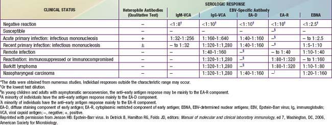
Endemic (African) Burkitt lymphoma, often found in the jaw, is the most common childhood cancer in equatorial East Africa and New Guinea (Chapter 490.2). The median age at onset is 5 yr. These regions are holoendemic for Plasmodium falciparum malaria and have a high rate of EBV infection early in life. The constant malarial exposure acts as a B-lymphocyte mitogen that contributes to the polyclonal B-lymphocyte proliferation with EBV infection, impairs T-lymphocyte surveillance of EBV-infected B lymphocytes, and increases the risk for development of Burkitt lymphoma. Approximately 98% of cases of endemic Burkitt lymphoma contain the EBV genome, compared with only 20% of cases of nonendemic (sporadic or American) Burkitt lymphoma. Individuals with Burkitt lymphoma have unusually and characteristically high levels of antibody to VCA and EA that correlate with the risk for developing tumor (see Table 246-1).

All cases of Burkitt lymphoma, including those that are EBV negative, are monoclonal and demonstrate chromosomal translocation of the c-myc proto-oncogene to the constant region of the immunoglobulin heavy-chain locus, t(8;14), to the κ constant light-chain locus, t(2;8), or to the λ constant light-chain locus, t(8;22). This translocation results in the deregulation and constitutive transcription of the c-myc gene with overproduction of a normal c-myc product that autosuppresses c-myc production on the untranslocated chromosome.

The incidence of Hodgkin disease peaks in childhood in developing countries and in young adulthood in developed countries. Levels of EBV antibodies are consistently elevated preceding development of Hodgkin disease; only a small minority of patients with the disease is seronegative for EBV. Infection with EBV appears to increase the risk for Hodgkin disease by a factor of 2-4. EBV is associated with more than 50% of cases of mixed cellularity Hodgkin disease and approximately 25% of cases of the nodular sclerosing subtype and is rarely associated with lymphocyte-predominant Hodgkin disease. Immunohistochemical studies have localized EBV to the Reed-Sternberg cells and their variants, the pathognomonic malignant cells of Hodgkin disease.

Failure to control EBV infection may result from host immunologic deficits. The prototype is the X-linked lymphoproliferative syndrome (Duncan syndrome), an X chromosome–linked recessive disorder of the immune system associated with severe, persistent, and sometimes fatal EBV infection (Chapter 118). Approximately two thirds of affected patients, who are male, die of disseminated and fulminating lymphoproliferation involving multiple organs at the time of primary EBV infection. Surviving patients acquire hypogammaglobulinemia, B-cell lymphoma, or both; most of these patients die within 10 yr.

Numerous congenital and acquired immunodeficiency syndromes are associated with an increased incidence of EBV-associated B-lymphocyte lymphoma, especially central nervous system lymphoma, and leiomyosarcoma. The incidence of lymphoproliferative syndromes parallels the degree of immunosuppression. A decline in T-cell function evidently permits EBV to escape from immune surveillance. Congenital immunodeficiencies predisposing to EBV-associated lymphoproliferation include the X-linked lymphoproliferative syndrome, common-variable immunodeficiency, ataxia-telangiectasia, Wiskott-Aldrich syndrome, and Chédiak-Higashi syndrome. Individuals with acquired immunodeficiencies resulting from anticancer chemotherapy, immunosuppression after solid organ or bone marrow transplantation, or HIV infection have a significantly increased risk for EBV-associated lymphoproliferation. The lymphomas may be focal or diffuse, and they are usually histologically polyclonal but may become monoclonal. Their growth is not reversed upon cessation of immunosuppression.

EBV is found intracellularly in all of the smooth muscle cells of leiomyosarcomas occurring in immunocompromised persons, including HIV-infected patients and transplant recipients, but not in leiomyosarcomas occurring in immunocompetent persons.

EBV is also associated with carcinoma of the salivary glands. Other tumors putatively associated with EBV include some T-lymphocyte lymphomas (including lethal midline), angioimmunoblastic lymphadenopathy-like lymphoma, thymomas and thymic carcinomas derived from thymic epithelial cells, supraglottic laryngeal carcinomas, lymphoepithelial tumors of the respiratory tract and gastrointestinal tract, and gastric adenocarcinoma. The precise contribution of EBV to these various malignancies is not well defined.

Clinical Manifestations

The incubation period of infectious mononucleosis in adolescents is 30-50 days. In children, it may be shorter. The majority of cases of primary EBV infection in infants and young children are clinically silent. In older patients, the onset of illness is usually insidious and vague. Patients may complain of malaise, fatigue, acute or prolonged (>1 wk) fever, headache, sore throat, nausea, abdominal pain, and myalgia. This prodromal period may last 1-2 wk. The sore throat and fever gradually worsen until patients seek medical care. Splenic enlargement may be rapid enough to cause left upper quadrant abdominal discomfort and tenderness, which may be the presenting complaint.

The classic physical examination findings are generalized lymphadenopathy (90% of cases), splenomegaly (50% of cases), and hepatomegaly (10% of cases). Lymphadenopathy occurs most commonly in the anterior and posterior cervical nodes and the submandibular lymph nodes and less commonly in the axillary and inguinal lymph nodes. Epitrochlear lymphadenopathy is particularly suggestive of infectious mononucleosis. Symptomatic hepatitis or jaundice is uncommon, but elevated liver enzyme values are common. Splenomegaly to 2-3 cm below the costal margin is typical; massive enlargement is uncommon.

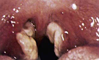
The sore throat is often accompanied by moderate to severe pharyngitis with marked tonsillar enlargement, occasionally with exudates (Fig. 246-1). Petechiae at the junction of the hard and soft palate are frequently seen. The pharyngitis resembles that caused by streptococcal infection. Other clinical findings may include rashes and edema of the eyelids.

Rashes are usually maculopapular and have been reported in 3-15% of patients. Up to 80% of patients with infectious mononucleosis experience “ampicillin rash” if treated with ampicillin or amoxicillin. This vasculitic rash is probably immune mediated and resolves without specific treatment. EBV is also associated with Gianotti-Crosti syndrome, a symmetric rash on the cheeks with multiple erythematous papules, which may coalesce into plaques, and persists for 15-50 days. The rash has the appearance of atopic dermatitis and may appear on the extremities and buttocks.

Diagnosis

The diagnosis of infectious mononucleosis implies primary EBV infection. A presumptive diagnosis may be made from the presence of typical clinical symptoms with atypical lymphocytosis in the peripheral blood. The diagnosis is usually confirmed by serologic testing, for either heterophile antibody or specific EBV antibodies.

Culture of EBV is tedious and requires 4-6 wk. The culture method is the transformation assay, which is performed by co-cultivation of oropharyngeal or genital secretions, peripheral blood (10-30 mL), or tumor with human umbilical cord lymphocytes. The cultures are observed for 6 wk for signs of cell transformation: proliferation and rapid growth, mitotic figures, large vacuoles, granular morphology, and cell aggregation. EBV immortalizes the umbilical cord cells, resulting in cell lines that harbor the EBV strain isolated from the patient and that can be maintained in vitro in perpetuity.

Laboratory Tests

In >90% of cases of EBV infection there is leukocytosis at 10,000-20,000 cells/mm3, of which at least two thirds are lymphocytes; atypical lymphocytes usually account for 20-40% of the total number. The atypical cells are mature T lymphocytes that have been antigenically activated. When compared with regular lymphocytes microscopically, atypical lymphocytes are larger overall, with larger, eccentrically placed indented and folded nuclei with a lower nucleus-to-cytoplasm ratio. Although atypical lymphocytosis may be seen with many of the infections usually causing lymphocytosis, the highest degree of atypical lymphocytes is classically seen with EBV infection. Other syndromes associated with atypical lymphocytosis include acquired cytomegalovirus infection (in contrast to congenital cytomegalovirus infection), toxoplasmosis, viral hepatitis, rubella, roseola, mumps, tuberculosis, typhoid, Mycoplasma infection, and malaria, as well as some drug reactions. Mild thrombocytopenia to 50,000-200,000 platelets/mm3 occurs in >50% of patients but only rarely is associated with purpura. Mild elevation of hepatic transaminase values occurs in approximately 50% of uncomplicated cases but is usually asymptomatic and not associated with jaundice.

Heterophile Antibody Test

Heterophile antibodies agglutinate cells from species different from those in the source serum. The transient heterophile antibodies seen in infectious mononucleosis, also known as Paul-Bunnell antibodies, are IgM antibodies detected by the Paul-Bunnell-Davidsohn test for sheep red blood cell (RBC) agglutination. The heterophile antibodies of infectious mononucleosis agglutinate sheep or, for greater sensitivity, horse RBCs but not guinea pig kidney cells. This adsorption property differentiates this response from the heterophile response found in patients with serum sickness, rheumatic diseases, and some normal individuals. Titers >1 : 28 or >1 : 40, depending on the dilution system used, after absorption with guinea pig kidney cells are considered positive.

Results of the sheep RBC agglutination test are often positive for several months after infectious mononucleosis; those of the horse RBC agglutination test may be positive for as long as 2 yr. The most widely used method is the qualitative rapid slide test using horse erythrocytes. It detects heterophile antibody in 90% of cases of EBV-associated infectious mononucleosis in older children and adults but in only up to 50% of cases in children <4 yr of age because they typically have a lower titer. From 5% to 10% of cases of infectious mononucleosis are not caused by EBV and are not uniformly associated with a heterophile antibody response. The false-positive rate is <10%, usually resulting from erroneous interpretation. If the heterophile test result is negative and an EBV infection is suspected, EBV-specific antibody testing is indicated.

Specific Epstein-Barr Virus Antibodies

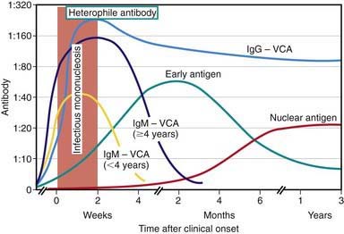
EBV-specific antibody testing is useful to confirm acute EBV infection, especially in heterophile-negative cases, or to confirm past infection and determine susceptibility to future infection. Several distinct EBV antigen systems have been characterized for diagnostic purposes (Fig. 246-2 and Table 246-1). The EBNA, EA, and VCA systems are most useful for diagnostic purposes. The acute phase of infectious mononucleosis is characterized by rapid IgM and IgG antibody responses to VCA in all cases and an IgG response to EA in most cases. The IgM response to VCA is transient but can be detected for at least 4 wk and occasionally for up to 3 mo. The laboratory must take steps to remove rheumatoid factor from the specimen, which may otherwise cause a false-positive IgM VCA result. The IgG response to VCA usually peaks late in the acute phase, declines slightly over the next several weeks to months, and then persists at a relatively stable level for life.

Anti-EA antibodies are usually detectable for several months but may persist or may be detected intermittently at low levels for many years. Antibodies to the diffuse-staining component of EA, EA-D, are found transiently in 80% of patients during the acute phase of infectious mononucleosis and reach high titers in patients with nasopharyngeal carcinoma. Antibodies to the cytoplasmic-restricted component of EA, EA-R, emerge transiently in the convalescence from infectious mononucleosis and often attain high titers in patients with EBV-associated Burkitt lymphoma, which in the terminal stage of the disease may be exceeded by antibodies to EA-D. High levels of antibodies to EA-D or EA-R may be found also in immunocompromised patients with persistent EBV infections and active EBV replication. Anti-EBNA antibodies are the last to develop in infectious mononucleosis, gradually appearing 3-4 mo after the onset of illness and remaining at low levels for life. Absence of anti-EBNA when other antibodies are present implies recent infection, whereas the presence of anti-EBNA implies infection occurring more than 3-4 mo previously. The wide range of individual antibody responses and the various laboratory methods used can occasionally make interpretation of an antibody profile difficult. The detection of IgM antibody to VCA is the most valuable and specific serologic test for the diagnosis of acute EBV infection and is generally sufficient to confirm the diagnosis.

Treatment

There is no specific treatment for infectious mononucleosis. Therapy with high doses of acyclovir, with or without corticosteroids, decreases viral replication and oropharyngeal shedding during the period of administration but does not reduce the severity or duration of symptoms or alter the eventual outcome. Rest and symptomatic treatments are the mainstays of management. Bed rest is necessary only when the patient has debilitating fatigue. As soon as there is definite symptomatic improvement, the patient should be allowed to begin to resume normal activities. Because blunt abdominal trauma may predispose patients to splenic rupture, it is customary and prudent to advise against participation in contact sports and strenuous athletic activities during the first 2-3 wk of illness or while splenomegaly is present.

Short courses of corticosteroids (<2 wk) may be helpful for complications of infectious mononucleosis, but this use has not been evaluated critically. Some appropriate indications include incipient airway obstruction, thrombocytopenia with hemorrhaging, autoimmune hemolytic anemia, seizures, and meningitis. A recommended regimen is prednisone 1 mg/kg/day (maximum: 60 mg/day) or equivalent for 7 days followed by a dosage taper over another 7 days. There are no controlled data showing efficacy of corticosteroids in any of these conditions. In view of the potential and unknown hazards of immunosuppression for a virus infection with oncogenic complications, corticosteroids should not be used in uncomplicated cases of infectious mononucleosis.

Complications

Very few patients with infectious mononucleosis experience complications. The most feared complication is subcapsular splenic hemorrhage or splenic rupture, which occurs most frequently during the second week of the disease at a rate of <0.5% of cases in adults; the rate in children is unknown but is probably much lower. Rupture is commonly related to trauma, which often may be mild and is rarely fatal. Swelling of the tonsils and oropharyngeal lymphoid tissue may be substantial and may cause airway obstruction that manifests as drooling, stridor, and interference with breathing. Airway compromise with progressive symptoms occurs in <5% of cases and is a common indication for hospitalization with infectious mononucleosis. It may be managed by elevation of the head of the bed and administration of intravenous hydration, humidified air, and systemic corticosteroids. Respiratory distress with incipient or actual airway occlusion should be managed by tonsilloadenoidectomy followed by endotracheal intubation for 12-24 hr in an intensive care setting.

Many uncommon and unusual neurologic conditions have been reported to be associated with EBV infectious mononucleosis. Headache is present in about half of cases, with severe neurologic manifestations, such as seizures and ataxia, in 1-5% of cases. Perceptual distortions of sizes, shapes, and spatial relationships, known as the Alice in Wonderland syndrome (metamorphopsia), may be a presenting symptom. There may be meningitis with nuchal rigidity and mononuclear cells in the cerebrospinal fluid, facial nerve palsy, transverse myelitis, and encephalitis. Most patients with confirmed EBV encephalitis, however, do not demonstrate the typical symptoms of infectious mononucleosis.

Guillain-Barré syndrome or Reye syndrome may follow acute illness. Hemolytic anemia, often with a positive Coombs test result and with cold agglutinins specific for RBC i antigen, occurs in 3% of cases. The onset is typically in the first 2 wk of illness and lasts <1 mo. Aplastic anemia is a rare complication that usually manifests 3-4 wk after the onset of illness, usually with recovery in 4-8 days, but some cases do require bone marrow transplantation. Mild thrombocytopenia and neutropenia are common, but severe thrombocytopenia (<20,000 platelets/µL) or severe neutropenia (<1,000 neutrophils/µL) is rare. Myocarditis or interstitial pneumonia may occur, and both resolve in 3-4 wk. Other rare complications are pancreatitis, parotitis, and orchitis.

Bibliography

Alpert G, Fleisher GR. Complications of infection with Epstein-Barr virus during childhood: a study of children admitted to the hospital. Pediatr Infect Dis. 1984;3:304-307.

Balfour HHJr, Holman CJ, Hokanson KM, et al. A prospective clinical study of Epstein-Barr virus and host interactions during acute infectious mononucleosis. J Infect Dis. 2005;192:1505-1512.

Crawford DH, Macsween KF, Higgins CD, et al. A cohort study among university students: identification of risk factors for Epstein-Barr virus seroconversion and infectious mononucleosis. Clin Infect Dis. 2006;43:276-282.

Crawford DH, Swedlow AJ, Higgins C, et al. Sexual history and Epstein-Barr virus infection. J Infect Dis. 2002;186:731-736.

Doja A, Bitnun A, Jones EL, et al. Pediatric Epstein-Barr virus-associated encephalitis: 10-year review. J Child Neurol. 2006;21:384-391.

Fafi-Kremer S, Morand P, Brion JP, et al. Long-term shedding of infectious Epstein-Barr virus after infectious mononucleosis. J Infect Dis. 2005;191:985-989.

Higgins CD, Swerdlow AJ, Macsween KF, et al. A study of risk factors for acquisition of Epstein-Barr virus and its subtypes. J Infect Dis. 2007;195:474-482.

Jenson H. Acute complications of Epstein-Barr virus infectious mononucleosis. Curr Opin Pediatr. 2000;12:263-268.

Jenson H, McIntosh K, Pitt J, et al. Natural history of primary Epstein-Barr virus infection in children of mothers infected with human immunodeficiency virus type 1. J Infect Dis. 1999;179:1395-1404.

Jenson HB, Leach CT, McClain KL, et al. Benign and malignant smooth muscle tumors containing Epstein-Barr virus in children with AIDS. Leuk Lymphoma. 1997;27:303-314.

Luzuriaga K, Sullivan JL. Infectious mononucleosis. N Engl J Med. 2010;362:1993-2000.

Njie R, Bell AI, Jia H, et al. The effects of acute malaria on Epstein-Barr virus (EBV) load and EBV-specific T cell immunity in Gambian children. J Infect Dis. 2009;199:31-38.

Sitki-Green D, Covington M, Raab-Traub N. Compartmentalization and transmission of multiple Epstein-Barr virus strains in asymptomatic carriers. J Virol. 2003;77:1840-1847.

Sitki-Green DL, Edwards RH, Covington MM, et al. Biology of Epstein-Barr virus during infectious mononucleosis. J Infect Dis. 2004;189:483-492.

Sokal EM, Hoppenbrouwers K, Vandermeulen C, et al. Recombinant gp350 vaccine for infectious mononucleosis: a phase 2, randomized, double-blind, placebo-controlled trial to evaluate the safety, immunogenicity, and efficacy of an Epstein-Barr virus vaccine in healthy young adults. J Infect Dis. 2007;196:1749-1753.

Straus SE, Tosato G, Armstrong G, et al. Persisting illness and fatigue in adults with evidence of Epstein-Barr virus infection. Ann Intern Med. 1985;102:7-16.

Sumaya CV, Ench Y. Epstein-Barr virus infectious mononucleosis in children I: clinical and general laboratory findings. Pediatrics. 1985;75:1003-1010.

Sumaya CV, Ench Y. Epstein-Barr virus infectious mononucleosis in children II: heterophile antibody and viral-specific responses. Pediatrics. 1985;75:1011-1019.

Thompson SK, Doerr TD, Hengerer AS. Infectious mononucleosis and corticosteroids. Arch Otolaryngol Head Neck Surg. 2005;131:900-904.

Tierney RJ, Edwards RH, Sitki-Green D, et al. Multiple Epstein-Barr virus strains in patients with infectious mononucleosis: comparison of ex vivo samples with in vitro isolates by use of heteroduplex tracking assays. J Infect Dis. 2006;193:287-297.

Torre D, Tambini R. Acyclovir for treatment of infectious mononucleosis: a meta-analysis. Scand J Infect Dis. 1999;31:543-547.

Tselis AC, Jenson HB, editors. Epstein-Barr virus. New York: Taylor & Francis, 2006.

Williams H, Macsween K, McAulay K, et al. Analysis of immune activation and clinical events in acute infectious mononucleosis. J Infect Dis. 2004;190:63-71.

Yoshida M, Tsuda N, Morihata T, et al. Five patients with localized facial eruptions associated with Gianotti-Crosti syndrome caused by primary Epstein-Barr virus infection. J Pediatr. 2004;145:843-844.