[level-membership-for-pediatrics-category]
Chapter 316 Eosinophilic Esophagitis and Non-GERD Esophagitis
Eosinophilic Esophagitis
The esophageal epithelium in eosinophilic esophagitis (EoE) is infiltrated by eosinophils, typically in a density exceeding 15 per high-power field (hpf). Presenting symptoms include vomiting, feeding problems, chest or epigastric pain, and dysphagia with occasional food impactions or strictures. Most patients are male. The mean age at diagnosis is 7 yr (range 1-17 yr), and the duration of symptoms is 3 yr. Most patients have other atopic diseases and associated food allergies; laboratory abnormalities can include peripheral eosinophilia and elevated immunoglobulin E (IgE) levels. Endoscopically, the esophagus presents a granular, furrowed, ringed, or exudative appearance (Fig. 316-1); esophageal histology reveals eosinophilia, with cut-points for diagnosis variably chosen at 15-20/hpf. Up to 30% children with EoE have grossly normal esophageal mucosa. EoE is differentiated from gastroesophageal reflux disease (GERD) by its general lack of erosive esophagitis, by its greater eosinophil density, and by its refractoriness to antireflux therapies. Gastroesophageal reflux disease may be an important coexisting diagnosis. Evaluation of EoE should include a thorough search for food and environmental allergies via skin prick (IgE mediated) and patch (non–IgE mediated) tests.
Abid S, Mumtaz K, Jafri W, et al. Pill induced esophagitis: endoscopic features and clinical outcomes. Endoscopy. 2005;37:740-744.
Atkins D, Kramer R, Capocelli K, et al. Eosinophilic esophagitis; the newest esophageal inflammatory disease. Nat Rev Gastroenterol Hepatol. 2009;6:267-278.
Canalejo E, Duran FG, Cabello N, et al. Herpes esophagitis in healthy adults and adolescents. Medicine. 2010;89(4):204-210.
Franciosi JP, Liacouras CA. Eosinophilic esophagitis. Immunol Allergy Clin N Am. 2009;29:19-27.
Kliemann DA, Pasqualotto AC, Falavigna M, et al. Candida esophagitis: species distribution and risk factors for infection. Rev Inst Med Trop Sao Paulo. 2008;50:261-263.
Lee B, Caddy G. A rare cause of dysphagia: herpes simplex esophagitis. World J Gastroenterol. 2007;13:2756-2757.
Lee JJ, Baker RD, Khan AR, Baker SS. Childhood esophagitis: then and now. J Pediatr Gastroenterol Nutr. 2009;48:37-40.
Pentiuk S, Putnam PE, Collinc MH, Rothenberg ME. Dissociation between symptoms and histological severity in pediatric eosinophilic esophagitis. J Pediatr Gastroenterol Nutr. 2009;48:152-160.
Spergel JM, Brown-Whitehorn TF, Beausoleil JL, et al. 14 years of eosinophilic esophagitis: clinical features and prognosis. J Pediatr Gastroenterol Nutr. 2009;48:30-36.
[/level-membership-for-pediatrics-category][not-level-membership-for-pediatrics-category]
Chapter 316 Eosinophilic Esophagitis and Non-GERD Esophagitis
Eosinophilic Esophagitis
The esophageal epithelium in eosinophilic esophagitis (EoE) is infiltrated by eosinophils, typically in a density exceeding 15 per high-power field (hpf). Presenting symptoms include vomiting, feeding problems, chest or epigastric pain, and dysphagia with occasional food impactions or strictures. Most patients are male. The mean age at diagnosis is 7 yr (range 1-17 yr), and the duration of symptoms is 3 yr. Most patients have other atopic diseases and associated food allergies; laboratory abnormalities can include peripheral eosinophilia and elevated immunoglobulin E (IgE) levels. Endoscopically, the esophagus presents a granular, furrowed, ringed, or exudative appearance (Fig. 316-1); esophageal histology reveals eosinophilia, with cut-points for diagnosis variably chosen at 15-20/hpf. Up to 30% children with EoE have grossly normal esophageal mucosa. EoE is differentiated from gastroesophageal reflux disease (GERD) by its general lack of erosive esophagitis, by its greater eosinophil density, and by its refractoriness to antireflux therapies. Gastroesophageal reflux disease may be an important coexisting diagnosis. Evaluation of EoE should include a thorough search for food and environmental allergies via skin prick (IgE mediated) and patch (non–IgE mediated) tests.
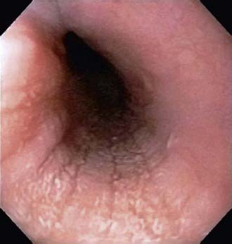
Figure 316-1 Endoscopic image of eosinophilic esophagitis with characteristic mucosal appearance of furrowing and white specks.
[/not-level-membership-for-pediatrics-category]
