Chapter 286 Enterobiasis (Enterobius vermicularis)
Diagnosis
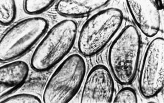
A history of nocturnal perianal pruritus in children strongly suggests enterobiasis. Definitive diagnosis is established by identification of parasite eggs or worms. Microscopic examination of adhesive cellophane tape pressed against the perianal region early in the morning frequently demonstrates eggs (Fig. 286-1). Repeated examinations increase the chance of detecting ova; a single examination detects 50% of infections, 3 examinations 90%, and 5 examinations 99%. Worms seen in the perianal region should be removed and preserved in 75% ethyl alcohol until microscopic examination can be performed. Digital rectal examination may also be used to obtain samples for a wet mount. Routine stool samples rarely demonstrate Enterobius ova.
Arca MJ, Gates RL, Groner JI, et al. Clinical manifestations of appendiceal pinworms in children: an institutional experience and a review of the literature. Pediatr Surg Int. 2004;20:372-375.
Girgikardesler N, Kurt A, Kilimciglu AA. Transmission of Dientamoeba fragilis: evaluation of the role of Enterobius vermicularis. Parasitol Int. 2008;57:72-75.
Kucik CJ, Martin GL, Sortor BV. Common intestinal parasites. Am Fam Physician. 2004;69:1161-1168.
Sizer AR, Nirmal DM, Shannon J, et al. A pelvic mass due to infestation of the fallopian tube with Enterobius vermicularis. J Obstet Gynaecol. 2004;24:462-463.
Tandan T, Pollard AJ, Money DM, et al. Pelvic inflammatory disease associated with Enterobius vermicularis. Arch Dis Child. 2002;86:439-440.


