Best modality for identifying intramural/intraluminal gallbladder (GB) gas (100% sensitive)


• Ultrasound


 in the gallbladder (GB) wall with “dirty” acoustic shadowing and “ring down” artifact
  , classic for emphysematous cholecystitis.
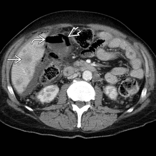
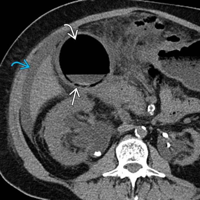
 . Gas is present within the GB lumen
  , and the GB wall appears to be perforated
  . Gangrenous perforation of the GB was seen at surgery, possibly related to the patient’s chemotherapy.
 within the wall and lumen corresponding to the echogenic foci identified on sonography, in keeping with emphysematous cholecystitis.
IMAGING
General Features


MR Findings
• Intraluminal and intramural gas appear as signal voids on all pulse sequences


Ultrasonographic Findings
• Gas in GB wall manifests as highly echogenic reflectors with dense posterior acoustic “dirty” shadowing


• Intraluminal gas bubbles should be mobile on real-time US and may rise up to nondependent portions of GB similar to bubbles of champagne (champagne sign)


DIFFERENTIAL DIAGNOSIS
Gangrenous Cholecystitis


Gas-Forming Hepatic Abscess


 and lumen
  of the GB, which has a thickened wall. Note the presence of significant surrounding fat stranding and ascites
  ,
,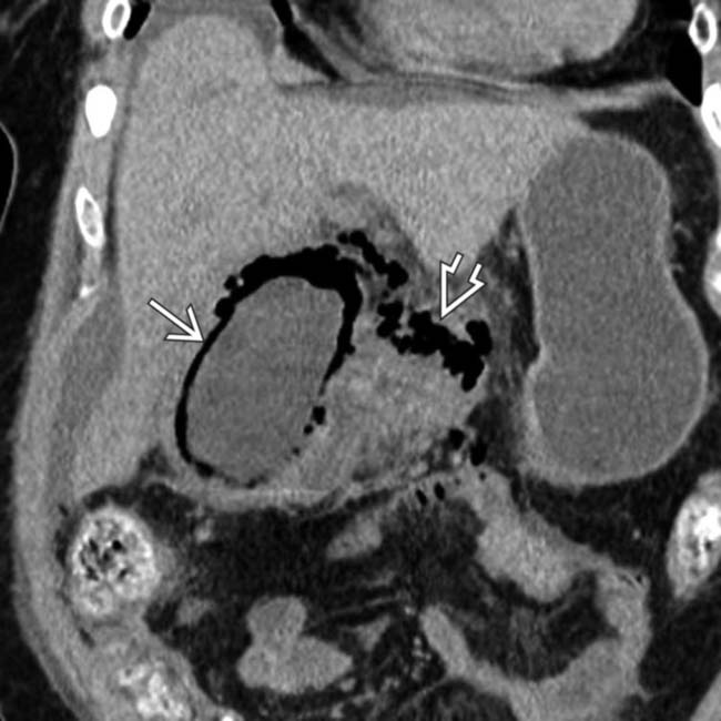
 , but also gas
  extending outside the GB, in keeping with perforation. Emphysematous cholecystitis carries a high risk of both wall gangrene and perforation.
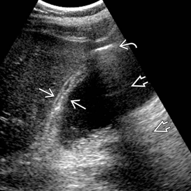
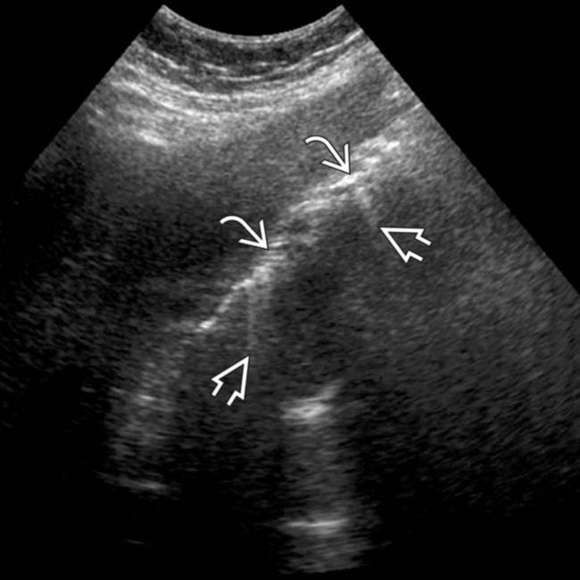
 and nondependent linear intramural echogenicity
  . Note the “dirty” posterior shadowing
  , an ultrasound artifact characteristic of gas.
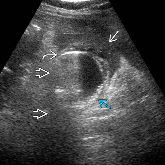
 and posterior “dirty” shadowing
  . Note the infiltrated omentum
  adjacent to the GB and fine, layering gallstones
  . Gangrenous cholecystitis was confirmed at pathology.
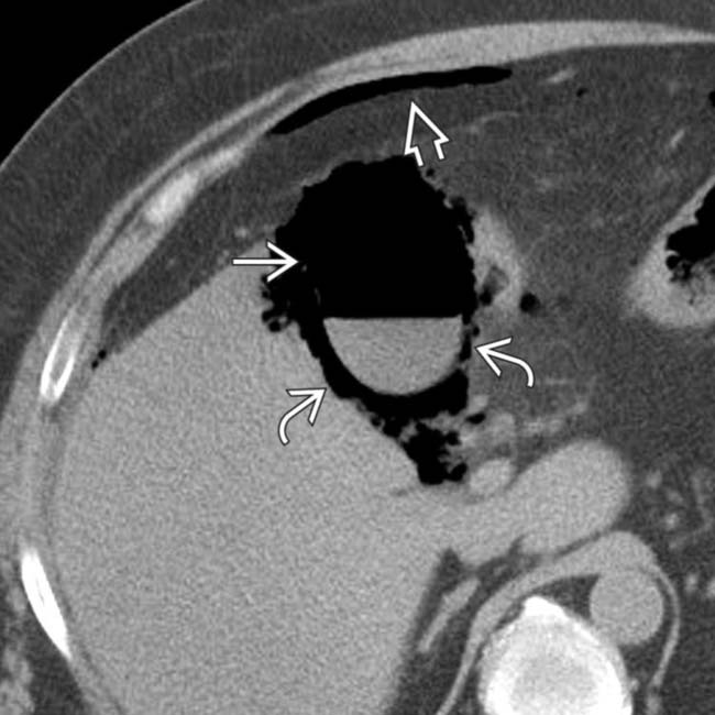
 and intramural
  GB gas. Note free intraperitoneal gas
  , a manifestation of GB perforation. Transmural GB necrosis and gram-negative rods were shown at pathology.
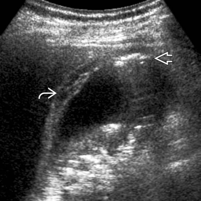
 and intramural gas
  , characteristic findings in the setting of emphysematous cholecystitis.
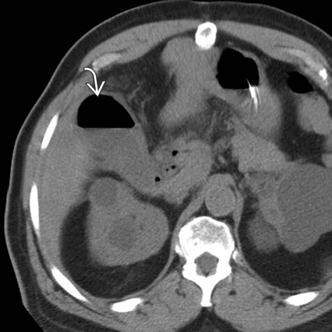
 , in keeping with emphysematous cholecystitis.
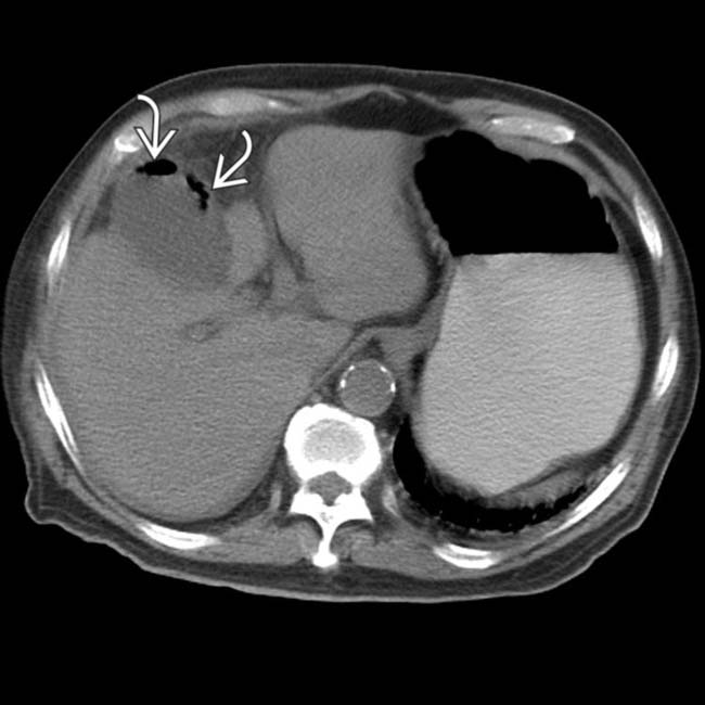
 within the wall of a distended GB, in keeping with emphysematous cholecystitis. A perforated gangrenous GB was found at surgery.
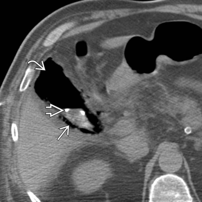
 and wall
  of the GB. At the request of the referring physicians, a percutaneous pigtail catheter
  was placed into the GB lumen with no benefit to the patient, who subsequently had an open cholecystectomy.
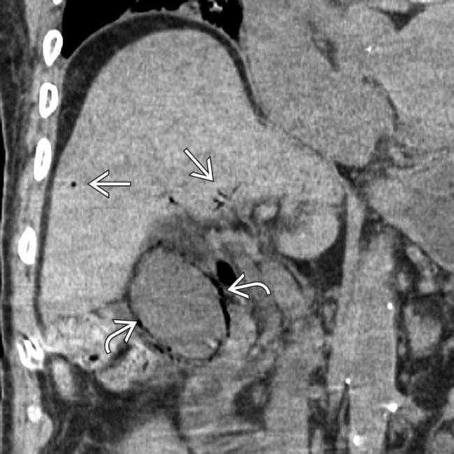
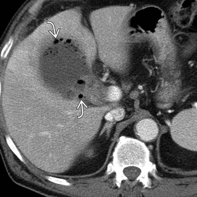
 . Note peripheral portal venous gas
  , a rare complication of emphysematous cholecystitis. The patient recovered after open cholecystectomy.
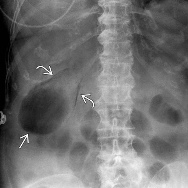
 and lumen
  of the GB.
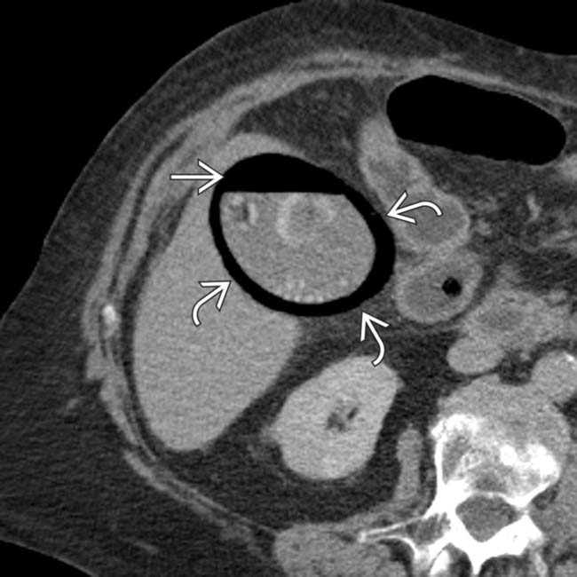
 and lumen
  of the GB.
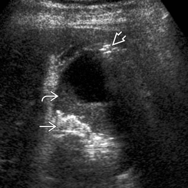
 and sludge
  in the GB, as well as intramural gas
  , which appears as “dirty” shadowing in the fundus.
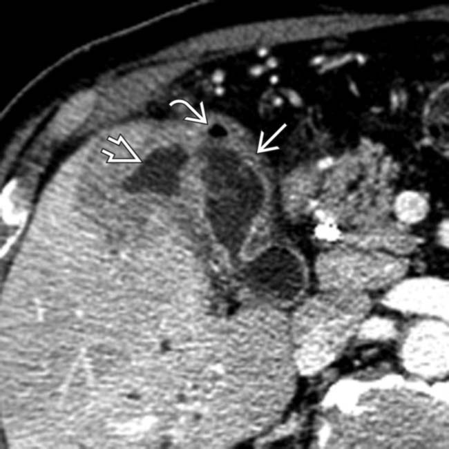
 and gas bubble
  in the GB wall. There is also an adjacent fluid collection
  , consistent with a pericholecystic abscess.
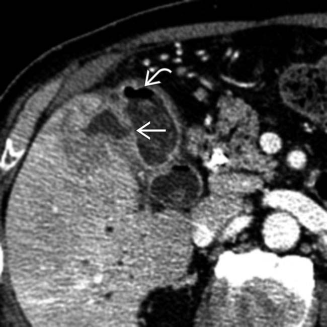
 and gas in the GB wall
  . GB perforation was confirmed at surgery.
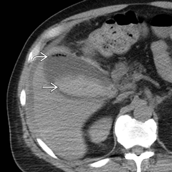
 and layering high-attenuation clot
  within the GB lumen. Hemorrhagic, gangrenous cholecystitis was confirmed at laparotomy.


