Embryology, Anatomy, and Function of the Esophagus

Published on 25/03/2015 by admin

Filed under Pediatrics

Last modified 22/04/2025

Print this page

rate 1 star rate 2 star rate 3 star rate 4 star rate 5 star
Your rating: none, Average: 0 (0 votes)

This article have been viewed 1339 times

Chapter 310 Embryology, Anatomy, and Function of the Esophagus

The esophagus is a hollow muscular tube, separated from the pharynx above and the stomach below by 2 tonically closed sphincters. Its primary function is to convey ingested material from the mouth to the stomach. Largely lacking digestive glands and enzymes, and exposed only briefly to nutrients, it has no active role in digestion.

Anatomy

The luminal aspect of the esophagus is covered by thick, protective, nonkeratinized stratified squamous epithelium, which abruptly changes to simple columnar epithelium at the stomach’s upper margin at the gastroesophageal junction (GEJ). This squamous epithelium is relatively resistant to damage by gastric secretions (in contrast to the ciliated columnar epithelium of the respiratory tract), but chronic irritation by gastric contents can result in morphometric changes (thickening of the basal cell layer and lengthening of papillary ingrowth into the epithelium) and subsequent metaplasia of the cells lining the lower esophagus from squamous to columnar. Deeper layers of the esophageal wall are composed successively of lamina propria, muscularis mucosae, submucosa, and the 2 layers of muscularis propria (circular surrounded by longitudinal). The 2 delimiting sphincters of the esophagus, the upper esophageal sphincter (UES) at the cricopharyngeus muscle and the LES at the GEJ, constrict the esophageal lumen at its proximal and distal boundaries. The muscularis propria of the upper third of the esophagus is predominantly striated, and that of the lower 2/3 is smooth muscle. Clinical conditions involving striated muscle (cricopharyngeal dysfunction, cerebral palsy) affect the upper esophagus, whereas those involving smooth muscle (achalasia, reflux esophagitis) affect the lower esophagus. The muscular LES and the mucosal “Z-line” of the GEJ may be discrepant up to several centimeters.

Function

The esophagus can be divided into 3 areas: the UES, the esophageal body, and the LES. At rest, the tonic LES pressure is normally ∼20 mm Hg; values <10 mm Hg are usually considered abnormal, although it seems that competence against retrograde flow of gastric material is maintained if the LES pressure is >5 mm Hg. The LES pressure rises during intragastric pressure amplifications, whether caused by gastric contractions, abdominal wall muscle contractions (“straining”), or external pressure applied to the abdominal wall. It also rises in response to cholinergic stimuli, gastrin, gastric alkalization, and certain drugs (bethanechol, metoclopramide, cisapride). The UES pressure is more variable and often higher than that of the LES; it decreases almost to zero during deep sleep and it increases markedly during stress and straining. The UES and LES relax briefly to allow material to pass through during swallowing, belching, reflux, and vomiting. They can contract in response to subthreshold levels of reflux (esophagoglottal closure reflex).

Swallowing is initiated by elevation of the tongue, propelling the bolus into the pharynx. The larynx elevates and moves anteriorly, pulling open the relaxing UES, while the opposed aryepiglottic folds close. The epiglottis drops back to cover the larynx and direct the bolus over the larynx and into the UES. The soft palate occludes the nasopharynx. The primary peristalsis thus initiated is a contraction originating in the oropharynx that clears the esophagus aborally (Fig. 310-1). The LES, tonically contracted as a barrier against gastroesophageal reflux (GER), relaxes as swallowing is initiated, at nearly the same time as the UES relaxation. The LES relaxation persists considerably longer, until the peristaltic wave traverses it and closes it. The normal esophageal peristaltic speed is ∼3 cm/sec; the wave takes ≥4 sec to traverse the 12 cm esophagus of a young infant and considerably longer in a larger child. Facial stimulation by a puff of air can induce swallowing and esophageal peristalsis in healthy young infants, a reflex termed the Santmyer swallow.

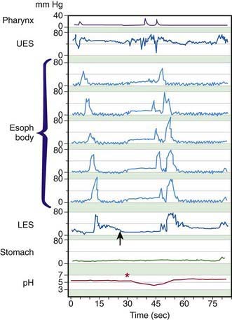
In addition to relaxing to move swallowed material past the GEJ into the stomach, the LES normally relaxes to vent swallowed air or to allow retrograde expulsion of material from the stomach. Perhaps as an extension of these functions, the normal LES also permits physiologic reflux episodes, brief events that occur approximately 5 times in the 1st postprandial hour, particularly in the awake state, but are otherwise uncommon. Transient LES relaxation, not associated with swallowing, is the major mechanism underlying pathologic reflux (see Fig. 310-1).

The close linkage of the anatomy of the upper digestive and respiratory tracts has mandated intricate functional protections of the respiratory tract during retrograde movement of gastric contents as well as during swallowing. The protective functions include the LES tone, the bolstering of the LES by the surrounding diaphragmatic crura, and the “backup protection” of the UES tone. Secondary peristalsis, akin to primary peristalsis but without an oral component, originates in the upper esophagus, triggered mainly by GER, and thereby also clears refluxed gastric contents from the esophagus. Another protective reflex is the “pharyngeal swallow” (initiated above the esophagus, but without lingual participation). Multiple levels of protection against aspiration include the rhythmic coordination of swallowing and breathing and a series of protective reflexes with esophagopharyngeal afferents and efferents that close the UES or larynx. These reflexes include the esophago-UES contractile reflex, the pharyngo-UES contractile reflex, the esophagoglottal closure reflex, and 2 pharyngoglottal adduction reflexes. The last 2 reflexes have chemoreceptors on the laryngeal surface of the epiglottis and mechanoreceptors on the aryepiglottic folds as their sites of stimulus. It is likely that interactions between the esophagus and the respiratory tract, which cause extraesophageal manifestations of gastroesophageal reflux disease (GERD), will be explained by subtle abnormalities in these protective reflexes.

310.1 Common Clinical Manifestations and Diagnostic Aids

Seema Khan and Susan R. Orenstein

Common Clinical Manifestations

Manifestations include pain, obstruction or difficulty swallowing, abnormal retrograde movement of gastric contents (reflux, regurgitation, or vomiting), or bleeding; esophageal disease can also engender respiratory symptoms. Pain in the chest unrelated to swallowing (heartburn) can be a sign of esophagitis, but similar pain might also represent cardiac, pulmonary, or musculoskeletal disease or visceral hyperalgesia. Pain during swallowing (odynophagia) localizes the disease more discretely to the pharynx and esophagus and often represents inflammatory mucosal disease. Complete esophageal obstruction can be produced acutely by esophageal foreign bodies, including food impactions; can be congenital, as in esophageal atresia; or can evolve over time as a peptic stricture occludes the esophagus. Difficulty swallowing (dysphagia) can be produced by incompletely occlusive esophageal obstruction (by extrinsic compression, intrinsic narrowing, or foreign bodies) but can also result from dysmotility of the esophagus (whether primary/idiopathic or secondary to systemic disease). Inflammatory lesions of the esophagus without obstruction or dysmotility are a 3rd cause of dysphagia; eosinophilic esophagitis, most often afflicting older boys, is relatively common.

The most common esophageal disorder in children is GERD, which is due to retrograde return of gastric contents into the esophagus. Esophagitis can be caused by GERD, by eosinophilic disease, by infection, or by caustic substances. Esophageal bleeding can result from severe esophagitis that produces erosions or ulcerations and can manifest as anemia or hemoccult-positive stools. More acute or severe bleeding can be due to ruptured esophageal varices. The resulting hematemesis must be differentiated from more distal bleeding (gastric ulcer) and from more proximal bleeding (a nosebleed or hemoptysis). Respiratory symptoms of esophageal disease can result from luminal contents incorrectly being directed into the respiratory tract or to reflexive respiratory responses to esophageal stimuli.

Diagnostic Aids

The esophagus can be evaluated by radiography, endoscopy, histology, scintigraphy, manometry, pH-metry (linked as indicated with other polysomnography), and multichannel intraluminal impedance (MII). Contrast (usually barium) radiographic study of the esophagus usually incorporates fluoroscopic imaging over time so that motility and anatomy can be assessed. Although most often requested to evaluate for GERD, it is neither sensitive nor specific for this purpose; it can detect complications of GERD (stricture or hiatal hernia) or conditions mimicking GERD (pyloric stenosis or malrotation with intermittent volvulus).

Barium fluoroscopy is optimal for evaluating for structural anomalies, such as duplications, strictures, or external esophageal compression by an aberrant blood vessel, or for causes of dysmotility, such as achalasia. Modifications of the routine barium fluoroscopic study are used in special situations. When an “H-type” tracheoesophageal fistula is suspected, the test is most sensitive if the radiologist, with the patient prone, distends the esophagus with barium via a nasogastric tube. The videofluoroscopic evaluation of swallowing performed with varying consistencies of barium (“modified barium swallow,” oropharyngeal videoesophagogram, or “cookie swallow”) optimally evaluates children with dysphagia by demonstrating incoordination of the pharyngeal and esophageal phases of swallowing and any associated aspiration.

In some centers, fiberoptic endoscopic evaluation of swallowing (FEES) uses nasopharyngeal endoscopy to visualize the pharynx and larynx during swallowing of dye-enhanced foods when dysphagia, laryngeal penetration, or aspiration are suspected. This is often combined with sensory testing (ST) of the laryngeal adductor reflex in response to a calibrated puff of air through the endoscope to the arytenoids, generating the composite FEESST that examines the mechanisms of any aspiration that is present. Endoscopy allows direct visualization of esophageal mucosa and helps therapeutically in the removal of foreign bodies and treatment of esophageal varices. Endoscopy also allows biopsy samples to be taken, thus improving the diagnosis of “endoscopy-negative” GERD, differentiating GERD from eosinophilic esophagitis, and identifying viral or fungal causes of esophagitis.

Radionuclide scintigraphy scans are helpful in evaluating the efficiency of peristalsis and demonstrating reflux episodes. They can be specific, although not very sensitive, for aspiration and can quantify gastric emptying, thus hinting at a cause for GERD. The related radionuclide salivagram can demonstrate aspiration of even minute amounts of saliva.

Esophageal manometry evaluates for dysmotility from the pharynx to the stomach; by synchronized quantitative pressure measurements along the esophagus, it detects and characterizes dysfunctions sometimes missed radiographically. Manometry is often challenging in young infants, and sphincters are optimally evaluated with special Dent sleeves, rather than the simple ports available for the esophageal body.

Extended pH monitoring of the distal esophagus is a sensitive test for acidic GER episodes that can quantify duration and degree of acidity, but not volume, of the reflux episodes. It is linked with polysomnography (a “pneumogram”) when GER is suspected to cause apnea or similar symptoms.

Multichannel intraluminal impedance is a method for pH-independent detection of bolus movements in the esophagus; with a pH probe incorporated, it can distinguish between acid and nonacid liquid and gaseous reflux, the proximal extent of reflux, and several aspects of esophageal function, such as direction of bolus flow, duration of bolus presence, and bolus clearance.