Echinococcosis (Echinococcus granulosus and Echinococcus multilocularis)

Published on 27/03/2015 by admin

Filed under Pediatrics

Last modified 22/04/2025

Print this page

rate 1 star rate 2 star rate 3 star rate 4 star rate 5 star
Your rating: none, Average: 0 (0 votes)

This article have been viewed 1604 times

Chapter 296 Echinococcosis (Echinococcus granulosus and Echinococcus multilocularis)

Etiology

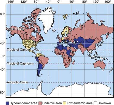
Echinococcosis (hydatid disease or hydatidosis) is the most widespread, serious human cestode infection in the world (Fig. 296-1). Two major Echinococcus species are responsible for distinct clinical presentations, E. granulosus (cystic hydatid disease) and the more malignant E. multilocularis (alveolar hydatid disease). The adult parasite is a small (2-7 mm) tapeworm with only 2-6 segments that inhabits the intestines of dogs, wolves, dingoes, jackals, coyotes, and foxes. These carnivores pass the eggs in their stool, which contaminates the soil, pasture, and water, as well as their own fur. Domestic animals such as sheep, goats, cattle, and camels ingest E. granulosus eggs while grazing. Humans are also infected by consuming food or water contaminated with eggs or by direct contact with infected dogs. The larvae hatch, penetrate the gut, and are carried by the vascular or lymphatic systems to the liver, lungs, and less commonly bones, brain, or heart.

image

Figure 296-1 Worldwide distribution of cystic echinococcosis.

(From McManus DP, Zhang W, Li J, et al: Echinococcosis, Lancet 362:1295–1304, 2003.)

E. granulosus shows high intraspecific variation. One distinct variant is found in a sylvatic wolf/moose cycle in North America and Siberia. The transmission cycle of E. multilocularis is similar to that of E. granulosus, except that this species is mainly sylvatic and uses small rodents as its natural intermediate hosts. The rodents are consumed by foxes, their natural predators, and sometimes by dogs and cats.

Pathogenesis

In areas endemic for E. granulosus, the parasite is often acquired in childhood, but liver cysts require many years to become large enough to detect or cause symptoms. In children, the lung is a common site, whereas in adults 70% of cysts develop in the right lobe of the liver. Cysts can also develop in bone, the genitourinary system, bowels, subcutaneous tissues, and brain. The host surrounds the primary cyst with a tough, fibrous capsule. Inside this capsule, the parasite produces a thick lamellar layer with the consistency of a soft-boiled egg white. This layer supports a thin germinal layer of cells responsible for production of thousands of juvenile-stage parasites (protoscolices) that remain attached to the wall or float free in the cyst fluid. Smaller internal daughter cysts may develop within the primary cyst capsule. The fluid in a healthy cyst is colorless, crystal clear, and watery. After medical treatment or with bacterial infection, it may become thick and bile stained.

Infection with E. multilocularis resembles a malignancy. The secondary reproductive units bud externally and are not confined within a single well-defined structure. Furthermore, the cyst tissues are poorly demarcated from those of the host, which makes these cysts unsuitable for surgical removal. The secondary cysts are also capable of distant metastatic spread. The growing cyst mass eventually replaces a significant portion of the liver and compromises adjacent tissues and structures.

Diagnosis

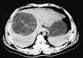
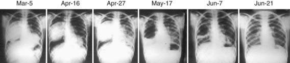
Subcutaneous nodules, hepatomegaly, or a palpable abdominal mass may be found. The parasite cannot be recovered from any easily accessible body fluid unless a lung cyst ruptures, after which protoscolices or layers of cyst wall may briefly be seen in sputum. Ultrasonography is the most valuable tool for both the diagnosis and treatment of cystic hydatid disease of the liver. The presence of internal membranes and falling echogenic cyst material (hydatid sand) observed in real time aid in the diagnosis. Alveolar disease is less cystic in appearance and resembles a diffuse solid tumor. CT findings (Fig. 296-2) are similar to those of ultrasonography and may at times be useful in distinguishing alveolar from cystic hydatid disease in geographic regions where both occur. CT or MRI is also important in planning a surgical intervention. Lung hydatid is usually apparent on chest x-ray (Fig. 296-3).

Serologic studies may be useful in confirming a diagnosis of cystic echinococcosis, but the false-negative rate may be >50%. Most patients with alveolar hydatidosis develop detectable antibody responses. Current tests use crude or partially purified antigens that can cross react in individuals infected with other parasitic infections, such as cysticercosis or schistosomiasis.

Treatment

For simple, accessible cysts surrounded by tissue, ultrasound- or CT-guided Percutaneous Aspiration, Instillation (hypertonic saline or another scolicidal agent) and Re-aspiration (PAIR) is the preferred therapy. Compared with surgical treatment alone, PAIR plus albendazole results in similar cyst disappearance with fewer adverse events and fewer days in the hospital. Compared to albendazole alone, PAIR with or without albendazole provides significantly better cyst reduction and symptomatic relief. Spillage with PAIR is surprisingly uncommon, but prophylactic albendazole therapy is routinely administered >1 wk prior to PAIR or surgery and continued for 1 mo thereafter. Albendazole treatment for 4 wk prior to any procedure is probably warranted, but may remove the diagnostic value of aspiration in some cases. PAIR is contraindicated in pregnancy and for bile-stained cysts, which should not be injected with a scolicidal agent because of increased risk for biliary complications.

Indications for surgery are the presence of large liver cysts with multiple daughter cysts, single superficially situated liver cysts that may rupture spontaneously or as the result of a trauma, infected cysts, cysts communicating with the biliary tree and/or exerting pressure on adjacent vital organs, and cysts in the lung, brain, kidney, or bones. For conventional surgery, the inner cyst wall (only laminate and germinal layers are of parasite origin) can be easily peeled from the fibrous layer, although some studies suggest that removal of the whole capsule has a better outcome. The cavity should then be topically sterilized and either closed or filled with omentum. Considerable care must be taken to avoid spillage of cyst contents, because cyst fluid contains viable protoscolices, each capable of producing secondary cysts wherever it lodges. An additional risk is anaphylaxis due to spilled cyst fluid, so it is therefore useful to employ a surgeon experienced in this surgery.

Nonpregnant patients with cysts not amenable to PAIR or surgery or with contraindications can be managed with albendazole (15 mg/kg/day divided bid PO for 1-6 mo, maximum 800 mg/day). A favorable response occurs in 40-60% of patients. Adverse effects include occasional alopecia, mild gastrointestinal disturbance, and elevated transaminases on prolonged use. Because of leukopenia, the U.S. Food and Drug Administration recommends that blood counts be monitored at the beginning and every 2 wk during therapy. Corticosteroids are not indicated unless patients show signs of immediate hypersensitivity. Ultrasonographic indications of successful therapy are reduction in diameter, a change in shape from spherical to elliptic or flat, progressive increase in echogenicity and density of cyst fluid, and detachment of membranes from the capsule (water lily sign).

Alveolar hydatidosis is frequently incurable by any modality, except by partial hepatectomy, lobectomy, or liver transplantation for limited disease. Medical therapy with albendazole may slow the progression of alveolar hydatidosis, and some patients have been maintained on long-term suppressive therapy, but the infection generally recurs if albendazole is stopped.