Dysmotility

Published on 22/03/2015 by admin

Filed under Pediatrics

Last modified 22/04/2025

Print this page

rate 1 star rate 2 star rate 3 star rate 4 star rate 5 star
Your rating: none, Average: 0 (0 votes)

This article have been viewed 1011 times

Chapter 313 Dysmotility

Upper Esophageal and Upper Esophageal Sphincter Dysmotility (Striated Muscle)

Cricopharyngeal achalasia signifies a failure of complete relaxation of the upper esophageal sphincter (UES), whereas cricopharyngeal incoordination implies full relaxation of the UES but incoordination of the relaxation with the pharyngeal contraction. These entities are usually detected on videofluoroscopic evaluation of swallowing (sometimes accompanied by visible cricopharyngeal prominence, termed a bar), but often the most precise definition of the dysfunction is obtained with manometry. A self-limited form of cricopharyngeal incoordination occurs in infancy and remits spontaneously in the 1st year of life if nutrition is maintained despite the dysphagia. In older children, idiopathic cricopharyngeal spasm is usually treated by myotomy of the UES. It is important, however, to evaluate such children thoroughly, including cranial MRI to detect Arnold Chiari malformations, which can manifest in this way but are best treated by cranial decompression, rather than esophageal surgery. Cricopharyngeal spasm may be severe enough to produce posterior pharyngeal (Zenker) diverticulum above the obstructive sphincter; this entity occurs rarely in children.

Systemic causes of swallowing dysfunction that can affect the oropharynx, UES, and upper esophagus include cerebral palsy, Arnold Chiari malformations, syringomyelia, bulbar palsy or cranial nerve defects (Möbius syndrome, transient infantile paralysis of the superior laryngeal nerve), transient pharyngeal muscle dysfunction, spinal muscular atrophy (including Werdnig-Hoffmann disease), muscular dystrophy, multiple sclerosis, infections (botulism, tetanus, poliomyelitis, diphtheria), inflammatory and autoimmune diseases (dermatomyositis, myasthenia gravis, polyneuritis, scleroderma), and familial dysautonomia. All of these can produce dysphagia. Medications (nitrazepam, benzodiazepines) and tracheostomy can adversely affect the function of the UES and thereby produce dysphagia.

Lower Esophageal and Lower Esophageal Sphincter Dysfunction (Smooth Muscle)

Causes of dysphagia due to more distal primary esophageal dysmotility include achalasia, diffuse esophageal spasm, nutcracker esophagus, and hypertensive LES; all but achalasia are rare in children. Secondary causes include Hirschsprung disease, pseudoobstruction, inflammatory myopathies, scleroderma, and diabetes.

Achalasia is a primary esophageal motor disorder of unknown etiology characterized by loss of LES relaxation and loss of esophageal peristalsis, both contributing to a functional obstruction of the distal esophagus. Degenerative, autoimmune (antibodies to Auerbach plexus), and infectious (Chagas disease due to Trypanosoma cruzi) factors are possible causes. In rare cases, achalasia is familial or part of the achalasia, alacrima, and corticotropin insensitivity, known as Allgrove syndrome. Pseudoachalsia refers to achalasia caused by various forms of cancer via obstruction of the gastroesophageal junction, infiltration of the submucosa and muscularis of the LES, or as part of the paraneoplastic syndrome with formation of anti-Hu antibodies. Pathologically, in achalasia, inflammation surrounds ganglion cells, which are decreased in number. There is selective loss of postganglionic inhibitory neurons that normally lead to sphincter relaxation, leaving postganglionic cholinergic neurons unopposed. This imbalance produces high basal LES pressures and insufficient LES relaxation. The loss of esophageal peristalsis can be a secondary phenomenon.

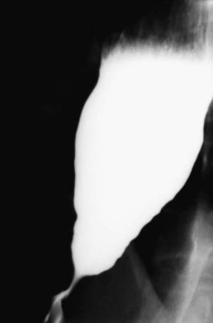
Achalasia manifests with regurgitation and dysphagia for solids and liquids and may be accompanied by undernutrition or respiratory symptoms; retained esophageal food can produce esophagitis. The mean age in children is 8.8 yr, with a mean duration of symptoms before diagnosis of 23 mo; it is uncommon before school age. Chest radiograph shows an air-fluid level in a dilated esophagus. Barium fluoroscopy reveals a smooth tapering of the lower esophagus leading to the closed LES, resembling a bird’s beak (Fig. 313-1). Loss of primary peristalsis in the distal esophagus with retained food and poor emptying are often present. Manometry is the most sensitive diagnostic test; it reveals the defining features of aperistalsis in the distal esophageal body and incomplete or absent LES relaxation, often accompanied by high pressure LES and low-amplitude esophageal body contractions.

The 2 most effective treatment options are pneumatic dilatation and surgical (Heller) myotomy. Pneumatic dilatation is the initial treatment of choice. Surgeons often supplement a myotomy with an antireflux procedure to prevent the gastroesophageal reflux disease that otherwise often ensues when the sphincter is rendered less competent. Calcium channel blockers (nifedipine) and phosphodiesterase inhibitors offer temporary relief of dysphagia. Endoscopic injection of the LES with botulinum toxin counterbalances the selective loss of inhibitory neurotransmitters by inhibiting the release of acetylcholine from nerve terminals and may be an effective therapy. Botulinum toxin is effective in 50-65% of patients and is expensive; half the patients might require a repeat injection within 1 yr. Most eventually require dilatation or surgery.

Diffuse esophageal spasm causes chest pain and dysphagia and affects adolescents and adults. It is diagnosed manometrically and can be treated with nitrates or calcium channel blocking agents.

Gastroesophageal reflux disease constitutes the most common cause of nonspecific abnormalities of esophageal motor function, probably through the effect of the esophageal inflammation on the musculature.