82% are spherical cysts: No communication with lumen
18% are tubular: Often communicate with lumen of adjacent gut
TOP DIFFERENTIAL DIAGNOSES
• Depends on location of cyst and imaging modality
Stomach or duodenum
– Pancreatic pseudocyst or cystic tumor
Ileum
– Meckel diverticulum
PATHOLOGY
• Usually lined by GI tract epithelium with smooth muscle in wall
Ectopic mucosa within cysts includes gastric, squamous, transitional, and ciliated mucosa
• Associated: Vertebral anomalies, esophageal atresia, other GI tract duplications
CLINICAL ISSUES
• Most are discovered in infancy or early childhood
Most common symptoms are related to luminal obstruction (tracheal compression, painful abdominal cramps, dysphagia, constipation)
Large cysts in neonates are easily palpable as masses through soft abdominal wall
• Symptoms: Usually low grade and chronic in adults (bleeding, pain, obstruction)
• Most cysts are discovered in early childhood
(Left) Spot film from esophagram in an elderly man with an esophageal duplication cyst shows deviation of the distal 1/3 of the esophagus , suggesting an extrinsic mass.
(Right) Axial CECT in the same patient shows a water-density mass indenting the wall of the distal esophagus . Most duplication cysts have a similar spherical or tubular morphology with near water density, nonenhancing contents.
(Left) Axial CECT in a 48-year-old man with chronic painful abdominal cramps shows a large mass in communication with the bowel, accounting for the air-fluid levels within it. The mass results in partial small bowel obstruction, accounting for the small bowel feces sign and dilation of upstream loops.
(Right) Gross pathology photograph of the same patient shows the opened cyst with a probe passing from the lumen of the ileum into the cyst, demonstrating its communication.
Axial CECT in the same patient shows a separate, more distal cystic lesion that is an esophageal duplication cyst.
Axial CECT in the same patient shows the bronchogenic cyst , with its typical subcarinal location.
Axial CECT in the same patient shows 2 separate water density masses, a larger lesion in the subcarinal region that is a proven bronchogenic cyst, which corresponds to the lesion seen on the chest film.


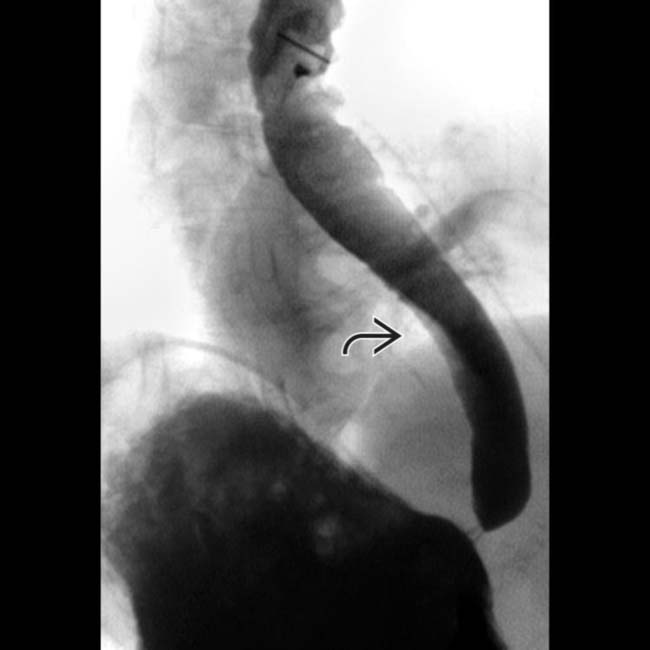
 , suggesting an extrinsic mass.
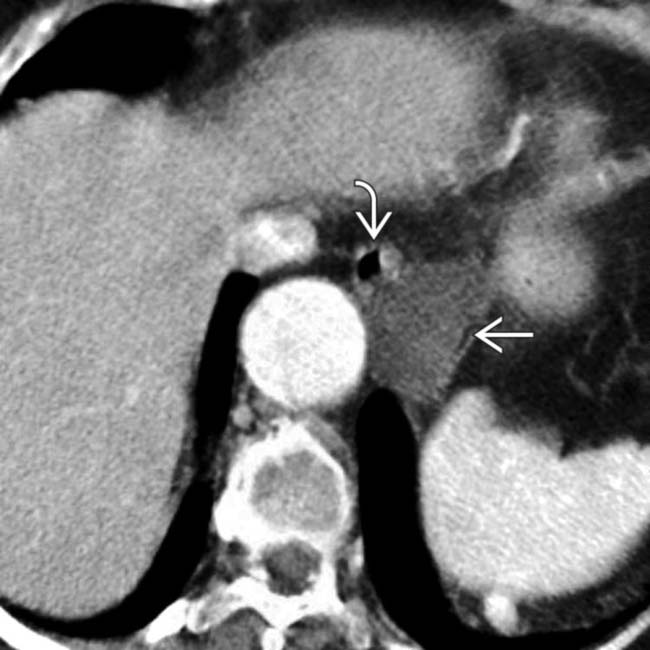
 indenting the wall of the distal esophagus
  . Most duplication cysts have a similar spherical or tubular morphology with near water density, nonenhancing contents.
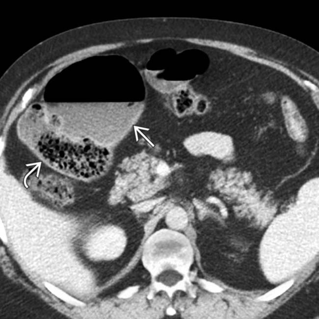
 in communication with the bowel, accounting for the air-fluid levels within it. The mass results in partial small bowel obstruction, accounting for the small bowel feces sign
  and dilation of upstream loops.
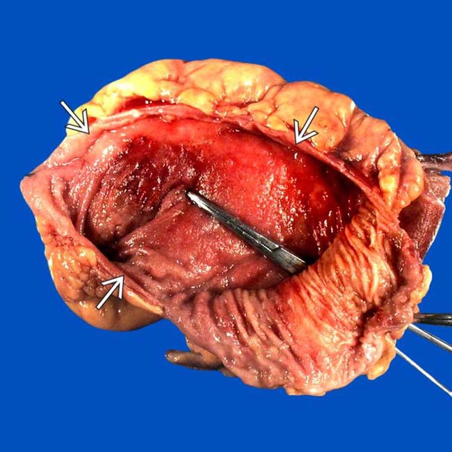
 with a probe passing from the lumen of the ileum into the cyst, demonstrating its communication.
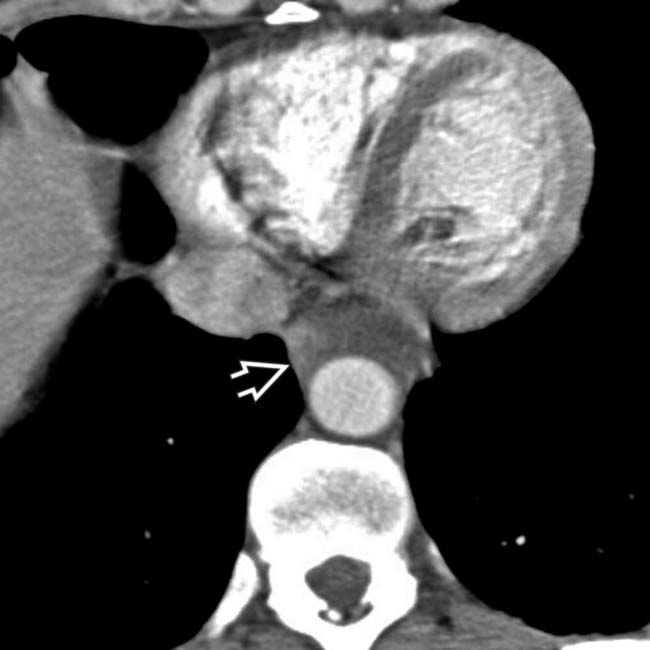
 that is an esophageal duplication cyst.
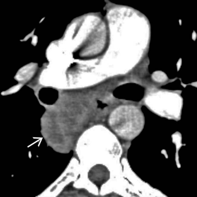
 , with its typical subcarinal location.
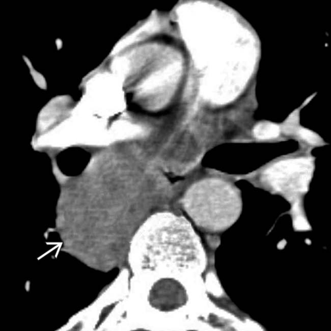
 that is a proven bronchogenic cyst, which corresponds to the lesion seen on the chest film.
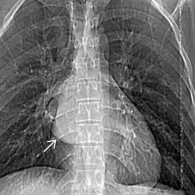
 .
.


