Metastases: Bull’s-eye or “target” lesion; submucosal or polypoid mass
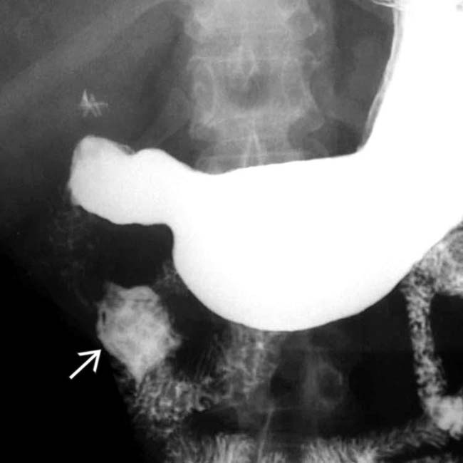
 arising from the 2nd portion of the duodenum. There is a persistent pooling of barium within the lesion after the remainder of the duodenum has cleared.
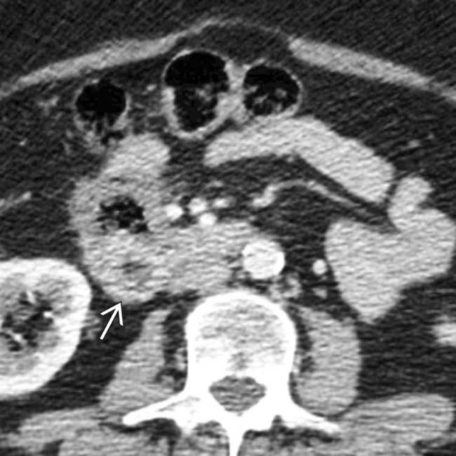
 within the wall of the 2nd duodenum. A metastatic tumor was confirmed at surgery with the same histology as the primary colon cancer.
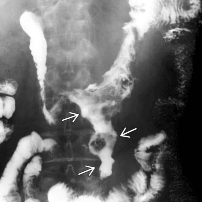
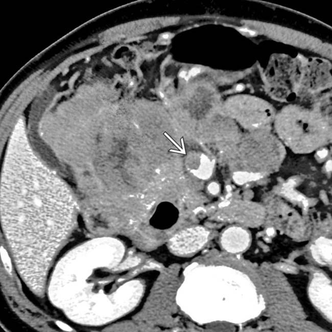
 apparently arising from, and in continuity with, the distal duodenum. There is no evidence of bowel obstruction.
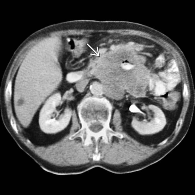
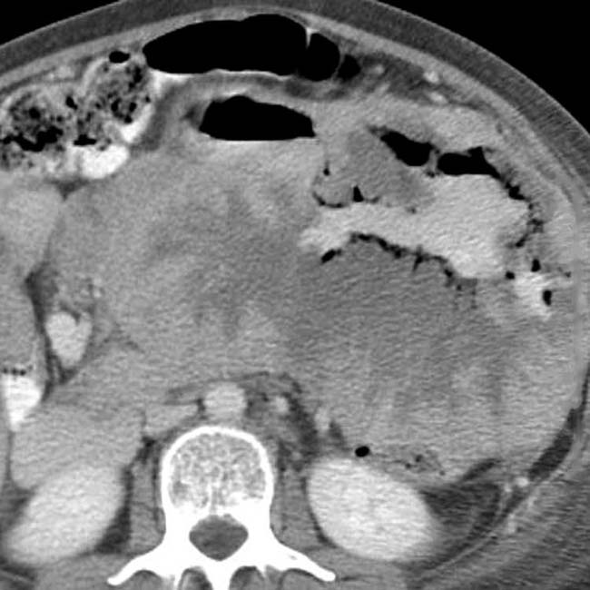
 arising from the distal duodenum. This is a good example of aneurysmal dilation of the bowel lumen caused by lymphoma.
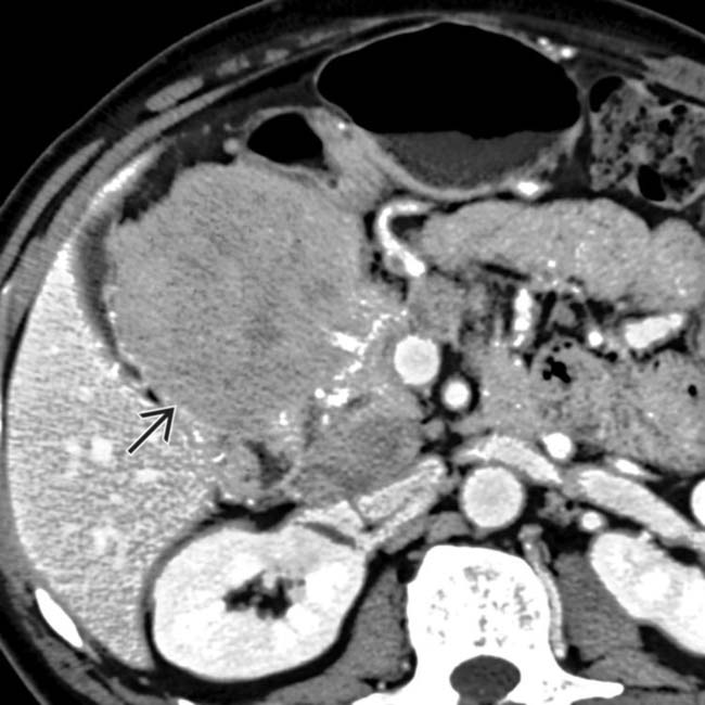
 .
.
 .
.
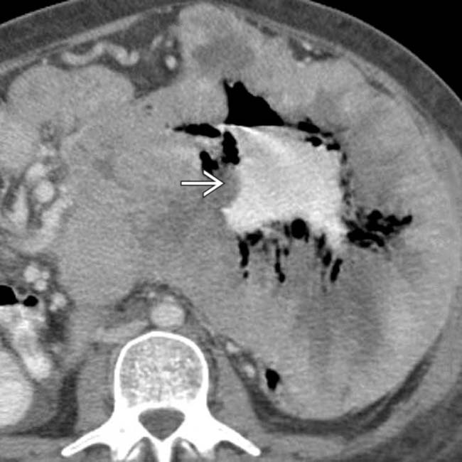
 appears to be dilated (aneurysmal dilation), but the bowel wall has been completely replaced by tumor.


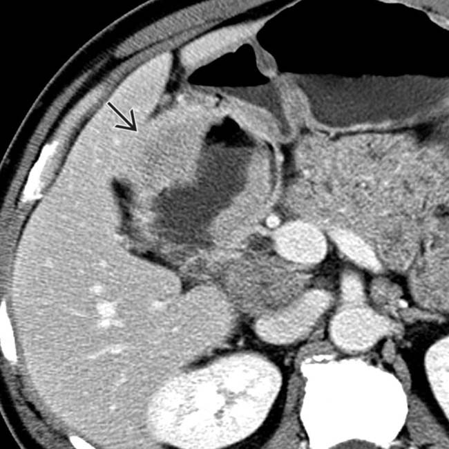
 due to lymphoma.
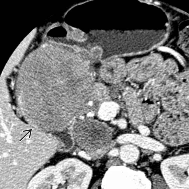
 . Biopsy revealed lymphoma.
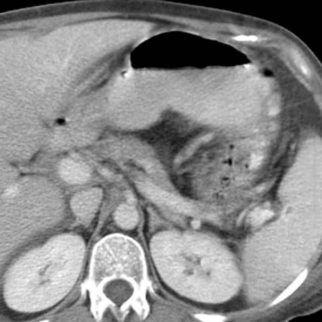
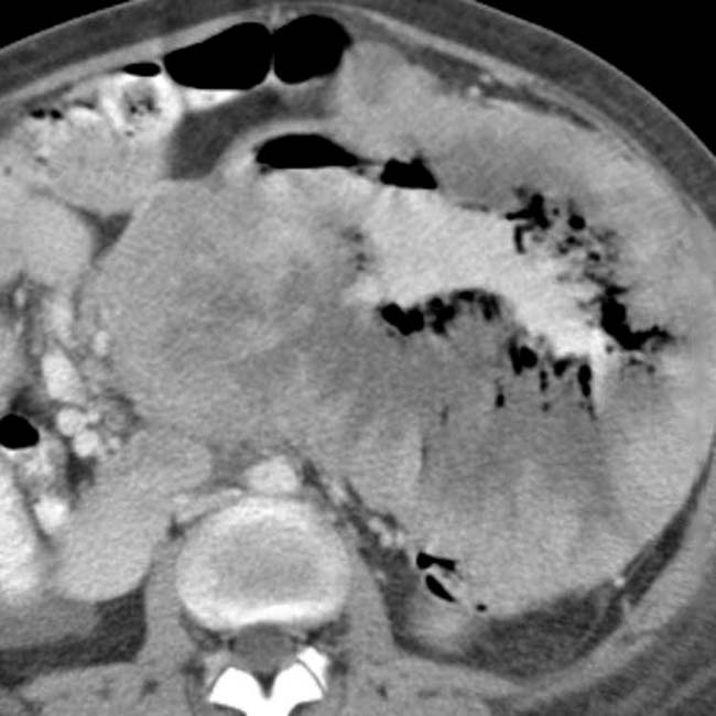
 .
.


