Acute angulation as duodenum becomes retroperitoneal, accentuates fold
• May even simulate ulcerated mass, with barium trapped between mucosal folds
• Changeable appearance on upper GI fluoroscopy
May disappear on upright or left decubitus positioning, or with compression
Normal peristalsis
• Can simulate polypoid mass on CT as well
CT has disadvantage of not allowing real-time evaluation of changes with peristalsis or different positions
Pseudolesion has same imaging characteristics (enhancement) as remainder of duodenal mucosa
• Best imaging test: Upper GI series with spot films and fluoroscopy
Characteristic location and changeable appearance are key observations
View in multiple obliquities: Upright and prone
View with and without compression
• Endoscopy can confirm diagnosis
Usually unnecessary
(Left) Axial CECT shows an apparent soft tissue density mass within the proximal duodenum.
(Right) Coronal CECT in the same patient shows the same apparent soft tissue density mass at the junction of the 1st and 2nd portions of the duodenum. This classic location should suggest the diagnosis. CT has the disadvantage of not allowing real-time evaluation.
(Left) Spot film from an upper GI series shows acute angulation of the duodenum at the junction of the apex of the bulb and the 2nd portion of the duodenum. The duodenal flexure creates folding and redundancy of the wall , accounting for the pseudotumor.
(Right) Overhead film from the upper GI series shows that the pseudolesion is redundant duodenal mucosa along the inner aspect of the flexure between the duodenal bulb and descending duodenum. Upper endoscopy confirmed normal duodenum.


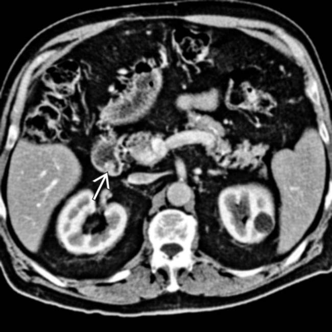
 within the proximal duodenum.
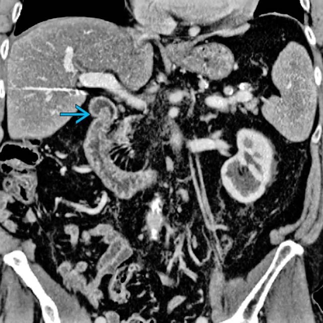
 at the junction of the 1st and 2nd portions of the duodenum. This classic location should suggest the diagnosis. CT has the disadvantage of not allowing real-time evaluation.
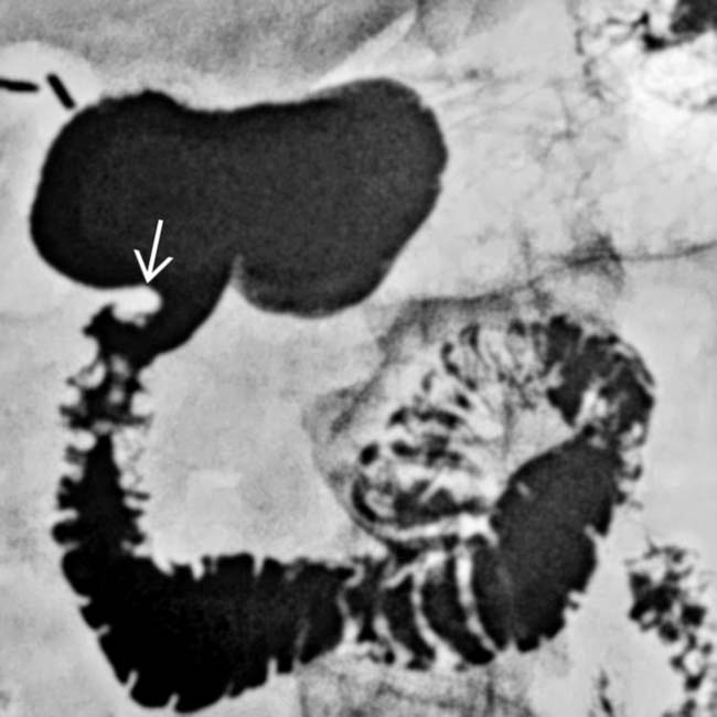
 , accounting for the pseudotumor.
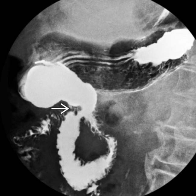
 is redundant duodenal mucosa along the inner aspect of the flexure between the duodenal bulb and descending duodenum. Upper endoscopy confirmed normal duodenum.

