Dubin-Johnson Syndrome

Published on 21/04/2017 by admin

Filed under Pathology

Last modified 22/04/2025

Print this page

rate 1 star rate 2 star rate 3 star rate 4 star rate 5 star
Your rating: none, Average: 3.3 (19 votes)

This article have been viewed 2885 times

Grossly Evident Pigment
Gross photograph of liver core biopsies embedded in the paraffin block show dark regions image 
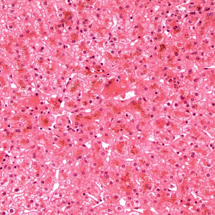
corresponding to the pigment within centrizonal hepatocytes.
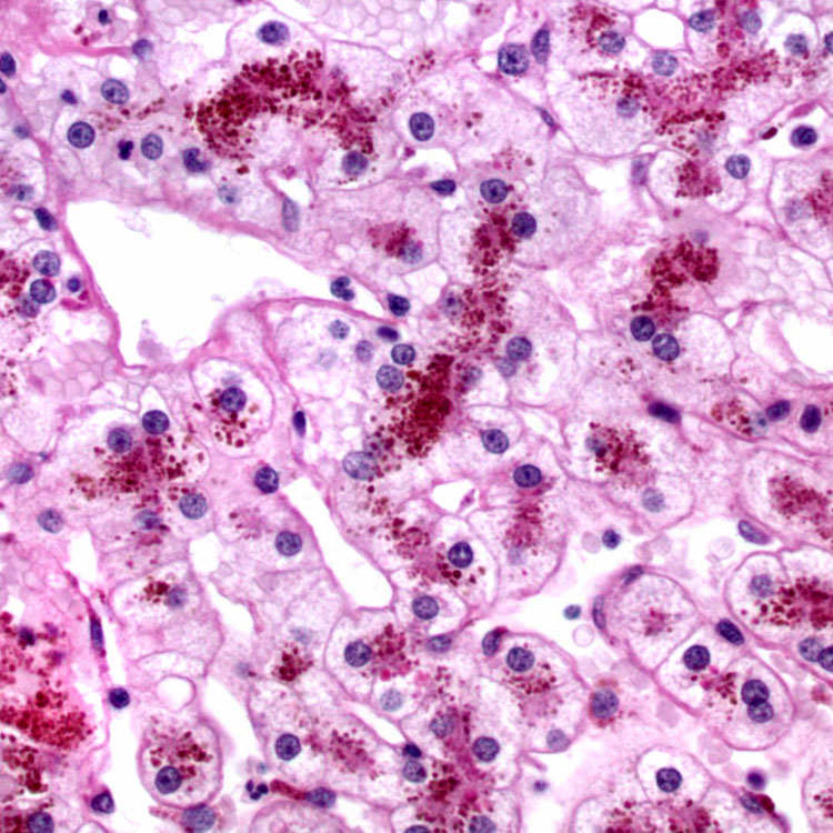
image
Cytoplasmic Pigment
H&E section shows coarse granular pigment deposition in centrizonal hepatocytes.
image
PAS Stain With Diastase Stain
PAS stain with diastase digestion accentuates the coarse pigment granules in the cytoplasm of centrizonal hepatocytes.
image
Fontana-Masson Stain
Fontana-Masson stain highlights the coarse pigment within centrizonal hepatocytes.

TERMINOLOGY

Definitions

• Defect in hepatocellular secretion of conjugated bilirubin

ETIOLOGY/PATHOGENESIS

Genetic Disorder

• Autosomal recessive

• Mutations in ABCC2 ( CMOAT / MRP2) gene, which codes for ATP-dependent organic anion transport localized to canalicular membrane
image Results in impaired biliary canalicular transport of organic anions including conjugated bilirubin
image Impaired glutathione excretion reduces bile salt-independent bile flow

CLINICAL ISSUES

Epidemiology

• Incidence

image Rare
• Age

image Develop jaundice in teenage years
• Sex

image M = F
• Ethnicity

image Prevalence highest among Moroccan and Iranian Jews (1:1,300)

Presentation

• Most patients asymptomatic
• Can present as chronic or intermittent jaundice or with mild right upper quadrant abdominal pain
• Serum bile acids are not increased, so pruritus is absent
• Urine may be darker than normal
• Some neonates present with cholestasis
• Jaundice can be precipitated by pregnancy or by drugs that decrease hepatic excretion of organic anions (e.g., oral contraceptives)

Laboratory Tests

• Measurement of urine coproporphyrin isomers shows shift from isomer III to isomer I
• Conjugated hyperbilirubinemia
• Normal alkaline phosphatase and γ-glutamyl transpeptidase

Treatment

• No treatment necessary

Prognosis

• Excellent

MACROSCOPIC

General Features

• Grossly, liver is darkly pigmented and can appear green, slate blue, dark gray, or black

MICROSCOPIC

Histologic Features

• Coarse granular pigment in centrizonal hepatocytes
• Pigment was earlier thought to be form of melanin or lipofuscin
• Likely composed of polymers of epinephrine metabolites
• No other histologic changes

Predominant Pattern/Injury Type

• Pigment accumulation

Predominant Cell/Compartment Type

• Centrizonal region

ANCILLARY TESTS

Histochemistry

• PAS with diastase digestion

image Accentuates cytoplasmic pigment
• Fontana-Masson

image Silver stain that stains cytoplasmic granules black

Immunohistochemistry

• MRP2: Absence of staining of canalicular membrane

image Available through referral centers
image Helpful in young children whose livers have not accumulated pigment

Electron Microscopy

• Membrane-bound, electron-dense lysosomal granules within cytoplasm of hepatocytes

DIFFERENTIAL DIAGNOSIS

Erythropoietic Protoporphyria

• Can also show grossly pigmented liver but has distinct clinical and histologic features

Gilbert Syndrome

• Pigment in centrizonal hepatocytes is not as coarse
• Unconjugated hyperbilirubinemia

Bilirubinostasis

• Inspissated bile in canaliculi
• Swelling (feathery degeneration) of hepatocytes in cholestatic area

Hemochromatosis

• Prussian blue (+) pigment in periportal hepatocytes

DIAGNOSTIC CHECKLIST

Pathologic Interpretation Pearls

• Coarse pigment in centrizonal hepatocytes in patient with isolated conjugated hyperbilirubinemia
• Pigment may disappear during episode of hepatitis and reaccumulate after recovery

SELECTED REFERENCES

1.Nisa, AU, et al. Dubin-Johnson syndrome. J Coll Physicians Surg Pak. 2008; 18(3):188–189.

2.Mor-Cohen, R, et al. Age estimates of ancestral mutations causing factor VII deficiency and Dubin-Johnson syndrome in Iranian and Moroccan Jews are consistent with ancient Jewish migrations. Blood Coagul Fibrinolysis. 2007; 18(2):139–144.

3.Jedlitschky, G, et al. Structure and function of the MRP2 (ABCC2) protein and its role in drug disposition. Expert Opin Drug Metab Toxicol. 2006; 2(3):351–366.

4.Lee, JH, et al. Neonatal Dubin-Johnson syndrome: long-term follow-up and MRP2 mutations study. Pediatr Res. 2006; 59(4 Pt 1):584–589.

5.Rastogi, A, et al. Dubin-Johnson syndrome–a clinicopathologic study of twenty cases. Indian J Pathol Microbiol. 2006; 49(4):500–504.

6.Sobaniec-Lotowska, ME, et al. Ultrastructure of Kupffer cells and hepatocytes in the Dubin-Johnson syndrome: a case report. World J Gastroenterol. 2006; 12(6):987–989.