Drugs Used in Renal Disease

Published on 27/02/2015 by admin

Filed under Anesthesiology

Last modified 22/04/2025

Print this page

rate 1 star rate 2 star rate 3 star rate 4 star rate 5 star
Your rating: none, Average: 0 (0 votes)

This article have been viewed 1607 times

Drugs Used in Renal Disease

DRUG CONSIDERATIONS IN PATIENTS WITH RENAL DYSFUNCTION

Influence of Renal Disease on Pharmacokinetics

Renal disease may affect drug pharmacokinetics through several mechanisms, especially effects on drug binding, distribution and elimination. Acidic drugs bind mainly to albumin. In renal failure, a decrease in serum albumin, an increase in serum urea, and the competition of endogenous substrates and drug metabolites for plasma protein binding sites lead to a decrease in the plasma protein binding of drugs. Highly protein-bound drugs have an increased unbound, active, free fraction. Under these circumstances, there may be an increase in the volume of distribution. Drugs are metabolized in the liver to water-soluble, inactive metabolites. Although uraemia has an effect on the intermediary metabolism of the liver, it does not seem to affect hepatic drug metabolism in humans.

The duration of action of most drugs administered by bolus or short-term infusion is dependent on redistribution and not elimination. It is usually not necessary to decrease the initial loading dose in patients with renal dysfunction, but subsequent maintenance doses may cause drug accumulation and should be reduced appropriately. The inactive water-soluble metabolites of drugs are eliminated by passive filtration at the glomerulus. A reduction in glomerular filtration in renal disease patients may lead to accumulation of these metabolites.

Influence of Drugs on Renal Function

All anaesthetic agents may cause a generalized depression of renal function which is transient and clinically insignificant. However, nephrotoxic drugs can impair renal function permanently. For example, they may lead to severe sodium and water depletion, reduction in renal blood supply, direct renal damage or renal obstruction (Table 10.1). Some drugs impair renal function by more than one mechanism.

TABLE 10.1

Mechanisms of Drug-Induced Renal Damage

Sodium and water depletion

Reduced renal perfusion

Direct renal toxicity

Urinary obstruction

Some of the fluorinated inhalation agents have well-recognized nephrotoxic effects, because they increase the serum inorganic fluoride concentration. Prolonged exposure of the renal tubules to fluoride ions impairs their ability to concentrate urine, leading to dehydration, hypernatraemia and increased plasma osmolarity. Experience with methoxyflurane (no longer in clinical use) has suggested that a plasma fluoride level of 50 μmol L−1 is potentially nephrotoxic. Although halothane and isoflurane do not seem to have a significant effect, prolonged administration of enflurane may lead to potentially nephrotoxic fluoride ion concentrations.

Sevoflurane undergoes approximately 5% metabolism and one of the primary metabolites is fluoride. There were initial concerns that sevoflurane may be similar to methoxyflurane and impair the ability of the kidneys to concentrate urine. However, after sevoflurane administration is stopped, there is a rapid decrease in plasma fluoride concentration because of its insolubility and rapid pulmonary elimination. Also, the metabolic production of fluoride within the kidney is much less with sevoflurane than with methoxyflurane. Though it would appear that sevoflurane renal toxicity is not a problem in clinical practice, prolonged administration of sevoflurane is not recommended in patients with significantly impaired renal function.

Aprotinin is a serine-protease inhibitor and an antifibrinolytic agent occasionally administered during major surgery to improve haemostasis. It undergoes active reabsorption by the proximal tubules and is metabolized by enzymes in the kidney. There is some controversy about its effect on renal function. Although some studies have shown a low incidence of reversible renal dysfunction, others have shown changes in biochemical markers of tubular damage without evidence of renal impairment.

Other drugs with potential for impairing renal function include aminoglycosides, NSAIDs, radiocontrast agents and various chemotherapeutic drugs. The potential for renal damage with these drugs is increased in the presence of hypovolaemia, dehydration and sepsis.

Several drugs have been investigated for protection against renal damage or dysfunction in patients at risk, for example those undergoing cardiac or aortic surgery, or ICU patients with sepsis. However, there is little convincing evidence that any specific drug effectively prevents perioperative renal dysfunction, and some of the drugs used may be harmful. These are discussed below.

VASOACTIVE DRUGS USED IN RENAL DYSFUNCTION

Dopamine

Dopamine is an endogenous catecholamine precursor of noradrenaline and adrenaline, and is a neurotransmitter in its own right. It has complicated dose-dependent pharmacodynamic effects, including positive inotropy, chronotropy, vasoconstriction, and renal and splanchnic vasodilatation. Dopamine is inactive orally and has to be administered as an intravenous infusion, because it is metabolized within minutes by the enzymes dopamine β-hydroxylase and monoamine oxidase (t1/2 < 2 min). Dopamine must be diluted before infusion.

Mechanism of Action

Dopamine acts on dopaminergic and adrenergic receptors in a dose-related fashion. Dopaminergic receptors are present in various sites in the body and have been classified into five subtypes. The two most important receptors in the peripheral cardiovascular and renal systems are DA1 and DA2.

The infusion of relatively low doses (< 2 μg kg–1 min–1) of dopamine activates postsynaptic DA1 receptors in splanchnic blood vessels and the renal tubules. Stimulation leads to vasodilatation and increases cortical renal blood flow, glomerular filtration rate (GFR), sodium excretion and urine output. There is also an increase in mesenteric blood flow. Activation of presynaptic DA2 receptors decreases intrarenal noradrenaline release, which leads to vasodilatation. It also causes inhibition of aldosterone secretion from the adrenal glands and a consequent decrease in sodium reabsorption. Theoretically, this should decrease renal oxygen consumption and improve the renal oxygen supply/demand relationship. At low infusion rates there is little change in cardiac output or heart rate. A reduction in arterial pressure may occur because of inhibition of the sympathetic nervous system by stimulation of the DA2 receptors, and by DA1-induced vasodilatation.

Increased infusion rates (2–5 μg kg–1 min–1) stimulate cardiac β1– and β2-adrenergic receptors, which causes an increase in myocardial contractility, stroke volume and cardiac output. At this infusion rate, the heart rate usually does not change.

Higher doses of dopamine (> 10 μg kg–1 min–1) lead to stimulation of the α-adrenergic receptors, causing vasoconstriction, an increase in peripheral vascular resistance and decreases in renal and splanchnic blood flow.

The dopamine infusion rates given above are guidelines and there is considerable intra- and inter-patient variation. The maximum dose at which dopamine affects only dopamine receptors is debatable. In addition, up- and downregulation of receptors occurs so the appropriate dose for a required effect may vary from hour to hour and doses must be individually titrated.

Clinical Uses

Dopamine has been used widely in ICU and surgical patients at risk of renal dysfunction, because of its effects on renal blood flow, diuretic and natriuretic effects and also because urine output is used as a surrogate marker of tissue perfusion. However, clinical studies have not demonstrated any benefit of ‘low-dose’ dopamine for the prevention and treatment of acute kidney injury in critically ill or surgical patients. In fact, it reduces regional redistribution of blood flow within the kidney by shunting blood away from the outer medulla to the cortex. This is potentially detrimental in acute kidney injury given that the outer medulla is very susceptible to ischaemic injury.

The use of higher doses of dopamine as a positive inotrope during cardiac failure, or a vasopressor during hypotension, is well established. Under these circumstances, it probably has a beneficial effect on renal function, but it is important to ensure that there is an adequate circulating blood volume.

Side-Effects

Side-effects of dopamine include tachyarrhythmias, vasoconstriction with acute hypertension, and nausea and vomiting because of a direct effect on receptors within the chemoreceptor trigger zone. Intravenous administration of dopamine does not result in central nervous system effects as dopamine does not cross the blood–brain barrier. Other potentially detrimental effects of low-dose dopamine in the critically ill patient include the following.

Dopexamine

Dopexamine is a synthetic catecholamine with structural and pharmacological similarities to dopamine; it is used for its increase in cardiac output and renal and splanchnic vasodilator effects. It is inactive orally and, because of its short half-life (~ 6 min), it is administered i.v. as an infusion. Infusion rate starts at 0.5 μg kg–1 min–1 and is titrated to a therapeutic response (up to 6 μg kg–1 min–1). Tolerance can occur and is usually associated with receptor downregulation. It is metabolized by the recognized pathways for all the catecholamines.

Dopexamine is an agonist at vascular and renal dopaminergic DA1 and DA2 receptors. It also stimulates cardiac and vascular β2-receptors, and has a limited indirect β1 effect. It therefore combines vasodilator, chronotropic and mild inotropic activity and is used in low cardiac output states where specific renal and hepatosplanchnic vasodilatation is considered beneficial. The heart rate is increased in a dose-related manner and it produces a natriuresis and diuresis. The protective effect of dopexamine on the kidneys is theoretical and an effect on outcome still has to be proven.

The most common side-effect is a tachycardia and ventricular ectopic beats when higher doses are used. Nausea and vomiting, probably caused by stimulation of DA2 receptors in the chemoreceptor trigger zone, have been reported.

Adenosine

Adenosine is a natural purine and is an important mediator in the control of renal blood flow and glomerular filtration. After i.v. administration it causes peripheral vasodilatation and decreased arterial pressure. Adenosine-induced arterial hypotension inhibits renin release by the juxtaglomerular cells and has an interesting effect on the renal vasculature, causing transient vasoconstriction of the afferent arterioles, combined with vasodilatation of the efferent arterioles. This results in decreases in renal blood flow and glomerular filtration pressure and rate. Adenosine A1 receptor antagonists increase GFR, urine production and sodium excretion without immediate effects on cardiac haemodynamics, and are being investigated for use in heart failure.

Adenosine also slows the heart rate and impairs atrioventricular conduction and is used in the treatment of supraventricular tachycardias (Ch 47).

Calcium Channel Blockers

Calcium channel blockers (CCBs) (Ch 8) are predominantly used in the treatment of hypertension. They act selectively on calcium channels in the cellular membrane of cardiac and vascular smooth muscle (VSM) cells. Free calcium within the VSM enhances vascular tone and contributes to vasoconstriction. CCBs reduce the transmembrane calcium influx in VSM cells, causing vasodilatation. In addition, CCBs have a direct diuretic effect that contributes to their long-term antihypertensive action. Nifedipine increases the urine volume and sodium excretion, and may inhibit aldosterone release. This diuretic action is independent of any change in renal blood flow or GFR.

Because of their vasodilator effects, CCBs are used in the management of conditions associated with pathological vasoconstriction, such as Prinzmetal’s angina, migraine and Raynaud’s disease.

In addition to renal vasodilatation, CCBs decrease calcium influx and production of oxygen free radicals in renal ischaemia. They therefore have theoretical benefits in patients undergoing renal transplantation. In transplant recipients, CCBs have been shown to improve intrarenal circulation, decrease the incidence of post-transplant acute tubular necrosis, and they may reduce the vasoconstrictive action of ciclosporin. However, despite these apparent benefits, CCBs have failed to improve graft survival. They may also cause hypotension and thereby decrease renal perfusion and there is no good clinical evidence that they have significant renal protective effects. CCBs may also enhance the effects of depolarizing and non-depolarizing neuromuscular blocking agents and this combination should be used with caution in patients with renal dysfunction.

Angiotensin-Converting Enzyme (ACE) Inhibitors

Most patients with heart failure and many with hypertension have increased activity of the renin–angiotensin–aldosterone system (RAAS). This leads to increased systemic vascular resistance, further decreases in cardiac output and renal perfusion, and more sodium and fluid retention. These patients are often receiving diuretics, which in itself triggers the RAAS. Angiotensin-converting enzyme (ACE) inhibitors (e.g. captopril, enalapril, lisinopril) are being used increasingly in this scenario, in place of, or in combination with, diuretics.

ACE inhibitors have a much greater affinity for the active site on the ACE than the natural substrate, angiotensin I. Consequently, the conversion of angiotensin I to angiotensin II is blocked. ACE is also responsible for the breakdown of bradykinin, a potent vasodilator. Therefore, ACE inhibitors lead not only to vasodilatation but, because of the decreases in aldosterone formation and sodium re-uptake, also have an indirect potassium-retaining diuretic effect. The combination of ACE inhibitors and the potassium-saving diuretics should be avoided because of the risk of hyperkalaemia.

Angiotensin II is important for the maintenance of an adequate glomerular filtration pressure in patients with decreased renal perfusion. In the presence of renal artery stenosis, the use of ACE inhibitors may lead to an impairment of renal function by decreasing renal perfusion pressure, caused by the decrease in arterial pressure together with dilatation of the efferent arteriole of the glomerulus. Underlying renal impairment should therefore always be excluded before using ACE inhibitors, and patients receiving these agents should be monitored carefully.

Antidiuretic Hormone (Vasopressin) and Desmopressin (DDAVP)

Antidiuretic hormone (ADH) is a naturally occurring hormone, produced in the hypothalamus, transported by nerve axons down to the posterior pituitary gland and thence secreted into the blood. The release of ADH is regulated by the osmolarity of the extracellular body fluids, changes in arterial pressure and intravascular volume, and the sympathetic nervous system. An increase in blood osmolarity and hypovolaemia stimulate the hypothalamic osmoreceptors and arterial baroreceptors as part of the stress response. ADH is released and acts primarily on receptors in the distal convoluted tubule and collecting ducts of the nephron to increase free water reabsorption and restore the plasma volume. The presence or absence of ADH determines to a large extent whether the kidney excretes a dilute or a concentrated urine. ADH is also termed vasopressin, because it has a very potent vasoconstrictor effect, even more powerful than that of angiotensin.

Vasopressin decreases splanchnic and renal blood flow and is sometimes used to treat bleeding oesophageal varices. There is some evidence that vasopressin may reduce progression to renal failure and mortality in ICU patients with septic shock.

Desmopressin (DDAVP, 1-desamino-8-D-arginine vasopressin) is a synthetic form of vasopressin that does not cause vasoconstriction. It is used in cases of central diabetes insipidus (i.e. spontaneous diuresis with a urine osmolarity < 200 mosm L–1).

DDAVP also has an influence on the coagulation system by increasing factor VIII von Willebrand (VIII:vWF), factor VIII coagulant (VIII:C), and factor VIII-related antigen (VIIIr:Ag) activity by stimulating their release from the storage sites. A dose of 0.3 μg kg–1 is often used in the treatment of haemorrhage in haemophiliacs.

Platelet dysfunction often occurs in renal disease when plasma urea concentrations are high. The increase in bleeding time (> 15 min), despite a normal platelet count (> 100 × 109 L–1), can be corrected before major surgery. The most appropriate treatment currently is the administration of DDAVP in the same dose as above. It has also been used prophylactically to reduce bleeding after cardiac surgery.

DDAVP cannot be used repeatedly because the endothelial storage sites of factor VIII:C become depleted, resulting in tachyphylaxis. Because it also seems to release tissue plasminogen activator, DDAVP enhances fibrinolysis and the simultaneous use of an antifibrinolytic agent has to be considered. Although vasopressin is a vasoconstrictor, rapid injection of DDAVP may cause acute hypotension as a result of vasodilatation.

DIURETICS

Diuretics increase the excretion of both water and sodium. They are widely prescribed for hypertension, heart failure and clinical situations associated with fluid overload. When used on a long-term basis, they not only change the body’s sodium and fluid balance, but also act as mild vasodilators. When diuretics are prescribed for the treatment of fluid retention and oedema, three important principles have to be kept in mind. First, although a dramatic diuretic response may be required in pulmonary oedema and acute cardiac failure, a mild sustained diuresis is more appropriate in the majority of patients and will reduce any adverse effects. Second, plasma potassium concentration and hydration status must always be monitored. Third, diuretic therapy only treats the symptoms and does not influence the underlying cause or change the outcome of a patient with oedema.

Some diuretic drugs have other potentially beneficial effects, e.g. reduction of renal tubular oxygen consumption and theoretical improvements in renal blood flow. In acute kidney injury (AKI), they may increase flow of solute through injured renal tubules, theoretically help maintain tubular patency and decrease tubular back-leak. It has also been believed that the prognosis of oliguric AKI is worse than non-oliguric AKI. For these reasons, diuretics have been used in the management of the oliguria in surgical patients or the critically ill, but studies have shown no clinical benefit. There is also no clear evidence that polyuric renal dysfunction has a better outcome than oliguric renal failure. Furthermore, the use of diuretics in oliguric patients may have adverse effects. These include adverse effects on distribution of intrarenal blood flow and inhibition of important feedback mechanisms; reduced circulating volume and renal perfusion pressure, with subsequent activation of the RAAS; and by maintaining urine flow, the clinician may delay the correction of hypovolaemia or optimization of cardiac output.

Diuretics are classified according to their mechanism and site of action on the nephron (see Fig. 10.1):
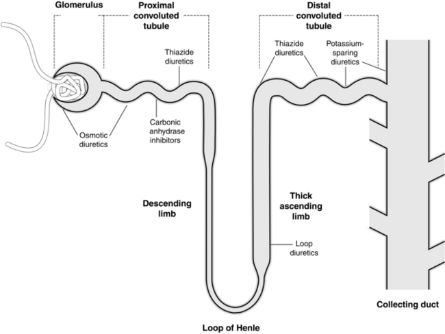
Carbonic Anhydrase Inhibitors

Acetazolamide

Acetazolamide is well absorbed, not metabolized, but excreted almost unchanged by the kidney within 24 h. Toxicity is very rare.

Acetazolamide is a carbonic anhydrase inhibitor. Under normal physiological conditions, the enzyme carbonic anhydrase is responsible for reabsorption of sodium and excretion of hydrogen ions in the proximal convoluted tubule of the nephron. Inhibition of carbonic anhydrase decreases hydrogen ion excretion and therefore sodium and bicarbonate ions stay in the renal tubule. This results in the production of alkaline urine with a high sodium bicarbonate content; the increased sodium excretion leads to a modest diuresis. Chloride ions are retained instead of bicarbonate to maintain an ionic balance. All these changes result in a hyperchloraemic metabolic acidosis.

Carbonic anhydrase inhibitors are seldom used as primary diuretics because of their weak diuretic effect. They may be used in the management of salicylate overdose to produce an alkaline diuresis as this increases the urinary elimination of weak acids. The most common use of acetazolamide is to reduce the intraocular pressure of glaucoma patients. The inhibition of carbonic anhydrase results in a decreased formation of ocular aqueous humour and cerebrospinal fluid. It is also valuable in the prevention and management of acute mountain sickness. When used in patients with familial periodic paralysis, the metabolic acidosis increases the potassium concentration in the skeletal muscles and improves symptoms.

Osmotic Diuretics

Mannitol

Mannitol is an alcohol produced by the reduction of mannose. It is absorbed unreliably from the gastrointestinal tract and therefore has to be given by i.v. injection; doses of 0.25–1 g kg–1 are used. Initially it stays within the intravascular space but is then slowly redistributed into the extravascular compartment. Mannitol does not undergo metabolism and is excreted unchanged through the kidneys.

Mannitol expands the intravascular volume and then undergoes free glomerular filtration with almost no reabsorption in the proximal tubule. It also decreases the energy-consuming process of sodium and water reabsorption in the proximal tubule. This leads to an osmotic force that retains water and sodium in the tubule with a consequent osmotic diuresis, i.e. increased urinary excretion of sodium, water, bicarbonate and chloride. Mannitol does not alter urinary pH. The raised renal blood flow reduces the rate of renin secretion which decreases the urine-concentrating capacity of the kidney.

Mannitol has often been used prophylactically to protect the kidneys against an ischaemic incident (e.g. cardiopulmonary bypass, aortic cross-clamping or hypotensive episodes) and subsequent acute renal failure. Its theoretical effects include reduced proximal tubular oxygen demand, scavenging of oxygen free radicals, reduced tubular endothelial cell swelling and increased tubular flow which might maintain tubular patency after ischaemic injury. However, there is no convincing evidence of a renal protective effect of mannitol in clinical practice. There is also little evidence that conversion from oliguric to non-oliguric renal failure decreases the mortality rate in critically ill patients. Nevertheless, mannitol is still used during renal transplantation to help ‘preserve’ the donor kidney.

Anaesthetists often administer mannitol to reduce intracranial pressure (Ch 32) and intraocular pressure.

Mannitol may precipitate pulmonary oedema in patients with compromised cardiac function. Occasionally, it may cause hypersensitivity reactions. If the blood–brain barrier is not intact after a head injury or neurosurgery, mannitol may enter the brain, draw water with it and cause rebound cerebral swelling.

Loop Diuretics

Loop diuretics act primarily on the medullary part of the ascending limb of the loop of Henle. After initial glomerular filtration and proximal tubular secretion, they inhibit the active reabsorption of chloride in the thick portion of the ascending limb. This leads to chloride, sodium, potassium and hydrogen ions remaining in the tubule to maintain electrical neutrality, and their increased excretion in the urine. The extent of the following diuresis is determined by the concentration of active drug in this part of the tubule. Because the ascending limb plays an important role in the reabsorption of sodium chloride in the kidney, these drugs produce a potent diuretic response. The decrease in sodium chloride reabsorption leads to a reduced urine-concentrating ability of the normally hypertonic medullary interstitium. Furosemide, bumetanide and torasemide are classified as loop diuretics because of their common site of action. Furosemide is the most commonly used.

Furosemide

Furosemide is usually administered intravenously (0.1–1 mg kg–1) or orally (0.75–3 mg kg–1). It is well absorbed orally and about 60% of the dose reaches the central circulation within a short period, with the peak effect after 1–1.5 h. Intravenous furosemide is usually started as a slow 20–40 mg injection in adults, but higher doses or even an infusion may be required in the case of elderly patients with renal failure or severe congestive cardiac failure. Typical doses in ICU patients are 2–5 mg per hour; i.v. infusions are associated with less toxicity than intermittent bolus doses. Approximately 90% of the drug is bound to plasma proteins and its volume of distribution is relatively low. Metabolism and excretion into the gastrointestinal tract contribute to about 30% of the elimination of a dose of furosemide. The remainder is excreted unchanged through glomerular filtration and tubular secretion. Impaired renal function affects the elimination process, but liver disease does not seem to influence this. The elimination half-life of furosemide is 1–1.5 h.

Furosemide increases renal artery blood flow if the intravascular fluid volume is maintained. It causes redistribution so that flow to the outer part of the cortex remains unchanged while inner cortex and medullary flow is increased. It leads to an improved renal tissue oxygen tension. This effect, together with the increased release of renin and the activation of the angiotensin–aldosterone axis, is mediated via prostaglandins. An advantage of loop diuretics is their high ceiling effect (i.e. increasing doses lead to increasing diuresis).

Furosemide is the diuretic of choice in acute pulmonary oedema or other states of fluid overload caused by cardiac, renal or liver failure. It reduces the intravascular fluid volume by promoting a rapid, powerful diuresis even in the presence of a low GFR. The pulmonary vascular bed and capacitance vessels are dilated by furosemide, and often a relief of dyspnoea and a reduction in pulmonary pressures may take place before the diuretic effect has occurred. In hypertensive patients, the vasodilatation and preload reduction lead to a decrease in arterial pressure.

Furosemide has been used widely in the management of the oliguric surgical or critically ill patient, but as with other diuretic drugs, studies have shown no clinical benefit. Furosemide should never be used to treat oliguria caused by a decreased intravascular fluid volume or dehydration, because the following diuresis could exaggerate hypovolaemia and renal ischaemic injury. It is important to restore the intravascular volume status first before any pharmacological intervention.

A raised intracranial pressure is often treated with furosemide. It mobilizes cerebral oedema fluid, decreases cerebrospinal fluid production and lowers intracranial pressure without changing plasma osmolarity. In contrast to mannitol, a disrupted blood–brain barrier does not influence the effect of furosemide on the intracranial pressure.

Excessive doses of furosemide often lead to fluid or electrolyte abnormalities. Severe hypokalaemia may precipitate dangerous cardiac arrhythmias, especially in the presence of high concentrations of digitalis. It may also enhance the effect of non-depolarizing neuromuscular blocking drugs. Hypovolaemia, dehydration and the consequent haemoconcentration may lead to changes in blood viscosity. Hyperuricaemia and prerenal uraemia may develop and may precipitate an acute gout attack in a patient with pre-existing gout.

Furosemide may cause high concentrations of aminoglycosides and cephalosporins in the kidneys and this may enhance their nephrotoxic effects. Prolonged high blood concentrations of furosemide may have a direct toxic action resulting in interstitial nephritis. It may also cause transient or permanent deafness because of changes in the endolymph electrolyte composition. Patients allergic to other sulphonamide drugs may have a cross-sensitivity, although idiosyncratic reactions are rare.

Bumetanide

The mechanism of action of bumetanide and its effects are similar to those of furosemide. The difference between these two drugs is the greater potency and bioavailability of bumetanide; smaller doses are needed. The normal adult dose is 0.5–3 mg i.v. over 1–2 min. The onset of diuresis is within 30 min and this usually lasts for about 4 h. The pharmacokinetics are similar to those of furosemide, with the exception that bumetanide is absorbed completely after oral administration and its rate of elimination is less dependent on renal function. Potassium loss is also a problem with bumetanide. Ototoxicity may be slightly less frequent than with furosemide, but renal toxicity is more of a problem. In clinical practice, there is no clear advantage or disadvantage over furosemide, providing equivalent doses are administered.

Thiazide Diuretics

Although thiazide diuretics are seldom used by anaesthetists, many patients scheduled for surgery are receiving these drugs for chronic hypertension or heart failure. There are a large number of thiazides available, all with a similar dose–response curve and diuretic effect. Bendroflumethiazide, chlorothiazide, hydrochlorothiazide and chlorthalidone are a few examples of the better known thiazide diuretics. The majority have a duration of action of 6–12 h. In comparison with loop diuretics, thiazides have a longer duration of action, act at a different site, have a low ‘ceiling’ effect and are less effective in renal failure.

Thiazide diuretics are administered orally, absorbed rapidly from the gastrointestinal tract and initiate a diuresis within 1–2 h. The major distinction between the available thiazides is their difference in elimination rate. They are distributed in the extracellular space and eliminated in the proximal tubule of the nephron by active secretion.

Thiazides inhibit the active pump for sodium and chloride reabsorption in the cortical ascending part of the loop of Henle and the distal convoluted tubule. Therefore, the urine-concentrating ability of the kidney is not impaired, as normally this area is responsible for less than 5% of sodium reabsorption. The diuresis achieved by the thiazides is therefore never as effective as that of the loop diuretics. It is mild but sustained. In contrast with loop diuretics, the excretion of calcium is decreased and hypercalcaemia may become a problem. In the presence of aldosterone activity, the increase in sodium delivery to the distal renal tubules is associated with increased potassium loss, similar to that of the loop diuretics. The reduced clearance of uric acid by thiazides may cause hyperuricaemia.

Thiazides are used extensively in low doses, and often combined with a low-sodium diet, for the management of essential hypertension. A reduction in extracellular fluid volume and mild peripheral vasodilatation are responsible for the sustained antihypertensive effect. The full antihypertensive effect may take up to 12 weeks to become established. Higher doses of thiazides are used for the management of congestive cardiac failure and other oedematous conditions such as nephrotic syndrome and liver cirrhosis.

The most common side-effects of the thiazides are probably dehydration and hypovolaemia. This may present as orthostatic hypotension. When administered chronically, these drugs lead typically to a diuretic-induced hypokalaemic, hypochloraemic, metabolic alkalosis. In combination with magnesium depletion, the hypokalaemia may trigger serious cardiac arrhythmias, in addition to digitalis toxicity, muscle weakness and the potentiation of non-depolarizing muscle relaxants.

Thiazides decrease the tubular secretion of urate, which may lead to hyperuricaemia and gout. They are sulphonamide derivatives and may therefore cause inhibition of insulin release from the pancreas and blockade of peripheral glucose utilization. This may precipitate hyperglycaemia or an increase in insulin requirements in a patient with diabetes mellitus. They also lead to an increase in total blood cholesterol.

Potassium-Sparing Diuretics

Only a small part of sodium reabsorption into the renal cells takes place via the sodium–potassium exchange mechanism in the distal tubules. The potassium-retaining diuretics act on the distal convoluted tubules and the collecting ducts and therefore cause only a limited diuresis. There are two subgroups in this category: drugs acting independently of the aldosterone mechanism (e.g. triamterene and amiloride) and aldosterone antagonists (e.g. spironolactone).

These drugs increase the urinary excretion of sodium, chloride and bicarbonate and lead to an increase in urinary pH. They prevent excessive loss of potassium that occurs with the loop and thiazide diuretics by reducing the sodium–potassium exchange. Potassium-sparing drugs do, however, augment the diuretic response of these drugs when given in combination.

Spironolactone

Aldosterone causes sodium reabsorption and potassium loss in the distal convoluted tubule. Spironolactone has a steroid molecular structure, acts as a competitive antagonist on the aldosterone receptors and inhibits sodium reabsorption and potassium loss. In the absence of aldosterone, it has no effect.

After oral absorption, spironolactone is immediately metabolized to a number of metabolites. Some of these are active and act for up to 15 h.

Spironolactone is the logical choice of diuretic in the management of liver cirrhosis, ascites and secondary hyperaldosteronism. Heart failure or hypertension in the presence of high mineralocorticoid levels (Conn’s syndrome or prednisone therapy) is another indication. Spironolactone is often combined with thiazides to maximize the diuretic effect and prevent potassium loss.

Hyperkalaemia may develop if spironolactone is used in the presence of renal dysfunction. If used in high doses, it may cause gynaecomastia and impotence.

ACUTE RENAL FAILURE, SEPSIS AND THE INTENSIVE CARE UNIT

Acute renal failure is a common complication of sepsis and septic shock; the combination is associated with a mortality of over 50%. The arterial vasodilatation that accompanies sepsis is mediated in part by cytokines that upregulate inducible nitric oxide synthase with a subsequent increased release of nitric oxide. The potent vasodilatory effect of nitric oxide is partly responsible for the vascular resistance to the pressor response to noradrenaline and angiotensin II. Early in sepsis-related acute renal failure, cytokines such as tumour necrosis factor alpha (TNF-α) cause vasoconstriction of the renal vasculature, though trials of monoclonal antibodies against TNF-α have not shown any improvement in survival.

This early vasoconstrictor phase is potentially reversible and some clinical studies have suggested that measures to optimize haemodynamics with fluids and vasoactive drugs can reduce the incidence of established acute kidney injury though more data are needed. The administration of vasopressin (see above) in patients with septic shock may help maintain arterial pressure despite the relative ineffectiveness of other vasopressors such as noradrenaline and angiotensin II. It constricts the glomerular efferent arteriole and therefore increases the filtration pressure and glomerular filtration rate. There is early evidence in patients who have septic shock and are at risk of acute kidney injury that vasopressin may reduce progression to renal failure, and mortality.

ACUTE KIDNEY INJURY AFTER CARDIAC OR MAJOR VASCULAR SURGERY

The incidence of acute kidney injury (AKI) after cardiac surgery is approximately 10% and is associated with a high morbidity and mortality. Clinical evidence suggests that preoperative renal insufficiency, diabetes mellitus, prolonged cardiopulmonary bypass (CPB) time and postoperative hypotension are all independent risk factors for AKI in the cardiac patient. International consensus statements have recently been drawn up regarding the pathophysiology and treatment of AKI in cardiac surgical patients. Six pathophysiological processes were found to be most likely to contribute to this: exogenous and endogenous toxins, metabolic factors, ischaemia-reperfusion injury, neurohormonal activation, inflammation, and oxidative stress, which are probably all interrelated.

The most effective way to prevent AKI in this setting is by adequate hydration and perfusion pressure during CPB, and the maintenance of cardiac output throughout the perioperative period. Data for the effects of hypothermia during CPB, pulsatile CPB flow or avoiding CPB altogether through off-pump cardiac surgical techniques on the incidence of AKI are not conclusive. Many drugs have been studied for renal protection during CPB. Low-dose dopamine is ineffective and may even be harmful. Although mannitol is used in routine pump prime in many cardiac units and furosemide potentially reduces renal medullary oxygen consumption, neither prevents AKI after cardiac surgery. Recent therapeutic trials to reduce acute kidney injury in cardiac surgery patients fall broadly into reactive oxygen molecule scavengers, anti-inflammatory agents, and anti-apoptotic agents (tetracyclines, minocycline, human recombinant erythropoietin). However the results have been inconsistent and disappointing and there is currently no pharmacological strategy known to reliably reduce the incidence of AKI.

Aprotinin has been used to reduce transfusion requirements in high risk cardiac surgical patients. Recent evidence has raised concerns that aprotinin is associated with an increased risk of AKI in cardiac surgery but the evidence is conflicting and the possibility of risk is not consistently supported by data from published, randomized, placebo-controlled clinical trials. Aprotinin is currently not recommended for routine use in cardiothoracic surgery.

DRUGS AND RENAL TRANSPLANTATION

The best treatment for end-stage renal failure is renal transplantation. Apart from optimizing the recipient’s general health (e.g. correction of anaemia, preoperative dialysis, etc.), immunosuppression plays an extremely important role in graft survival.

Erythropoietin

Erythropoietin is a circulating hormone secreted by the kidneys. It stimulates the bone marrow to produce red blood cells. The ability of the kidney to secrete erythropoietin deteriorates as excretory function decreases. Patients with severe chronic renal failure are unable to produce adequate quantities of erythropoietin, which leads to diminished red blood cell production. The retention of toxic substances also contributes to bone marrow depression. In addition, red cell survival is reduced by 50% in advanced renal failure. Therefore these patients almost always develop a chronic anaemia.

Long-term administration of recombinant human erythropoietin (rHUEPO) in chronic renal failure patients results in global stimulation of the bone marrow, increasing red blood cell differentiation and maintaining cell viability, thereby improving anaemia. It also decreases bleeding by increasing platelet adhesion in haemodialysed uraemic patients. A side-effect of rHUEPO is the development of hypertension or exacerbation of existing hypertension.

Immunosuppression

Prednisolone and Azathioprine

Corticosteroids were the first drugs to be used as immunosuppressive agents. Initially, very high doses were used, producing the typical steroid side-effects, e.g. Cushingoid appearance, hypertension, hyperglycaemia and osteoporosis. Experience and research showed that large doses were not necessary and that better results and fewer side-effects were possible with lower doses.

The ‘modern era’ of immunosuppression started with the discovery of azathioprine. For a long period of time, the combination of azathioprine and corticosteroids was the ‘gold standard’ in transplant surgery. Azathioprine is a derivative of 6-mercaptopurine and is metabolized to its active form in the liver. It affects the synthesis of DNA and RNA and is broken down by the enzyme xanthine oxidase. Co-administration of allopurinol (xanthine oxidase inhibitor) is contraindicated because it may result in bone marrow suppression, agranulocytosis and leucopaenia. Patients receiving azathioprine are prone to develop viral warts or malignancies of the skin and hepatic dysfunction.

Ciclosporin A and Ciclosporin-Neoral

The next major advance in transplant surgery was the discovery of ciclosporin A. This fungal peptide prevents the proliferation and clonal expansion of T lymphocytes. The chance of acute rejection is reduced significantly by administration of this drug. In spite of the large number of side-effects of ciclosporin A, it has been accepted as the new standard against which all other immunosuppressants are judged. Ciclosporin A is lipophilic and incompletely absorbed in the small bowel. Serious side-effects such as gingival hypertrophy, hepatotoxicity and nephrotoxicity make this a less than perfect drug. For a long time, classic triple therapy consisted of prednisolone, azathioprine and ciclosporin A.

Recently, ciclosporin was released in a new form under the tradename Neoral. This form is a microemulsion that enhances the bioavailability of ciclosporin through improved absorption. Ciclosporin (Neoral) is equipotent to the parent drug and most renal transplant patients are presently receiving this agent.

Mycophenolate Mofetil

Mycophenolate mofetil (MMF) is a new immunosuppressant licensed for use in renal transplantation. It inhibits a key enzyme in the purine synthesis pathway and therefore has a specific effect on B and T lymphocytes. The synthesis of adhesion molecules is also inhibited by MMF. It seems as though MMF effectively prevents chronic rejection in renal transplant patients. MMF is neither nephrotoxic nor hepatotoxic.

There are a variety of substances with potent immunosuppressant properties which are usually used in combination with each other. Despite their powerful immunosuppressant effects, it is unlikely that any of the new or existing drugs may be used as monotherapy. The calcineurin inhibitors (CNIs), ciclosporin and tacrolimus, have revolutionized the overall success of renal transplantation through reduction in early immunological injury and acute rejection rates. However, the CNIs have a significant adverse effect on renal function and cardiovascular disease, and extended long-term graft survival has not been achieved. CNI minimization using mycophenolate mofetil or sirolimus may be associated with a modest increase in creatinine clearance and a decrease in serum creatinine in the short term. A typical perioperative regimen for renal transplantation is shown in Table 10.2.

TABLE 10.2

A Typical Perioperative Regimen for Renal Transplant

Preoperative Induction immunosuppression (ciclosporin-Neoral, FK506/tacrolimus, rapamycin, or sirolimus and mycophenolate mofetil (MMF)
Heparin 5000 units subcutaneously
Ranitidine 150 mg orally (stress ulcer prophylaxis)
Nifedipine 20 mg orally (vasodilator, free radical scavenger)
At induction Antibiotic prophylaxis – co-amoxiclav (augmentin) 1.2 g intravenously methylprednisolone 0.5 g i.v.
During vascular anastomosis Mannitol 0.5 g kg–1 intravenously ± dopamine 3–5 μg kg–1 min–1
Postoperative Antibiotic prophylaxis
Heparin 5000 units subcutaneously, twice daily
Aspirin 150 mg orally
Ranitidine 150 mg orally, twice daily
Co-trimoxazole 480 mg orally, daily (prophylaxis against Pneumocystis carinii)
Immunosuppression (‘triple therapy’)

The role of drugs inhibiting antigen presentation and the use of monoclonal antibodies are still being defined.

FURTHER READING

Fischereder, M., Kretzler, M. New immunosuppressive strategies in renal transplant recipients. J. Nephrol. 2004;17:9–18.

Flechner, S.M., Kobashigawa, J., Klintmalm, G. Calcineurin inhibitor-sparing regimens in solid organ transplantation: focus on improving renal function and nephrotoxicity. Clin. Transplant. 2008;22(1):1–15.

Garwood, S. Cardiac surgery-associated acute renal injury: new paradigms and innovative therapies. J. Cardiothorac. Vasc. Anesth. 2010;24(6):990–1001.

Gordon, A.C., Russell, J.A., Whalley, K.R., et al. The effects of vasopressin on acute kidney injury in septic shock. Intensive Care Med. 2010;36(1):83–91.

Gottlieb, S.S. Adenosine A1 antagonists and the cardiorenal syndrome. Curr. Heart Fail. Rep. 2008;5(2):105–109.

Guyton, A.C., Hall, J.E. Diuretics, kidney diseases (Ch 31). In: Guyton A.C., Hall J.E., eds. Textbook of Medical Physiology. eleventh ed. Philadelphia: WB Saunders; 2011:397–409.

Huang, D.T., Clermont, G., Dremsizov, T.T., et al. Implementation of early goal-directed therapy for severe sepsis and septic shock: a decision analysis. Crit. Care Med. 2007;35(9):2090–2100.

Lindvall, G., Sartipy, U., Ivert, T., et al. Aprotinin is not associated with postoperative renal impairment after primary coronary surgery. Ann. Thorac. Surg. 2008;86:13–19.

Schrier, R.W., Wang, W. Acute renal failure and sepsis. N. Engl. J. Med. 2004;351:59–69.