Biphosphonates (to prevent bone loss; can cause severe, longer segment esophageal ulceration)
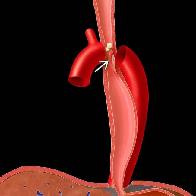
(Left) Graphic shows medication pills stuck at the level of the aortic arch with focal spasm and ulceration .
(Right) Esophagram shows broad, shallow ulceration at the aortic arch level. The patient had odynophagia and recent tetracycline ingestion, and the symptoms resolved spontaneously. Physiological points of esophageal narrowing, such as at the aortic arch and the retrocardiac portion of the esophagus, are the most commonly cited for pill-induced esophagitis.
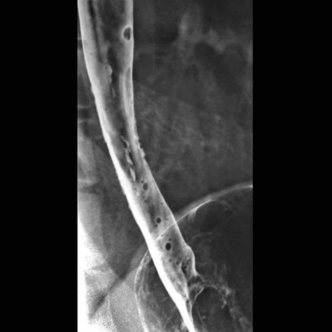
(Left) Double-contrast barium esophagram in a 50-year-old woman with odynophagia while taking tetracycline shows multiple ulcerations and a subtle stricture or spasm of the distal esophagus.
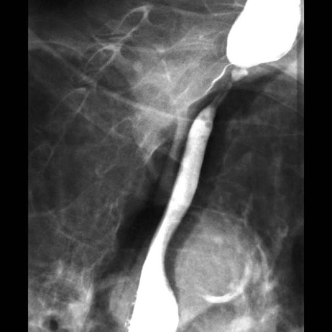
(Right) This 70-year-old woman with cardiac disease awoke with severe odynophagia the morning after taking her quinidine at bedtime. Barium esophagram demonstrates a long stricture or focal spasm from the thoracic inlet to the aortic arch without definite ulceration.
Double-contrast esophagram shows a cluster of ulcers in the distal esophagus.
In this elderly patient with heart disease taking quinidine and other medications, stricture and ulceration are seen at the thoracic inlet; an unusual site for drug-induced esophagitis.
[/level-membership-for-radiology-category][not-level-membership-for-radiology-category] Aortic arch, left main bronchus, retrocardiac
• Findings on esophagram (double contrast)
Solitary or localized cluster of tiny ulcers distributed circumferentially on normal background mucosa
Punctate, linear, stellate, serpiginous, or ovoid; collections of barium on esophageal surface
Longer areas of ulceration with potassium chloride, quinidine, biphosphonates, and in patients with cardiomegaly
Mass effect surrounding ulcer due to edema and inflammation; can mimic ulcerated carcinoma
• Superficial ulceration
Giant, flat ulcers are uncommonly seen
TOP DIFFERENTIAL DIAGNOSES
• Reflux esophagitis
• Viral esophagitis
• Esophageal carcinoma
• Barrett esophagus
Buy Membership for Radiology Category to continue reading. Learn more here
 Solitary or localized cluster of tiny ulcers distributed circumferentially on normal background mucosa


 .
.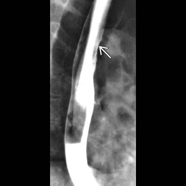
 at the aortic arch level. The patient had odynophagia and recent tetracycline ingestion, and the symptoms resolved spontaneously. Physiological points of esophageal narrowing, such as at the aortic arch and the retrocardiac portion of the esophagus, are the most commonly cited for pill-induced esophagitis.
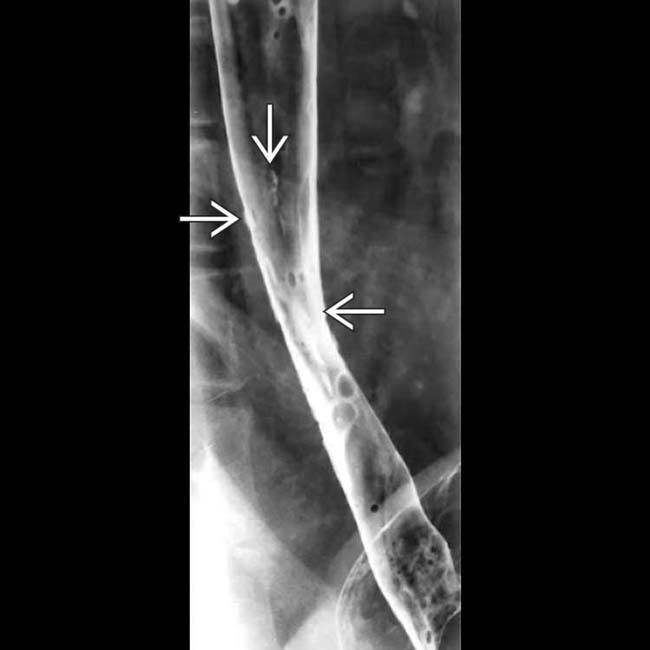
 and a subtle stricture or spasm of the distal esophagus.
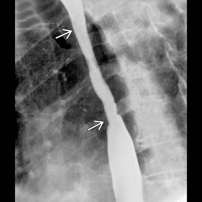
 from the thoracic inlet to the aortic arch without definite ulceration.