23 Distal Trigeminal Block
Placement
Anatomy
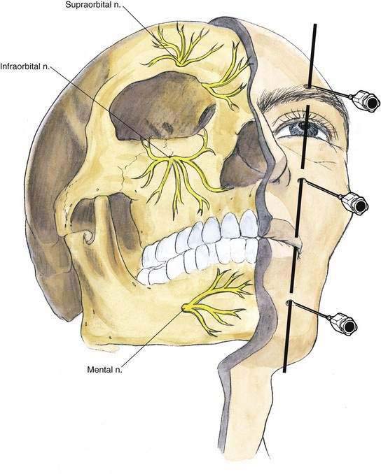
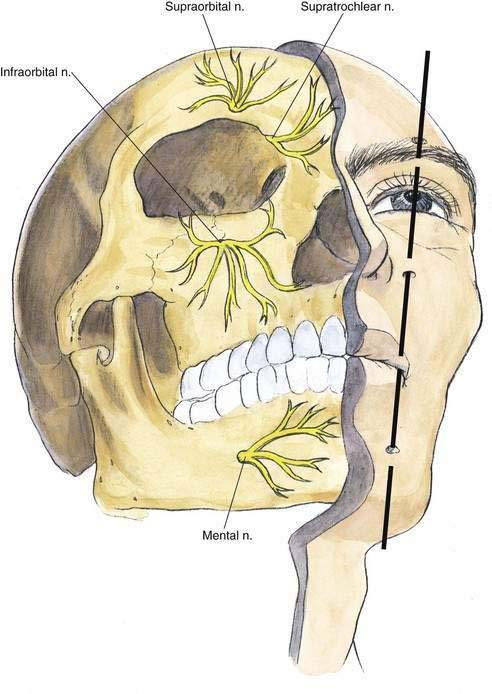
The distal branches of the three divisions of the trigeminal nerve—ophthalmic (supraorbital), maxillary (infraorbital), and mandibular (mental)—exit from the skull through their respective foramina on a line that runs almost vertically through the pupil (Fig. 23-1).
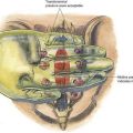
Needle Puncture
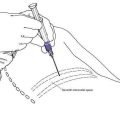
For this block, as illustrated in Figure 23-2, once the respective foramina are identified by palpation, a short, 25-gauge needle is inserted in a cephalomedial direction near each foramen, and approximately 2 to 3 mL of local anesthetic is injected at each site. If a paresthesia is obtained, the local anesthetic can be deposited at that point.