Disorders of the Conjunctiva

Published on 25/03/2015 by admin

Filed under Pediatrics

Last modified 22/04/2025

Print this page

rate 1 star rate 2 star rate 3 star rate 4 star rate 5 star
Your rating: none, Average: 0 (0 votes)

This article have been viewed 1451 times

Chapter 618 Disorders of the Conjunctiva

Conjunctivitis

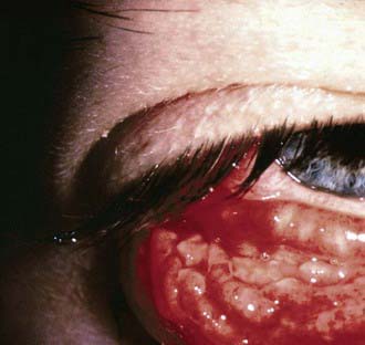
The conjunctiva reacts to a wide range of bacterial and viral agents, allergens, irritants, toxins, and systemic diseases. Conjunctivitis is common in childhood and may be infectious or noninfectious. The differential diagnosis of a red-appearing eye includes conjunctival as well as other ocular sites (Table 618-1).

Ophthalmia Neonatorum

This form of conjunctivitis, occurring in infants younger than 4 wk of age, is the most common eye disease of newborns. Its many different causal agents vary greatly in their virulence and outcome. Silver nitrate instillation may result in a mild self-limited chemical conjunctivitis, whereas Neisseria gonorrhoeae and Pseudomonas are capable of causing corneal perforation, blindness, and death. The risk of conjunctivitis in newborns depends on frequencies of maternal infections, prophylactic measures, circumstances during labor and delivery, and postdelivery exposure to microorganisms.

Clinical Manifestations

The clinical manifestations of the various forms of ophthalmia neonatorum are not specific enough to allow an accurate diagnosis. Although the timing and character of the signs are somewhat typical for each cause of this condition, there is considerable overlap and physicians should not rely solely on clinical findings. Regardless of its cause, ophthalmia neonatorum is characterized by redness and chemosis (swelling) of the conjunctiva, edema of the eyelids, and discharge, which may be purulent.

Neonatal conjunctivitis is a potentially blinding condition. The infection may also have associated systemic manifestations that require treatment. Therefore, any newborn infant who develops signs of conjunctivitis needs a prompt and comprehensive systemic and ocular evaluation to determine the agent causing the infection and the appropriate treatment.

The onset of inflammation caused by silver nitrate drops usually occurs within 6-12 hr after birth, with clearing by 24-48 hr. The usual incubation period for conjunctivitis due to N. gonorrhoeae is 2-5 days, and for that due to C. trachomatis, it is 5-14 days. Gonococcal infection may be present at birth or be delayed beyond 5 days of life owing to partial suppression by ocular prophylaxis. Gonococcal conjunctivitis may also begin in infancy after inoculation by the contaminated fingers of adults. The time of onset of disease with other bacteria is highly variable.

Gonococcal conjunctivitis begins with mild inflammation and a serosanguineous discharge. Within 24 hr, the discharge becomes thick and purulent, and tense edema of the eyelids with marked chemosis occurs. If proper treatment is delayed, the infection may spread to involve the deeper layers of the conjunctivae and the cornea. Complications include corneal ulceration and perforation, iridocyclitis, anterior synechiae, and rarely panophthalmitis. Conjunctivitis caused by C. trachomatis (inclusion blennorrhea) may vary from mild inflammation to severe swelling of the eyelids with copious purulent discharge. The process involves mainly the tarsal conjunctivae; the corneas are rarely affected. Conjunctivitis due to Staphylococcus aureus or other organisms is similar to that produced by C. trachomatis. Conjunctivitis due to Pseudomonas aeruginosa is uncommon, acquired in the nursery, and a potentially serious process. It is characterized by the appearance on days 5-18 of edema, erythema of the lids, purulent discharge, pannus formation, endophthalmitis, sepsis, shock, and death.

Prognosis and Prevention

Before the institution of topical ophthalmic prophylaxis at birth, gonococcal ophthalmia was a common cause of blindness or permanent eye damage. If properly applied, this form of prophylaxis is highly effective unless infection is present at birth. Drops of 0.5% erythromycin or 1% silver nitrate are instilled directly into the open eyes at birth using wax or plastic single-dose containers. Saline irrigation after silver nitrate application is unnecessary. Silver nitrate is ineffective against active infection and may have limited use against Chlamydia. Povidone-iodine (2% solution) may also be an effective prophylactic agent.

Identification of maternal gonococcal infection and appropriate treatment has become a standard element of routine prenatal care. An infant born to a woman who has untreated gonococcal infection should receive a single dose of ceftriaxone, 50 mg/kg (maximum 125 mg) IV or IM, in addition to topical prophylaxis. The dose should be reduced for premature infants. Penicillin (50,000 U) should be used if the mother’s gonococcal isolate is known to be penicillin sensitive.

Neither topical prophylaxis nor topical treatment prevents the afebrile pneumonia that occurs in 10-20% of infants exposed to C. trachomatis. Although chlamydial conjunctivitis is often a self-limiting disease, chlamydial pneumonia may have serious consequences. It is important that infants with chlamydial disease receive systemic treatment. Treatment of colonized pregnant women with erythromycin may prevent neonatal disease.

Vernal Conjunctivitis

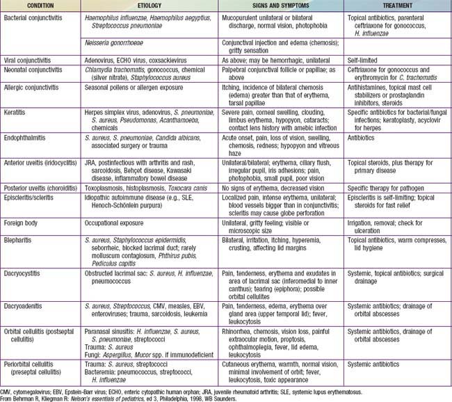
This usually begins in the prepubertal years and may recur for many years. Atopy appears to have a role in its origin, but the pathogenesis is uncertain. Extreme itching and tearing are the usual complaints. Large, flattened, cobblestone-like papillary lesions of the palpebral conjunctivae are characteristic (Fig. 618-1). A stringy exudate and a milky conjunctival pseudomembrane are frequently present. Small elevated lesions of the bulbar conjunctiva adjacent to the limbus (limbal form) may be found. Smear of the conjunctival exudate reveals many eosinophils. Topical corticosteroid therapy and cold compresses afford some relief. Topical mast cell stabilizers or prostaglandin inhibitors are useful when long-term control is needed. The long-term use of corticosteroids should be avoided.

Parinaud Oculoglandular Syndrome

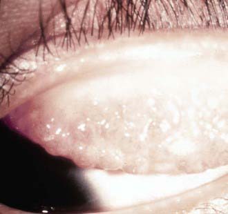
This represents a form of cat-scratch disease and is caused by Bartonella henselae, which is transmitted from cat to cat by fleas (Chapter 201). Kittens are more likely than adult cats to be infected. Humans can become infected when they are scratched by a cat. In addition, bacteria may pass from a cat’s saliva to its fur during grooming. The bacteria can then be deposited on the conjunctiva after rubbing one’s eyes after handling the cat. Lymphadenopathy and conjunctivitis are hallmarks of the disease. Conjunctival granulomas may develop (Fig. 618-2). The course is generally self-limited, but antibiotics may be used in some cases.

Other Conjunctival Disorders

Subconjunctival hemorrhage is manifested by bright or dark red patches in the bulbar conjunctiva and may result from injury or inflammation. It commonly occurs spontaneously. It may occasionally result from severe sneezing or coughing. Rarely, it may be a manifestation of a blood dyscrasia. Subconjunctival hemorrhages are self-limiting and require no treatment.

Pinguecula is a yellowish-white, slightly elevated mass on the bulbar conjunctiva, usually in the interpalpebral region. It represents elastic and hyaline degenerative changes of the conjunctiva. No treatment is required except for cosmetic reasons, in which case simple excision suffices.

Pterygium is a fleshy triangular conjunctival lesion that may encroach on the cornea. It typically occurs in the nasal interpalpebral region. The pathologic findings are similar to those of a pinguecula. The development of pterygia is related to exposure to ultraviolet light, and it therefore is more commonly found among people who live near the equator. Removal is suggested when the lesion encroaches far onto the cornea. Recurrence after removal is common.

Dermoid cyst and dermolipoma are benign lesions, clinically similar in appearance. They are smooth, elevated, round to oval lesions of various sizes. The color varies from yellowish white to fleshy pink. The most frequent site is the upper outer quadrant of the globe; they also commonly occur near or straddling the limbus. Dermolipoma is composed of adipose and connective tissue. Dermoid cysts may also contain glandular tissue, hair follicles, and hair shafts. Excision for cosmetic reasons is feasible. Dermolipomas are often connected to the extraocular muscles, making their complete removal impossible without sacrificing ocular motility.

Conjunctival nevus is a small, slightly elevated lesion that may vary in pigmentation from pale salmon to dark brown. It is usually benign, but careful observation for progressive growth or changes suggestive of malignancy is advised.

Symblepharon is a cicatricial adhesion between the conjunctiva of the lid and the globe; the lower lid is usually affected. It follows operation or injuries, especially burns from lye, acids, or molten metals. It is a serious complication of Stevens-Johnson syndrome. It may interfere with motion of the eyeball and may cause diplopia. The adhesions should be separated and the raw surfaces kept from uniting during healing. Grafts of oral mucous membrane may be necessary.

Bibliography

Atik B, Thanh TTK, Luong VQ, et al. Impact of annual targeted treatment of infectious trachoma and susceptibility to reinfection. JAMA. 2006;296:1488-1497.

Bremond-Gignac D, Mariani-Kurkdjian P, Beresniak A, et al. Efficacy and safety of azithromycin 1.5% eye drops for purulent bacterial conjunctivitis in pediatric patients. Pediatr Infect Dis J. 2010;29:222-226.

Centers for Disease Control and Prevention. Acute hemorrhagic conjunctivitis outbreaks caused by coxsackievirus A24v—Uganda and Southern Sudan, 2010. MMWR Morb Mortal Wkly Rep. 2010;59:1024.

Centers for Disease Control and Prevention. Outbreak of bacterial conjunctivitis at a college—New Hampshire, January-March, 2002. MMWR Morb Mortal Wkly Rep. 2002;51:205-208.

Centers for Disease Control and Prevention. Acute hemorrhagic conjunctivitis outbreak caused by coxsackievirus A24—Puerto Rico, 2003. MMWR Morb Mortal Wkly Rep. 2004;53:632.

Chang DC, Grant GB, O’Donnell K, et al. Multistate outbreak of fusarium keratitis associated with use of a contact lens solution. JAMA. 2006;296:953-963.

Everitt HA, Little PS, Smith PWF. A randomised controlled trial of management strategies for acute infective conjunctivitis in general practice. BMJ. 2006;333:321-324.

House JI, Ayele B, Porco TC, et al. Assessment of herd protection against trachoma due to repeated mass antibiotic distributions: a cluster-randomised trial. Lancet. 2009;373:1111-1118.

The Medical Letter. Ophthalmic besifloxacin (Besivance). Med Lett. 2009;51:101-102.

The Medical Letter. Ophthalmic azithromycin (Azasite). Med Lett. 2008;50:1279-1280.

Meltzer JA, Kunkov S, Crain EF. Identifying children at low risk for bacterial conjunctivitis. Arch Pediatr Adolesc Med. 2010;164:263-267.

O’Hara MA. Ophthalmia neonatorum. Pediatr Clin North Am. 1993;40:715.

Porco TC, Gebre T, Ayele B, et al. Effect of mass distribution of azithromycin for trachoma control on overall mortality in Ethiopian children. JAMA. 2009;302:962-968.

Rietveld RP, ter Riet G, Bindels PJE, et al. Predicting bacterial cause in infectious conjunctivitis: cohort study on informativeness of combinations of signs and symptoms. Br Med J. 2004;329:206-208.

Rose PW, Hamden A, Brueggemann AB, et al. Chloramphenicol treatment for acute infective conjunctivitis in children in primary care: a randomized double-blind placebo-controlled trial. Lancet. 2005;366:37-42.

Shiuey Y, Ambati BK, Adamis AP, et al. A randomized, double-masked trial of topical ketorolac versus artificial tears for treatment of viral conjunctivitis. Ophthalmology. 2000;107:1512-1517.

Weiss A, Brinser JH, Nazar-Stewart V. Acute conjunctivitis in childhood. J Pediatr. 1993;122:10-14.