Disorders of the Complement System

Published on 22/03/2015 by admin

Filed under Pediatrics

Last modified 22/04/2025

Print this page

rate 1 star rate 2 star rate 3 star rate 4 star rate 5 star
Your rating: none, Average: 0 (0 votes)

This article have been viewed 1744 times

Chapter 128 Disorders of the Complement System

128.1 Evaluation of the Complement System

Testing for total hemolytic complement activity (CH50) effectively screens for most of the diseases of the complement system. A normal result in this assay depends on the ability of all 11 components of the classical pathway and membrane attack complex to interact and lyse antibody-coated sheep erythrocytes. The dilution of serum that lyses 50% of the cells determines the end-point. In congenital deficiencies of C1 through C8, the CH50 value is 0 or close to 0; in C9 deficiency, the value is approximately half-normal. Values in the acquired deficiencies vary with the type and severity of the underlying disorder. This assay does not detect deficiency of mannose-binding lectin (MBL), factors D or B of the alternative pathway, or properdin. Deficiency of factors I or H permits consumption of C3, with partial reduction in the CH50 value. When clotted blood or serum sits at room temperature or warms, CH50 activity begins to decline, which leads to values that are falsely low but not zero. It is important to separate the serum and freeze it at −70°C by no more than an hour after blood draw.

In hereditary angioedema, depression of C4 and C2 during an attack significantly reduces the CH50. Typically, C4 is low and C3 normal or slightly decreased. Concentrations of C1 inhibitor protein will be normal in 15% of cases; but C1 acts as an esterase, and the diagnosis can be established by showing increased capacity of patients’ sera to hydrolyze synthetic esters.

A decrease in serum concentration of both C4 and C3 suggests activation of the classical pathway by immune complexes. Decreased C3 and normal C4 levels suggest activation of the alternative pathway. This difference is particularly useful in distinguishing nephritis secondary to immune complex deposition from that due to NeF (nephritic factor). In the latter condition and in deficiency of factor I or H, factor B is consumed and C3 serum concentration is low. Alternative pathway activity can be measured with a relatively simple and reproducible hemolytic assay that depends on the capacity of rabbit erythrocytes to serve as both an activating (permissive) surface and a target of alternative pathway activity. This assay (AP50) detects deficiency of properdin, factor D, and factor B. Immunochemical methods can be used to quantify individual components of all 3 pathways, guided by results of the screening hemolytic assays. It is possible to analyze the genes encoding many of the components.

A defect of complement function should be considered in any patient with recurrent angioedema, autoimmune disease, chronic nephritis, hemolytic-uremic syndrome (HUS), or partial lipodystrophy, or with recurrent pyogenic infections, disseminated meningococcal or gonococcal infection, or a second episode of bacteremia at any age. A previously well adolescent or young adult with meningococcal meningitis due to an uncommon serotype (not A, B, or C) should undergo screening for a late-component or alternative pathway deficiency with CH50 and AP50 assays.

128.2 Genetic Deficiencies of Complement Components

Congenital deficiencies of all 11 components of the classical-membrane attack pathway and of factor D and properdin of the alternative pathway are described (Table 128-1). All of the components of the classical and alternative pathways except properdin are inherited as autosomal recessive co-dominant traits. Each parent transmits a gene that codes for synthesis of half the serum level of the component. Deficiency results from inheritance of 1 null gene from each parent; the hemizygous parents typically have CH50 levels that are low normal. Properdin deficiency is transmitted as an X-linked trait.

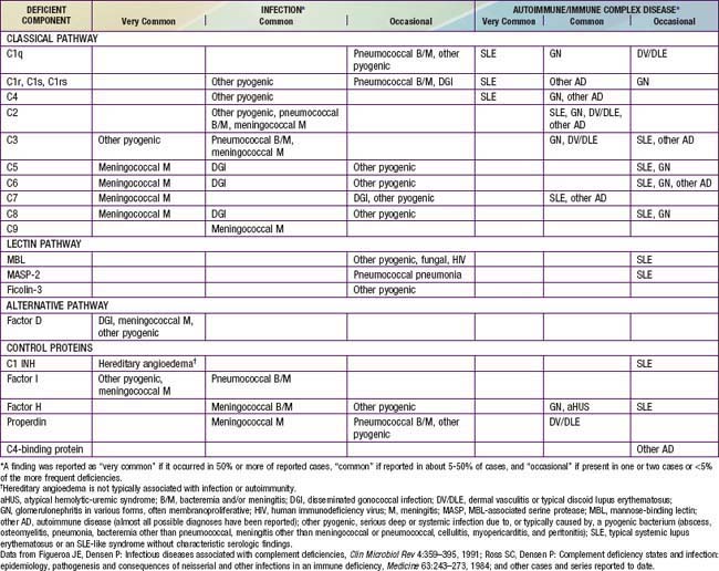
Most patients with primary C1q deficiency have systemic lupus erythematosus (SLE), an SLE-like syndrome without typical SLE serology, a chronic rash that has shown an underlying vasculitis on biopsy, or membranoproliferative glomerulonephritis (MPGN). Some C1q-deficient children have serious infections, including septicemia and meningitis. Individuals with C1r, C1s, combined C1r/C1s, C4, C2, or C3 deficiency also have a high incidence of autoimmune syndromes (see Table 128-1), especially SLE or an SLE-like syndrome in which antinuclear antibody level is not elevated.

C4 is encoded by 2 genes, termed C4A and C4B. C4 deficiency represents absence of both gene products. Complete deficiency of only C4A, present in about 1% of the population, also predisposes to SLE, though C4 levels are only partially reduced. Patients with only C4B deficiency may be predisposed to infection. A few patients with C5, C6, C7, or C8 deficiency have SLE, but recurrent meningococcal infections are much more likely to be the major problem.

The reason for the concurrence of deficiencies of complement components, especially C1, C4, C2, or C3 deficiency, and autoimmune-immune complex diseases is not entirely clear, but deposition of C3 on autoimmune complexes facilitates their removal from the circulation through binding to complement receptor 1 (CR1) on erythrocytes and transport to the spleen and liver. The early components, particularly C1q and C3, expedite the clearance of necrotic and apoptotic cells, which are sources of autoantigens. Inefficiency of either or both of these processes might explain the concurrence of autoimmune disease and complement deficiency.

Individuals with C2 deficiency are predisposed to life-threatening septicemic illnesses, most commonly due to pneumococci. Most have not had problems with increased susceptibility to infection, presumably because of the protective function of the alternative pathway. The genes for C2, factor B, and C4 are situated close to each other on chromosome 6, and a partial depression of factor B levels can occur in conjunction with C2 deficiency. Persons with a deficiency of both proteins may be at particular risk.

Because C3 can be activated by C142 or by the alternative pathway, a defect in the function of either pathway can be compensated for, at least to some extent. Without C3, however, opsonization of bacteria is inefficient, and the chemotactic fragment from C5 (C5a) is not generated. Some organisms must be well opsonized in order to be cleared, and genetic C3 deficiency has been associated with recurrent, severe pyogenic infections due to pneumococci and meningococci.

More than half of the individuals reported to have congenital C5, C6, C7, or C8 deficiency have had meningococcal meningitis or extragenital gonococcal infection. Patients with C9 deficiency retain about one-third normal CH50 titers; some of these patients have also had Neisseria disease. In studies of patients ≥10 yr of age with systemic meningococcal disease, 3-15% have had a genetic deficiency of C5, C6, C7, C8, C9, or properdin. Among patients with infections caused by the uncommon Neisseria meningitidis serogroups (X, Y, Z, W135, 29E, or nongroupable; not A, B, or C), 33-45% have an underlying complement deficiency. It is not clear why patients with a deficiency of 1 of the late-acting components suffer a particular predisposition to Neisseria infections. It may be that serum bacteriolysis is uniquely important in defense against this organism. Many persons with such a deficiency have no significant illness.

A few individuals have been identified with deficiency of factor D of the alternative pathway, all with recurrent infections, most often neisserial. Hemolytic complement activity and C3 levels in their serum were normal, but alternative pathway activity was markedly deficient or absent. Complete factor B deficiency has not been described.

Mutations in the structural gene encoding MBL or polymorphisms in the promoter region of the gene result in pronounced interindividual variation in the level of circulating MBL. More than 90% of individuals with MBL deficiency do not express a predisposition to infection. Those with a very low level of MBL have a predisposition to recurrent respiratory infections in infancy and to serious pyogenic and fungal infections if there is another underlying defect of host defense. MASP-2 deficiency has been reported with SLE-like symptoms and recurrent pneumococcal pneumonia. Homozygous ficolin-3 deficiency has been associated with repeated pneumonia since early childhood, cerebral abscesses, and bronchiectasis.

128.3 Deficiencies of Plasma, Membrane, or Serosal Complement Control Proteins

Congenital deficiencies of 5 plasma complement control proteins have been described (see Table 128-1). Factor I deficiency was reported originally as a deficiency of C3 resulting from hypercatabolism. The 1st patient described had suffered a series of severe pyogenic infections similar to those associated with agammaglobulinemia or congenital deficiency of C3. Factor I is an essential regulator of both pathways. Its deficiency permits prolonged existence of C3b in the C3 convertase of the alternative pathway, C3bBb, resulting in constant activation of the alternative pathway and cleavage of more C3 to C3b, in circular fashion. Intravenous infusion of plasma or purified factor I induced a prompt rise in serum C3 concentration in the patient and a return to normal of in vitro C3-dependent functions such as opsonization.

The effects of factor H deficiency are like those of factor I deficiency because factor H assists in dismantling the alternative pathway C3 convertase. Levels of C3, factor B, total hemolytic activity, and alternative pathway activity have been low or undetectable in these patients. Patients have sustained systemic infections due to pyogenic bacteria, particularly N. meningitidis. Many have had glomerulonephritis or atypical HUS (aHUS) (Chapter 512). Mutations in genes encoding membrane cofactor protein (MCP), factors I or B, C3, or the endothelial anti-inflammatory protein thrombomodulin, or autoantibodies to factor H, have also been associated with aHUS. The few patients thus far reported as having C4-binding protein deficiency have about 25% of the normal levels of the protein and no typical disease presentation, although one had angioedema and Behçet disease.

Persons with properdin deficiency have a striking predisposition to N. meningitidis meningitis. All reported patients have had been male. The predisposition to infection in these patients demonstrates clearly the need for the alternative pathway in defense against bacterial infection. Serum hemolytic complement activity is normal in these patients, and if the patient has specific antibacterial antibody, the need for the alternative pathway and properdin is greatly reduced. Several patients have had dermal vasculitis or discoid lupus.

Hereditary angioedema occurs in persons unable to synthesize normal levels of active C1 inhibitor (C1 INH). In 85% of affected families, the patient has markedly reduced concentrations of inhibitor, averaging 30% of normal; the other 15% have normal or elevated concentrations of an immunologically cross-reacting but nonfunctional protein. Both forms of the disease are transmitted as autosomal dominant traits. C1 INH suppresses the complement proteases C1rs and MASP-2 and the activated proteases of the contact and fibrinolysis systems. In doing so, C1 INH, is consumed as a “suicide inhibitor.” Thus, in the absence of full C1 INH function, activation of any of these proteases tips the balance toward the protease. This activation leads to uncontrolled C1 and kallikrein activity with breakdown of C4 and C2 and release of bradykinin, which interacts with vascular endothelial cells to cause vasodilation and localized, nonpitting edema. The biochemical triggers that induce attacks of angioedema in these patients are not clearly defined.

Swelling of the affected part progresses rapidly, without urticaria, itching, discoloration, or redness and often without severe pain. Swelling of the intestinal wall, however, can lead to intense abdominal cramping, sometimes with vomiting or diarrhea. Concomitant subcutaneous edema is often absent, and patients have undergone abdominal surgery or psychiatric examination before the true diagnosis was established. Laryngeal edema can be fatal. Attacks last 2-3 days and then gradually abate. They may occur at sites of trauma, especially dental, after vigorous exercise, or with menses, fever, or emotional stress. Attacks can begin in the 1st two years of life but are usually not severe until late childhood or adolescence. Acquired C1 INH deficiency can occur in association with B-cell cancer or autoantibody to C1 INH. SLE and glomerulonephritis have been reported in patients with the congenital disease.

Three of the membrane complement control proteins—CR1, membrane cofactor protein (CD46), and decay-accelerating factor (DAF)—prevent the formation of the full C3-cleaving enzyme, C3bBb, which is triggered by C3b deposition. CD59 (membrane inhibitor of reactive lysis) prevents the full development of the membrane attack complex that creates the “hole.” Paroxysmal nocturnal hemoglobinuria (PNH) is a hemolytic anemia that occurs when DAF and CD59 are not expressed on the erythrocyte surface. The condition is acquired as a somatic mutation in a hematopoietic stem cell of the PIG-A gene on the X chromosome. The product of this gene is required for normal synthesis of a glycosyl-phosphatidylinositol molecule that anchors about 20 proteins to cell membranes, including DAF and CD59. One patient with genetic isolated CD59 deficiency had a mild PNH-like disease in spite of normal expression of membrane DAF. In contrast, genetic isolated DAF deficiency has not resulted in hemolytic anemia.

128.4 Secondary Disorders of Complement

Partial deficiency of C1q has occurred in patients with severe combined immunodeficiency disease or hypogammaglobulinemia, apparently secondary to the deficiency of IgG, which normally binds reversibly to C1q and prevents its rapid catabolism.

Chronic membranoproliferative glomerulonephritis (MPGN) can be due to nephritic factor (NeF), an IgG autoantibody to the C3-cleaving enzyme of the alternative pathway, C3bBb, which protects the enzyme from inactivation and promotes overactivation of the alternative pathway. The result is increased consumption of C3 and decreased concentration of serum C3. Pyogenic infections, including meningitis, may occur if the serum C3 level drops to <10% of normal. This disorder has been found in children and adults with partial lipodystrophy. Adipocytes are the main source of factor D and synthesize C3 and factor B; exposure to NeF induces their lysis. An IgG nephritic factor that binds to and protects C42, the classical pathway C3 convertase, has been described in acute postinfectious nephritis and in SLE. The consumption of C3 that characterizes poststreptococcal nephritis and SLE could be due to this factor, to complement activation by immune complexes, or to both.

Newborn infants have mild to moderate reductions in all plasma components of the complement system. Opsonization and generation of chemotactic activity in serum from full-term newborns can be markedly deficient through either the classical or alternative pathway. Complement activity is even lower in preterm infants. Patients with severe chronic cirrhosis of the liver, hepatic failure, malnutrition, or anorexia nervosa can have significant deficiency of complement components and functional activity. Synthesis of components is depressed in these conditions, and serum from some patients with malnutrition also contains immune complexes that could accelerate depletion.

Patients with sickle cell disease have normal activity of the classical pathway, but some have defective function of the alternative pathway in opsonization of pneumococci, in bacteriolysis and opsonization of Salmonella, and in lysis of rabbit erythrocytes. Deoxygenation of erythrocytes from patients with sickle cell disease alters their membranes to increase exposure of phospholipids that can activate the alternative pathway and consume its components. This activation is accentuated during painful crisis. An alternative pathway defect has been described in about 10% of individuals who have undergone splenectomy, and in some patients with β-thalassemia major. The underlying mechanism for this defect in these last 2 conditions is not known. Children with nephrotic syndrome may have decreased serum levels of factors B and D and subnormal serum opsonizing activity.

Immune complexes initiated by microorganisms or their by-products can induce complement consumption. Activation occurs primarily through fixation of C1 and initiation of the classical pathway. Formation of immune complexes and consumption of complement have been demonstrated in lepromatous leprosy, bacterial endocarditis, infected ventriculojugular shunts, malaria, infectious mononucleosis, dengue hemorrhagic fever, and acute hepatitis B. Nephritis or arthritis can develop as a result of deposition of immune complexes and activation of complement in these infections. In SLE, immune complexes activate C142, and C3 is deposited at sites of tissue damage, including kidneys and skin; depressed synthesis of C3 is also noted. The syndrome of recurrent urticaria, angioedema, eosinophilia, and hypocomplementemia secondary to activation of the classical pathway may be due to autoantibody to C1q and circulating immune complexes. Circulating immune complexes and decreased C3 have been reported in some patients with dermatitis herpetiformis, celiac disease, primary biliary cirrhosis, and Reye syndrome.

Circulating bacterial products in sepsis or tissue factors released after severe trauma can initiate activation of the classical and alternative pathways, leading to respiratory distress syndrome and multiple organ failure. Intravenous injection of iodinated roentgenographic contrast medium can trigger a rapid and significant activation of the alternative pathway, which may explain the occasional reactions that occur in patients undergoing this procedure.

Burns can induce massive activation of the complement system, especially the alternative pathway, within a few hours after injury. Resulting generation of C3a and C5a stimulates neutrophils and induces their sequestration in the lungs, leading to shock lung. Cardiopulmonary bypass, ECMO, plasma exchange, or hemodialysis using cellophane membranes may be associated with a similar syndrome due to activation of plasma complement, with release of C3a and C5a. In patients with erythropoietic protoporphyria or porphyria cutanea tarda, exposure of the skin to light of certain wavelengths activates complement, generating chemotactic activity. This chemotactic activity results in lysis of capillary endothelial cells, mast cell degranulation, and the appearance of neutrophils in the dermis.

Some tumor cells can avoid complement-mediated lysis by overexpressing DAF, MCP, CD59, CR1, or factor H or by secreting proteases that cleave tumor-bound C3b. Microorganisms have evolved similar evasive mechanisms; for example, HIV-1 particles budding from infected cells acquire the membrane proteins DAF and CD59.

128.5 Treatment of Complement Disorders

Richard B. Johnston, Jr.

No specific therapy is available at present for genetic deficiencies of the complement system except hereditary angioedema, but much can be done to protect patients with any of these disorders from serious complications. Management of hereditary angioedema starts with avoidance of precipitating factors, usually trauma. Infusion of C1 INH concentrate or a kallikrein inhibitor are approved in the USA for use in adolescents and adults for long-term prophylaxis, preparation of surgery or dental procedures, or treatment of acute attacks. The synthetic androgen oxandrolone increases the level of functional C1 INH severalfold and is approved for cautious use in children. Antihistamines, adrenalin, and corticosteroids have no effect. Agents that block the bradykinin receptor or inhibit kallikrein are under development.

Effective supportive management is available for other primary diseases of the complement system, and identification of a specific defect in the complement system can have an important impact on management. Concern for the associated complications such as autoimmune disease and infection should encourage vigorous diagnostic efforts and earlier institution of therapy. Individuals with SLE and a complement defect generally respond as well to therapy as those without complement deficiency. With the onset of unexplained fever, cultures should be obtained and antibiotic therapy instituted more quickly and with less stringent indications than in a normal child. The parent or patient should be given letters describing any predisposition to systemic bacterial infection or autoimmune disease associated with the patient’s deficiency, along with the recommended approach to management, for possible use by school, camp, or emergency room physicians. The patient and close household contacts should be immunized against Haemophilus influenzae, Streptococcus pneumoniae, and N. meningitidis. High titers of specific antibody might opsonize effectively without the full complement system, and immunization of household members could reduce the risk of exposing patients to these particularly threatening pathogens. Repeat immunization of patients is advisable since complement deficiency can be associated with a blunted or shorter-lived antibody response than normal.

Considering the many conditions in which complement is a central mediator of disease, there is an intensive effort to develop therapeutic complement inhibitors. These include soluble CR1 and inhibitors of C5 convertase and C3a and C5a binding. Heparin, which inhibits both classical and alternative pathways, has been used to prevent “post-pump syndrome.”