Disorders of Sex Development

Published on 25/03/2015 by admin

Filed under Pediatrics

Last modified 22/04/2025

Print this page

rate 1 star rate 2 star rate 3 star rate 4 star rate 5 star
Your rating: none, Average: 1 (1 votes)

This article have been viewed 6156 times

Chapter 582 Disorders of Sex Development

Sexual Differentiation (Chapter 576)

In normal differentiation, the final form of all sexual structures is consistent with normal sex chromosomes (either XX or XY). A 46,XX complement of chromosomes as well as genetic factors such as DAX1 and the signaling molecule WNT-4 are necessary for the development of normal ovaries. Development of the male phenotype is even more complex. It requires a Y chromosome and, specifically, an intact SRY gene, which, in association with other genes such as SOX9, SF1, and WT1 and others (Chapter 576), directs the undifferentiated gonad to become a testis. Aberrant recombinations may result in X chromosomes carrying SRY, resulting in XX males, or Y chromosomes that have lost SRY, resulting in XY females.

Antimüllerian hormone (AMH) causes the müllerian ducts to regress; in its absence, they persist as the uterus, fallopian tubes, cervix, and upper vagina. AMH activation in the testes may require the SF1 gene for activation. By about 8 wk of gestation, the Leydig cells of the testis begin to produce testosterone. During this critical period of male differentiation, testosterone secretion is stimulated by placental human chorionic gonadotropin (hCG), which peaks at 8-12 wk. In the latter half of pregnancy, lower levels of testosterone are maintained by luteinizing hormone secreted by the fetal pituitary. Testosterone produced locally initiates virilization of the ipsilateral Wolffian duct into the epididymis, vas deferens, and seminal vesicle. Development of the external genitals also requires dihydrotestosterone (DHT), an active metabolite of testosterone. DHT produced from circulating testosterone is necessary to fuse the genital folds to form the penis and scrotum. A functional androgen receptor, produced by an X-linked gene, is required for testosterone and DHT to induce these virilizing changes.

In the XX fetus with normal long and short arms of the X chromosome, the bipotential gonad develops into an ovary by about the 10th-11th wk. This occurs only in the absence of SRY, testosterone, and AMH and requires a normal gene in the DSS locus DAX1, and the WNT-4 molecule. A female phenotype develops in the absence of fetal gonads. However, the male phenotype development requires androgen production and action. Estrogen is unnecessary for normal prenatal sexual differentiation, as demonstrated by 46,XX patients with aromatase deficiency and by mice without estradiol receptors.

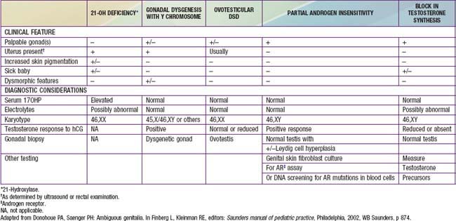
Chromosomal aberrations may result in ambiguity of the external genitalia. Conditions of aberrant sex differentiation may also occur with the XX or XY genotype. The appropriate term for what was previously called intersex is disorders of sex development (DSD). This term defines a condition “in which development of chromosomal, gonadal or anatomical sex is atypical.” It is becoming more preferable to use the term “atypical genitalia” rather than “ambiguous genitalia.” Comparison with the previous terms and a new etiologic classification are seen in Tables 582-1 and 582-2. Some of the genes involved in disorders of sex development are listed in Table 576-1.

Table 582-1 REVISED NOMENCLATURE

PREVIOUS CURRENTLY ACCEPTED
Intersex Disorders of sex development (DSD)
Male pseudohermaphrodite 46,XY DSD
Undervirilization of an XY male 46,XY DSD
Undermasculinization of an XY male 46,XY DSD
46,XY intersex 46,XY DSD
Female pseudohermaphrodite 46,XX DSD
Overvirilization of an XX female 46,XX DSD
Masculinization of an XX female 46,XX DSD
46,XX intersex 46,XX DSD
True hermaphrodite Ovotesticular DSD
Gonadal intersex Ovotesticular DSD
XX male or XX sex reversal 46,XX testicular DSD
XY sex reversal 46,XY complete gonadal dysgenesis

From Lee PA, Houk CP, Ahmed SF, et al: Consensus statement on management of intersex disorders, Pediatrics 118:e488–e500, 2006.

Table 582-2 ETIOLOGIC CLASSIFICATION OF DISORDERS OF SEX DEVELOPMENT (DSD)

46,XX-DSD

Androgen Exposure

Disorder of Ovarian Development

Undetermined Origin

Associated with genitourinary and gastrointestinal tract defects

46,XY DSD

Defects in Testicular Development

Deficiency of Testicular Hormones

Defect in Androgen Action

Ovotesticular DSD

Sex Chromosome DSD

From Lee PA, Houk CP, Ahmed SF, et al: Consensus statement on management of intersex disorders, Pediatrics 118:e488–e500, 2006.

The definition of atypical or ambiguous genitalia, in a broad sense, is any case in which the external genitalia do not appear completely male or completely female. Although there are standards for genital size dimensions, variations in size of these structures do not always constitute ambiguity.

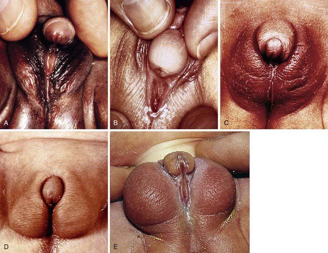
Development of the external genitalia begins with the potential to be either male or female (Fig. 582-1). Virilization of a female, the most common form of DSD, results in varying phenotypes (Fig. 582-2), which start from the basic genital appearances of the embryo (see Fig. 582-1).

image

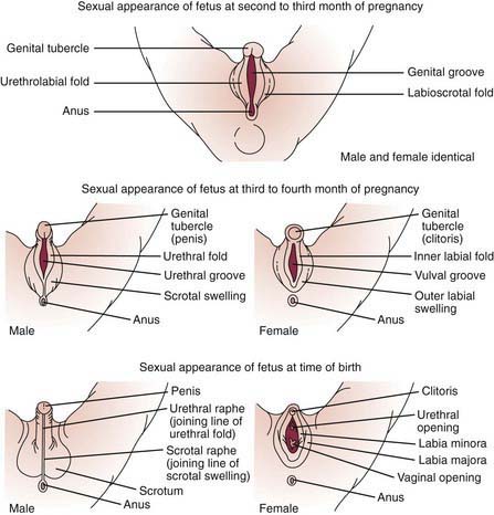
Figure 582-1 Schematic demonstration of differentiation of normal male and female genitalia during embryogenesis.

(From Zitelli BJ, Davis HW: Atlas of pediatric physical diagnosis, ed 4, St Louis, 2002, Mosby, p 328.)

Diagnostic Approach to the Patient with Atypical or Ambiguous Genitalia

The appearance of the external genitalia is rarely diagnostic of a particular disorder, and thus does not often allow distinction among the various forms of DSD. The most common forms of 46,XX DSD are virilizing forms of congenital adrenal hyperplasia (CAH). It is important to note that in 46,XY DSD, the specific diagnosis is not found in up to 50% of cases. At 1 center with a large experience, the etiologies of DSD in 250 patients over 25 yr were compiled. The 6 most common diagnoses accounted for 50% of the cases. These included virilizing CAH (14%), androgen insensitivity syndrome (10%), mixed gonadal dysgenesis (8%), clitoral/labial anomalies (7%), hypogonadotropic hypogonadism (6%), and 46,XY small-for-gestational age males with hypospadias (6%).

This potential source of error in diagnosis and management emphasizes the need for careful diagnostic evaluation including biochemical characterization of possible steroidogenic enzymatic defects in each patient with genital ambiguity. The parents need counseling about the complex nature of the baby’s condition, and guidance as to how to deal with their well-meaning but curious friends and family members. The evaluation and management should be carried out by a multidisciplinary team of experts that include pediatric endocrinology, pediatric surgery/urology, pediatric radiology, newborn medicine, genetics, and psychology. Once the sex of rearing has been agreed on by the family and team, treatment can be organized. Genetic counseling should be offered when the specific diagnosis is established.

After a complete history and physical exam, the common diagnostic approach includes multiple steps, described in the following outline. These steps are usually performed simultaneously rather than waiting for results of 1 test prior to performing another, due to the sensitive and sometimes urgent nature of the condition. Careful attention to the presence of physical features other than the genitalia is crucial, to determine if a diagnosis of a particular multisystem syndrome is possible. These are described in more detail in Chapters 582.1, 582.2, and 582.3. A summary of many features of commonly encountered causes of DSD is provided in Table 582-3.

Diagnostic tests include the following:

582.1 46,XX DSD

In this condition, the genotype is XX and the gonads are ovaries, but the external genitalia are virilized. Because there is no significant AMH production—the gonads are ovaries—the uterus, fallopian tubes, and cervix develop. The varieties and causes of this condition are relatively few. Most instances result from exposure of the female fetus to excessive exogenous or endogenous androgens during intrauterine life. The changes consist principally of virilization of the external genitalia (clitoral hypertrophy and labioscrotal fusion).

Congenital Adrenal Hyperplasia (Chapter 570.1)

This is the most common cause of genital ambiguity and of 46,XX DSD. Females with the 21-hydroxylase and 11-hydroxylase defects are the most highly virilized, although minimal virilization also occurs with the type II 3β-hydroxysteroid dehydrogenase defect (see Fig. 582-1). Salt losers tend to have greater degrees of virilization than do non–salt-losing patients. Masculinization may be so intense that a complete penile urethra results, and the condition may mimic a male with bilateral cryptorchidism.

Bibliography

Bose HS, Pescovitz OH, Miller WL. Spontaneous feminization in a 46,XX female patient with congenital lipoid adrenal hyperplasia due to a homozygous frameshift mutation in the steroidogenic acute regulatory protein. J Clin Endocrinol Metab. 1997;82:1511-1515.

Diamond M, Sigmundson K. Sex reassignment at birth. Arch Pediatr Adolesc Med. 1997;151:298-304.

Diamond DA, Mitchell C, Lamb K, et al. Sex assignment for newborns with ambiguous genitalia and exposure to fetal testosterone: attitudes and practices of pediatric urologists. J Pediatr. 2006;148:445-449.

Frade Costa EM, Bilharinho Mendonca B, Inacio M, et al. Management of ambiguous genitalia in pseudohermaphrodites: new perspectives on vaginal dilation. Fertil Steril. 1997;67:229-232.

Mendonca BB, Leite MV, DeCastro M, et al. Female pseudohermaphroditism caused by a novel homozygous mutation of the GR gene. J Clin Endocrinol Metab. 2002;87:1805-1809.

Moisan AM, Ricketts ML, Tardy V, et al. New insight into the molecular basis of 3β-hydroxysteroid dehydrogenase deficiency: identification of eight mutations in the HSD3 gene in eleven patients from seven new families and comparison of the functional properties of twenty-five mutant enzymes. J Clin Endocrinol Metab. 1999;84:4410-4425.

Morishima A, Grumbach MM, Simpson ER, et al. Aromatase deficiency in male and female siblings caused by a novel mutation and the physiological role of estrogens. J Clin Endocrinol Metab. 1995;80:3689-3698.

Mullis PE, Yoshimura N, Kuhlmann B, et al. Aromatase deficiency in a female who is compound heterozygote for two new point mutations in the P450arom gene: impact of estrogens on hypergonadotropic hypogonadism, multicystic ovaries, and bone densitometry in childhood. J Clin Endocrinol Metab. 1997;82:1739-1745.

Parisi MA, Ramsdell LA, Burns MW, et al. A gender assessment team: experience with 250 patients over a period of 25 years. Genet Med. 2007;9:348-357.

Saenger P. New developments in congenital lipoid adrenal hyperplasia and steroidogenic acute regulatory protein. Pediatr Clin North Am. 1997;44:397-421.

Wallien MS, Cohen-Kettenis PT. Psychosexual outcome of gender-dysphoric children. J Am Acad Child Adolesc Psychiatry. 2008;47:1413-1423.

582.2 46,XY DSD

In this condition, the genotype is XY, but the external genitalia are either not completely virilized, ambiguous (atypical), or completely female. When gonads can be found, they invariably contain testicular elements; their development ranges from rudimentary to normal. Because the process of normal virilization in the fetus is so complex, it is not surprising that there are many varieties and causes of 46,XY DSD.

Defects in Testicular Differentiation

The 1st step in male differentiation is conversion of the indifferent gonad into a testis. In the XY fetus, if there is a deletion of the short arm of the Y chromosome or of the SRY gene, male differentiation does not occur. The phenotype is female; müllerian ducts are well developed because of the absence of AMH, but gonads consist of undifferentiated streaks. By contrast, even extreme deletions of the long arm of the Y chromosome (Yq-) have been found in normally developed males, most of whom are azoospermic and have short stature. This indicates that the long arm of the Y chromosome normally has genes that prevent these manifestations. In many syndromes in which the testes fail to differentiate, Y chromosomes are morphologically normal.

XY Gonadal Agenesis Syndrome (Embryonic Testicular Regression Syndrome)

In this rare syndrome, the external genitalia are slightly ambiguous but more nearly female. Hypoplasia of the labia; some degree of labioscrotal fusion; a small, clitoris-like phallus; and a perineal urethral opening are present. No uterus, no gonadal tissue, and usually no vagina can be found. At the age of puberty, no sexual development occurs and gonadotropin levels are elevated. Most children have been reared as females. In several patients with XY gonadal agenesis in whom no gonads could be found on exploration, significant rises in testosterone followed stimulation with hCG, indicating Leydig cell function somewhere. Siblings with the disorder are known.

It is presumed that testicular tissue was active long enough during fetal life for AMH to inhibit development of müllerian ducts but not long enough for testosterone production to result in virilization. In 1 patient, no deletion of the Y chromosome was found by means of Y-specific DNA probes. Testicular degeneration seems to occur between the 8th and the 12th fetal wk. Regression of the testis before the 8th wk of gestation results in Swyer syndrome; between the 14th and the 20th wk of gestation, it results in the rudimentary testis syndrome; and after the 20th wk, it results in anorchia.

In bilateral anorchia, testes are absent, but the male phenotype is complete; it is presumed that tissue with fetal testicular function was active during the critical period of genital differentiation but that sometime later it was damaged. Bilateral anorchia in identical twins and unilateral anorchia in identical twins and in siblings suggest a genetic predisposition. Coexistence of anorchia and the gonadal agenesis syndrome in a sibship is evidence for a relationship between the disorders. SRY defects have not yet been reported for patients with anorchia.

A retrospective review of urologic explorations revealed absent testes in 21% of 691 testes. Of those, 73% had blind-ending cord structures with the suggested site of the vanishing testes being the inguinal canal (59%), the abdomen (21%), superficial inguinal ring (18%), and scrotum (2%). It was suggested that the presence of cord structures on laparoscopy should prompt inguinal exploration because viable testicular tissue was found in 4 of these children. No hormonal data (hCG stimulation tests, AMH levels) were reported.

This condition is sometimes referred to as vanishing testes syndrome.

Defects in Testicular Hormones

Five genetic defects have been delineated in the enzymatic synthesis of testosterone by fetal testis, and a defect in Leydig cell differentiation has been described. These defects produce 46,XY males with inadequate masculinization (see Fig. 576-1). Because levels of testosterone are normally low before puberty, an hCG stimulation test may be needed in children to assess the ability of the testes to synthesize testosterone.

Lipoid Adrenal Hyperplasia (Chapter 570)

The most severe form of congenital adrenal hyperplasia derives its name from the appearance of the enlarged adrenal glands resulting from accumulation of cholesterol and cholesterol esters. The rate-limiting process in steroidogenesis is the transport of free cholesterol through the cytosol to the inner mitochondrial membrane, where P450SCC (CYP11A1) acts. Cholesterol transport into mitochondria is mediated by the steroidogenetic acute regulatory protein (StAR) whose synthesis occurs via cAMP through a cyclic AMP response element–binding protein (CREB). StAR is a 30 kDa protein essential for steroidogenesis and is encoded by a gene on chromosome 8p11.2. The mitochondrial content of StAR increases between 1 and 5 hr after ACTH stimulation, long after the acute ACTH-induced increase in steroidogenesis. This has led some to suggest that extramitochondrial StAR might also be involved in the acute response to ACTH.

All serum steroid levels are low or undetectable, whereas corticotropin and plasma renin levels are quite elevated. The phenotype is female in both genetic females and males. Genetic males have no müllerian structures because the testes can produce normal AMH but no steroid hormones. These children present with acute adrenal crisis and salt wasting in infancy. Most patients are 46,XY. In a few patients, ovarian steroidogenesis is present at puberty.

The regulatory role of StAR-independent steroidogenesis is illustrated by 46,XX 4 mo old twins with lipoid adrenal hyperplasia. One died at 15 mo because of cardiac complications related to coarctation of the aorta. The adrenal glands had characteristic lipid deposits. The surviving twin had spontaneous puberty with feminization at 11.5 yr and menarche at 13.8 yr. When restudied at the age of 15 yr, a homozygous frameshift-inactivating mutation in her StAR gene was discovered. This and the fact that she survived as an infant until 4 mo of age without replacement therapy with detectable serum aldosterone levels supports the hypothesis that StAR-independent steroidogenesis was able to proceed until enough intracellular lipid accumulated to destroy steroidogenic activity. Partial defects in only partially virilized males and delayed onset of salt wasting have been described. Complete P450scc defects may be incompatible with life because only this enzyme can convert cholesterol to pregnenolone, which then becomes progesterone, a hormone essential for the maintenance of normal mammalian pregnancy. Heterozygous mutation in P450scc was described in a 4 yr old with 46,XY sex reversal and late-onset form of lipoid adrenal hyperplasia. At 6-7 wk of gestation, when maternal corpus luteum progesterone synthesis stops, the placenta, which does not express StAR, produces progesterone by StAR-independent steroidogenesis using the P450scc enzyme system.

3β-Hydroxysteroid Dehydrogenase Deficiency

Males with this form of congenital adrenal hyperplasia (Chapter 570) have various degrees of hypospadias, with or without bifid scrotum and cryptorchidism and, rarely, a complete female phenotype. Affected infants usually develop salt-losing manifestations shortly after birth. Incomplete defects, occasionally seen in boys with premature pubarche, as well as late-onset nonclassic forms have been reported. These children have point mutations of the gene for type II 3β-hydroxysteroid enzyme, resulting in impairment of steroidogenesis in the adrenals and gonads; the impairment may be unequal between adrenals and gonads. Normal pubertal changes in some boys could be explained by the normally present type I 3β-hydroxysteroid dehydrogenase present in many peripheral tissues. Infertility is frequent. There is no correlation between degree of salt wasting and degree of phenotypic abnormality.

Deficiency of 17-Hydroxylase/17,20 Lyase

A single enzyme (P450C17 or CYP17) encoded by a single gene on chromosome 10q24.3 has both 17-hydroxylase and 17,20 lyase activities in adrenal and gonadal tissues (Chapter 570). Many different genetic lesions have been reported. Genetic males usually have a complete female phenotype or, less often, various degrees of undervirilization from labioscrotal fusion to perineal hypospadias and cryptorchidism. Pubertal development fails to occur in both genetic sexes.

In the classical disorder, there is decreased synthesis of cortisol by the adrenals and of sex steroids by the adrenals and gonads. Levels of deoxycorticosterone (DOC) and corticosterone are markedly increased and lead to the hypertension and hypokalemia characteristic of this form of male DSD. Although levels of cortisol are low, the elevated corticotropin and corticosterone levels maintain a eucorticoid state. The renin-aldosterone axis is suppressed because of the strong mineralocorticoid effect of elevated DOC. Virilization does not occur at puberty; levels of testosterone are low, and those of gonadotropins are increased. Because fetal production of AMH is normal, no müllerian duct remnants are present. In phenotypic XY females, gonadectomy and replacement therapy with hydrocortisone and sex steroids are indicated.

The defect follows autosomal recessive inheritance. Affected XX females are usually not detected until young adult life, when they fail to experience normal pubertal changes and are found to have hypertension and hypokalemia. This condition should be suspected in patients presenting with primary amenorrhea and hypertension whose chromosomal complement is either 46,XX or 46,XY.

Deficiency of 17-Ketosteroid Reductase

This enzyme, also called 17β-hydroxysteroid dehydrogenase (17β-HSD), catalizes the final step in testosterone biosynthesis. It is necessary to convert androstenedione to testosterone and also dehydroepiandrosterone to androstenediol and estrone to estradiol. Enzymatic defects in the fetal testis give rise to males with complete or near-complete female phenotype in 46,XY males. Müllerian ducts are absent, and a shallow vagina is present. The diagnosis is based on the ratio of androstenedione to testosterone; in prepubertal children, stimulation with hCG may be necessary to make the diagnosis.

The defect is inherited in an autosomal recessive fashion. At least 4 different types of 17β-HSD are recognized, each coded by a different gene or different chromosomes. Type III is the enzyme defect that is especially common in a highly inbred Arab population in Gaza. The gene for the disorder is at 9q22 and is expressed only in the testes, where it converts androstenedione to testosterone. Most patients are diagnosed at puberty because of the failure to menstruate and of virilization. Testosterone levels at puberty may approach normal, presumably as a result of peripheral conversion of androstenedione to testosterone; at this time, some patients spontaneously adopt a male gender role.

Type I 17β-HSD, encoded by a gene on chromosome 17q21, converts estrone to estradiol and is found in placenta, ovary, testis, liver, prostate, adipose tissue, and endometrium. Type II, whose gene is on chromosome 16q24, has activities that are opposite to those of types I and III (convert testosterone to androstenedione and estrone to estradiol). Type IV is similar in action to type II. A late-onset form of 17-ketosteroid reductase deficiency presents as gynecomastia in young adult males.

Defects in Androgen Action

In the following group of disorders, fetal synthesis of testosterone is normal and defective virilization results from inherited abnormalities in androgen action.

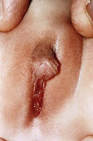
5α-Reductase Deficiency

Decreased production of dihydrotestosterone (DHT) in utero results in marked ambiguity of external genitalia of affected males. Biosynthesis and peripheral action of testosterone are normal.

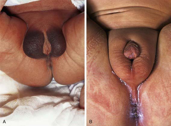
The phenotype most commonly associated with this condition results in boys who have a small phallus, bifid scrotum, urogenital sinus with perineal hypospadias, and a blind vaginal pouch (Fig. 582-3). Testes are in the inguinal canals or labioscrotal folds and are normal histologically. There are no müllerian structures. Wolffian structures—the vas deferens, epididymis, and seminal vesicles—are present. Most affected patients have been identified as females. At puberty, virilization occurs; the phallus enlarges, the testes descend and grow normally, and spermatogenesis occurs. There is no gynecomastia. Beard growth is scanty, acne is absent, the prostate is small, and recession of the temporal hairline fails to occur. Virilization of the Wolffian duct is caused by the action of testosterone itself, although masculinization of the urogenital sinus and external genitals depends on the action of DHT during the critical period of fetal masculinization. Growth of facial hair and of the prostate also appears to be DHT dependent.

image

Figure 582-3 5α-Reductase deficiency.

(From Wales JKH, Wit JM, Rogol AD: Pediatric endocrinology and growth, ed 2, Philadelphia, 2003, Elsevier/Saunders, p 165.)

The adult height reached is close to that of the father and other male siblings. There is significant phenotypic heterogeneity. This has led to a classification of such patients into 5 types of steroid 5α-reductase deficiency (SRD).

Several different gene defects leading to SRD have been identified in the 5α-reductase type 2 gene, located on the short arm of chromosome 2, in patients from throughout the world. Familial clusters have been reported from the Dominican Republic, Turkey, Papua New Guinea, Brazil, Mexico, and the Middle East. There is no reliable correlation between genotype and phenotype.

The disorder is inherited as an autosomal recessive trait but is limited to males; normal homozygous females with normal fertility indicate that in females DHT has no role in sexual differentiation or in ovarian function later in life. The clinical diagnosis should be made as early as possible in infancy. It is important to distinguish this from partial androgen insensitivity syndrome (PAIS), as patients with PAIS are far less sensitive to androgen than are patients with SRD. The biochemical diagnosis of SRD is based on finding normal serum testosterone levels, normal or low DHT levels with markedly increased basal and especially hCG-stimulated testosterone : DHT ratios (>17), and high ratios of urinary etiocholanolone to androsterone. Children with androgen insensitivity have normal hepatic 5α reduction and, thus, a normal ratio of tetrahydrocortisol to 5α-tetrahydrocortisol, as opposed to those with SRD.

It is important to note that most but not all children with SRD reared as females in childhood have changed to male around the time of puberty. It appears that exposures to testosterone in utero, neonatally, and at puberty have variable contributions to the formation of male gender identity. Much more needs to be learned about the influences of hormones such as androgens as well as the influences of cultural, social, psychologic, genetic, and other biologic factors in gender identity and behavior. Infants with this condition should be reared as boys whenever practical. Treatment of male infants with DHT results in phallic enlargement.

Androgen Insensitivity Syndromes

The AISs are the most common forms of male DSD, occurring with an estimated frequency of 1/20,000 genetic males. This group of heterogeneous X-linked recessive disorders is due to more than 150 different mutations in the androgen receptor gene, located on Xq11-12: single point mutations resulting in amino acid substitutions or premature stop codons, frameshift and premature terminations, gene deletions, and splice site mutations.

Clinical Manifestations

The clinical spectrum of patients with AISs, all of whom have a 46,XY chromosomal complement, range from phenotypic females (in complete AIS) to males with various forms of ambiguous genitalia and undervirilization (partial AIS, or clinical syndromes such as Reifenstein syndrome) to phenotypically normal-appearing males with infertility. In addition to normal 46,XY chromosomes, the presence of testes and normal or elevated testosterone and LH levels are common to all such children (Figs. 582-4 and 582-5).

In complete AIS, an extreme form of failure of virilization, genetic males appear female at birth and are invariably reared accordingly. The external genitalia are female. The vagina ends blindly in a pouch, and the uterus is absent due to the normal production and effect of AMH by the testes. In about one third of patients, unilateral or bilateral fallopian tube remnants are found. The testes are usually intra-abdominal but may descend into the inguinal canal; they consist largely of seminiferous tubules. At puberty, there is normal development of breasts, and the habitus is female, but menstruation does not occur and sexual hair is absent. Adult heights of these women are commensurate with those of normal males despite profound congenital deficiency of androgenic effects.

The testes of affected adult patients produce normal male levels of testosterone and DHT. Failure of normal male differentiation during fetal life reflects defective response to androgens at that time. The absence of androgenic effects is caused by a striking resistance to the action of endogenous or exogenous testosterone at the cellular level.

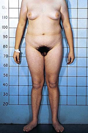
Prepubertal children with this disorder are often detected when inguinal masses prove to be testes or when a testis is unexpectedly found during herniorrhaphy in a phenotypic female. About 1-2% of girls with an inguinal hernia prove to have this disorder. In infants, elevated gonadotropin levels should suggest the diagnosis. In adults, amenorrhea is the usual presenting symptom. In prepubertal children, the condition must be differentiated from other types of XY under virilized males in which there is complete feminization. These include XY gonadal dysgenesis (Swyer syndrome), true agonadism, Leydig cell aplasia including LH receptor defects, and 17-ketosteroid reductase deficiency; all these conditions, unlike complete AIS, are characterized by low levels of testosterone as neonates and during adult life and by failure to respond to hCG during the prepubertal years. Although patients with complete AIS have unambiguously female external genitals at birth, those with partial AIS have a wide variety of phenotypic presentations ranging from perineoscrotal hypospadias, bifid scrotum, and cryptorchidism to extreme under virilization appearing as clitoromegaly and labial fusion. Some forms of partial AIS have been known as specific syndromes. Patients with Reifenstein syndrome have incomplete virilization characterized by hypogonadism, severe hypospadias, and gynecomastia (see Fig. 582-5). Gilbert-Dreyfus and Lubs are additional syndromes classified as partial AIS. In all cases, abnormalities in the androgen receptor gene have been identified.

Treatment and Prognosis

In patients with CAIS whose sexual orientation is unambiguously female, the testes should be removed as soon as they are discovered. Laparoscopic removal of Y chromosome–bearing gonads has been performed in patients with AIS and in those with gonadal dysgenesis. In one third of patients, malignant tumors, usually seminomas, develop by 50 yr of age. Several teenage girls have acquired seminomas. Replacement therapy with estrogens is indicated at the age of puberty.

Normal breasts develop in affected girls who have not had their testes removed by the age of puberty. In these individuals, production of estradiol results from aromatase activity on testicular testosterone. The absence of androgenic activity also contributes to the feminization of these women.

The psychosexual and surgical management of patients with partial AIS is extremely complex and depends in large part on the presenting phenotype. Osteopenia is recognized as a late feature of AIS.

Molecular analyses have suggested that phenotype may depend in part on somatic mosaicism of the androgen receptor gene. This was based on the case of a 46,XY patient who had a premature stop codon in exon 1 of the AR gene but who also had evidence of virilization (pubic hair and clitoral enlargement) explained by the discovery of the wild-type alleles on careful examination of the sequencing gel. The presence of mosaicism shifts the phenotype to a higher degree of virilization than expected from the genotype of the mutant allele alone.

Genetic counseling is difficult in families with androgen receptor gene mutation. In addition to lack of genotype-phenotype correlations, there is a high rate (27%) of de novo mutations in families.

Sex hormone–binding globulin reduction after exogenous androgen administration (stanozolol) has been shown to correlate with the severity of the receptor defect and may become a useful clinical tool. Successful therapy with supplemental androgens has been reported in patients with partial AIS and various mutations of the androgen receptor in the DNA-binding domain and the ligand-binding domain.

Mutated androgen receptors are also reported in patients with spinal and bulbar muscular atrophy in whom clinical manifestations including testicular atrophy, infertility, gynecomastia, and elevated LH, FSH, and estradiol levels usually manifest between the 3rd and 5th decades of life. Androgen receptor mutations have also been described in patients with prostate cancer.

Undetermined Causes

Other XY undervirilized males display great variability of the external and internal genitalia and various degrees of phallic and müllerian development. Testes may be histologically normal or rudimentary, or there may only be 1. Even the newer techniques may find no recognized cause in a up to 50% of children with 46,XY DSD. Some ambiguity of the genitalia is associated with a wide variety of chromosomal aberrations, which must always be considered in the differential diagnosis, the most common being the 45,X/46,XY syndrome (Chapter 580.1). It may be necessary to karyotype several tissues to establish mosaicism. Other complex genetic syndromes, many resulting from single gene mutations, are associated with varying degrees of ambiguity of the genitalia, particularly in the male. These entities must be identified on the basis of the associated extragenital malformations.

Smith-Lemli-Opitz syndrome is an autosomal recessive disorder caused by mutations in the sterol Δ7-reductase gene located on chromosome 11q12-q13. It is characterized by prenatal and postnatal growth retardation, microcephaly, ptosis, anteverted nares, broad alveolar ridges, syndactyly of the 2nd-3rd toes, and severe mental retardation (Chapter 80.3). Its incidence is 1/20,000 to 1/60,000; 70% are male. Genotypic males usually have genital ambiguity and, occasionally, partial sex reversal with female genital ambiguity or complete sex reversal with female external genitals. Müllerian duct derivatives are usually absent. Affected 46,XX patients have normal genitalia. Two types of Smith-Lemli-Opitz syndrome have been recognized. The classical form (type I) described earlier and the acrodysgenital syndrome, which is usually lethal within 1 yr and is associated with severe malformations, postaxial polydactyly, and extremely abnormal external genitalia (type II). Pyloric stenosis is associated with Smith-Lemli-Opitz syndrome type I and Hirschsprung disease with type II. Cleft palate, skeletal abnormalities, and 1 case of a lipoma of the pituitary gland have been seen in type II cases. Some authors believe in a spectrum of disease severity rather than in the above classification. Low plasma cholesterol with elevated 7-dehydrocholesterol, its precursor, are found in types 1 and 2, and the levels do not correlate with severity. Maternal apolipoprotein E values do seem to correlate with severity. The most common prenatal expression of Smith-Lemli-Opitz syndrome is intrauterine growth retardation (see Chapter 80.3 for treatment).

46,XY DSD subjects also have been described in siblings with the α-thalassemia/mental retardation syndrome.

Bibliography

Brinkmann AO. Molecular basis of androgen insensitivity. Mol Cell Endocrinol. 2001;179:105-1099.

Canto P, Vilchis F, Chavez B, et al. Mutations of the 5α-reductase type 2 gene in eight Mexican patients from six different pedigrees with 5α-reductase-2 deficiency. Clin Endocrinol. 1997;46:155-160.

Chu J, Zhang R, Zhao Z, et al. Male fertility is compatible with an Arg840 Cys substitution in the AR in a large Chinese family affected with divergent phenotypes of AR insensitivity syndrome. J Clin Endocrinol Metab. 2002;87:347-351.

Diamond DA, Mitchell C, Lamb K, et al. Sex assignment for newborns with ambiguous genitalia and exposure to fetal testosterone: attitudes and practices of pediatric urologists. J Pediatr. 2006;148:445-449.

Diamond M, Sigmundson K. Sex reassignment at birth. Arch Pediatr Adolesc Med. 1997;151:298-304.

Frade Costa EM, Bilharinho Mendonca B, Inacio M, et al. Management of ambiguous genitalia in pseudohermaphrodites: new perspectives on vaginal dilation. Fertil Steril. 1997;67:229-232.

Geissler WM, Davis DL, Wu L, et al. Male pseudohermaphroditism caused by mutation of testicular 17β-hydroxysteroid dehydrogenase 3. Nat Genet. 1994;7:34-39.

Gul D. Third case of WAGR syndrome with severe obesity and constitutional deletion of chromosome (11)(p12p14). Am J Med Genet. 2002;107:70-71.

Hiort O, Sinnecker GHG, Holterbus PM, et al. Inherited and de novo androgen receptor gene mutations: investigation of single-case families. J Pediatr. 1998;132:939-943.

Hochberg Z, Chayen R, Reiss N, et al. Clinical, biochemical and genetic findings in a large pedigree of male and female patients with 5α-reductase 2 deficiency. J Clin Endocrinol Metab. 1996;81:2821-2827.

Kremer H, Karaaij R, Toledo SPA, et al. Male pseudohermaphroditism due to a homozygous missense mutation of the luteinizing hormone receptor gene. Nat Genet. 1995;9:160-164.

McPherson EW, Clemens MM, Gibbons RJ, et al. X-linked α-thalassemia/mental retardation (ATR-X) syndrome: a new kindred with severe genital anomalies and mild hematologic expression. Am J Med Genet. 1995;55:302-306.

Mebarki F, Sanchez R, Rheaumes E, et al. Non-salt-losing male pseudohermaphroditism due to the novel homozygous N100S mutation in the type II 3 β-hydroxysteroid dehydrogenase gene. J Clin Endocrinol Metab. 1995;80:2127-2134.

Mendonca BB, Inacio M, Costa EMF, et al. Male pseudohermaphroditism due to steroid 5α-reductase 2 deficiency. Medicine. 1996;75:64-76.

Moisan AM, Ricketts ML, Tardy V, et al. New insight into the molecular basis of 3β-hydroxysteroid dehydrogenase deficiency: identification of eight mutations in the HSD3 gene in eleven patients from seven new families and comparison of the functional properties of twenty-five mutant enzymes. J Clin Endocrinol Metab. 1999;84:4410-4425.

Mongan NP, Jaaskelainen J, Green K, et al. Two de novo mutations in the AR gene cause the complete androgen insensitivity syndrome in a pair of monozygotic twins. J Clin Endocrinol Metab. 2002;87:1057-1061.

Mueller RF. The Denys-Drash syndrome. J Med Genet. 1994;31:471-477.

Parisi MA, Ramsdell LA, Burns MW, et al. A gender assessment team: experience with 250 patients over a period of 25 years. Genet Med. 2007;9:348-357.

Quigley CA, French FS. Androgen insensitivity syndromes. Curr Ther Endocrinol Metab. 1994;5:342-351.

Reardon W, Gibbons RJ, Winter RM, et al. Male pseudohermaphroditism in sibs with the α-thalassemia/mental retardation (ATR-X) syndrome. Am J Med Genet. 1995;55:285-287.

Saenger P. New developments in congenital lipoid adrenal hyperplasia and steroidogenic acute regulatory protein. Pediatr Clin North Am. 1997;44:397-421.

Sarafoglou K, Ostrer H. Clinical Review 111—familial sex reversal: a review. J Clin Endocrinol Metab. 2000;85:483-493.

Tajima T, Fujieda K, Kouda N, et al. Heterozygous mutation in the cholesterol side chain cleavage enzyme (P450scc) gene in a patient with 46,XY sex reversal and adrenal insufficiency. J Clin Endocrinol Metab. 2001;86:3820-3825.

Wallien MS, Cohen-Kettenis PT. Psychosexual outcome of gender-dysphoric children. J Am Acad Child Adolesc Psychiatry. 2008;47:1413-1423.

Weidemann W, Peters B, Romalo G, et al. Response to androgen treatment in a patient with partial androgen insensitivity and a mutation in the deoxyribonucleic acid-binding domain of the androgen receptor. J Clin Endocrinol Metab. 1998;83:1173-1176.

582.3 Ovotesticular DSD

In ovotesticular DSD, both ovarian and testicular tissues are present, either in the same or in opposite gonads. Affected patients have ambiguous genitalia, varying from normal female with only slight enlargement of the clitoris to almost normal male external genitalia (see Fig. 582-2A).

About 70% of all patients have a 46,XX karyotype. Ninety-seven percent of affected patients of African descent are 46,XX. Fewer than 10% of persons with ovotesticular DSD are 46,XY. About 20% have 46,XX/46,XY mosaicism. Half of these are derived from more than 1 zygote and are chimeras (chi 46,XX/46,XY). The presence of paternal and both maternal alleles for some blood groups is demonstrated. An ovotesticular DSD chimera, 46,XX/46,XY, was reported as resulting from embryo amalgamation after in vitro fertilization. Each embryo was derived from an independent, separately fertilized ovum.

Examination of 46,XX ovotesticular DSD patients with Y-specific probes has detected fewer than 10% with a portion of the Y chromosome including the SRY gene. Ovotesticular DSD is usually sporadic, but a number of siblings have been reported. The cause of most cases of ovotesticular DSD is unknown.

The most frequently encountered gonad in ovotesticular DSD is an ovotestis, which may be bilateral. If unilateral, the contralateral gonad is usually an ovary but may be a testis. The ovarian tissue is normal, but the testicular tissue is dysgenetic. The presence and function of testicular tissue can be determined by measuring basal and hCG-stimulated testosterone levels as well as AMH levels. Patients who are highly virilized and have had adequate testicular function with no uterus are usually reared as males. If a uterus exists, virilization is often mild and testicular function minimal; assignment of female sex may be indicated. Selective removal of gonadal tissue inconsistent with sex of rearing may be indicated. In a few families, 46,XY ovotesticular DSD subjects and 44,XX males have been described in the same sibship.

Pregnancies with living offspring have been reported in 46,XX ovotesticular DSD individuals reared as females, but very few males with ovotesticular DSD have fathered children. About 5% of patients will develop gonadoblastomas, dysgerminomas, or seminomas.

Diagnosis and Management

In the neonate, ambiguity of the genitals requires immediate attention to decide on the sex of rearing as early in life as possible. The family of the infant needs to be informed of the child’s condition as early, completely, compassionately, and honestly as possible. Caution must be used to avoid feelings of guilt, shame, and discomfort. Guidance needs to be provided to alleviate both short-term and long-term concerns and to allow the child to grow up in a completely supportive environment. The initial care is best provided by a team of professionals that include neonatologists and pediatric specialists, endocrinologists, radiologists, urologists, psychologists, and geneticists, all of whom remain focused foremost on the needs of the child. Management of the potential psychologic upheaval that these disorders can generate in the child or the family is of paramount importance and requires physicians and other health care professionals with sensitivity, training, and experience in this field.

While awaiting the results of chromosomal analysis, pelvic ultrasonography is indicated to determine the presence of a uterus and ovaries. Presence of a uterus and absence of palpable gonads usually suggests a virilized XX female. A search for the source of virilization should be undertaken; this includes studies of adrenal hormones to rule out varieties of congenital adrenal hyperplasia, and studies of androgens and estrogens occasionally may be necessary to rule out aromatase deficiency. Virilized XX females are generally (but not always) reared as females even when highly virilized.

The absence of a uterus, with or without palpable gonads, almost always indicates an under virilized male and an XY karyotype. Measurements of levels of gonadotropins, testosterone, AMH, and DHT are necessary to determine whether testicular production of androgen is normal. Under virilized males who are totally feminized may be reared as females. Certain significantly feminized infants, such as those with 5α-reductase deficiency, may be reared as males because these children virilize normally at puberty. Sixty percent of individuals with 5α-reductase deficiency assigned as female in infancy live as males. An infant with a comparable degree of feminization resulting from an androgen receptor defect, such as CAIS, is best reared as a female.

When receptor disorders are suspected in the XY male with a small phallus (micropenis), a course of 3 monthly intramuscular injections of testosterone enanthate (25-50 mg) may assist in the differential diagnosis of androgen insensitivity, as well as in treatment.

In some mammals, the female exposed to androgens prenatally or in early postnatal life exhibits nontraditional sexual behavior in adult life. Most, but not all, girls who have undergone fetal masculinization from congenital adrenal hyperplasia or from maternal progestin therapy have female sexual identity, although during childhood they may appear to prefer male playmates and activities over female playmates and feminine play with dolls in mothering roles.

In the past it was thought that surgical treatment of ambiguous genitalia to create a female appearance, particularly when a vagina is present, was more successful than construction of male genitalia. Considerable controversy exists regarding these decisions. Sexual functioning is to a large extent more dependent on neurohormonal and behavioral factors than the physical appearance and functional ability of the genitalia. Similarly, controversy exists regarding the timing of the performance of invasive and definitive procedures, such as surgery. Whenever possible without endangering the physical or psychologic health of the child, an expert multidisciplinary team should consider deferring elective surgical repairs and gonadectomies until the child can participate in the informed consent for the procedure. One study (n = 59 boys and 18 girls) with gender dysphoria but without documentation of genomic or enzymologic abnormalities indicated that most of these children no longer have gender dysphoria after completion of puberty. Among those who do, homosexuality and bisexuality are the most frequent diagnoses.

The pediatrician, pediatric endocrinologist, and psychologist, along with the appropriate additional specialists, should provide ongoing compassionate, supportive care to the patient and the patient’s family throughout childhood, adolescence, and adulthood. Support groups are available for families and patients with many of the conditions discussed.

Bibliography

Cameron FJ, Montalto J, Byrt E, et al. Gonadal dysgenesis: associations between clinical features and sex of rearing. Endocr J. 1997;44:95-104.

Creighton S, Minto C. Managing intersex. Br Med J. 2001;323:1264-1265.

Daaboul J, Frader J. Ethics and the management of the patient with intersex: a middle way. J Pediatr Endocrinol Metab. 2001;14:1575-1583.

Diamond DA, Mitchell C, Lamb K, et al. Sex assignment for newborns with ambiguous genitalia and exposure to fetal testosterone: attitudes and practices of pediatric urologists. J Pediatr. 2006;148:445-449.

Diamond M, Sigmundson K. Sex reassignment at birth. Arch Pediatr Adolesc Med. 1997;151:298-304.

Frade Costa EM, Bilharinho Mendonca B, Inacio M, et al. Management of ambiguous genitalia in pseudohermaphrodites: new perspectives on vaginal dilation. Fertil Steril. 1997;67:229-232.

Houk CP, Hughes IA, Ahmed SF, et al. Summary of consensus statement on intersex disorders and their management. Pediatrics. 2006;118:753-757.

Lee PA, Houk CP, Ahmed SF, et al. Consensus statement on management of intersex disorders. Pediatrics. 2006;118:e488-e500.

MacLaughlin DT, Donahoe PK. Sex determination and differentiation. N Engl J Med. 2004;350:367-378.

Parisi MA, Ramsdell LA, Burns MW, et al. A gender assessment team: experience with 250 patients over a period of 25 years. Genet Med. 2007;9:348-357.

Smith EP, Boyd J, Frank GR, et al. Estrogen resistance caused by a mutation in the estrogen-receptor gene in a man. N Engl J Med. 1994;331:1056-1061.