Diseases of the Thoracic Cage and Respiratory Muscles

Published on 06/06/2015 by admin

Filed under Pulmolory and Respiratory

Last modified 22/04/2025

Print this page

rate 1 star rate 2 star rate 3 star rate 4 star rate 5 star
Your rating: none, Average: 0 (0 votes)

This article have been viewed 3573 times

Chapter 64 Diseases of the Thoracic Cage and Respiratory Muscles

Dyspnea and alveolar hypoventilation occur in patients with restrictive lung disease as a consequence of an imbalance between the respiratory muscle load and respiratory muscle capacity. Subsequent modification of the neural respiratory drive occurs, which directly reflects this imbalance. This chapter begins with an overview of the anatomy of the respiratory muscle pump and includes a detailed explanation of the pathophysiology of chronic respiratory failure. It provides a comprehensive guide to the physician of how best to assess and investigate a patient with suspected respiratory compromise from respiratory muscle weakness, chest wall disease, or obesity. The second part of the chapter focuses on specific neurologic/neuromuscular and chest wall diseases associated with respiratory impairment.

Respiratory Muscle Pump

The respiratory system is made up of two main components: the respiratory muscle pump, which facilitates airflow, thereby enabling ventilation, and the lungs, which support pulmonary gas exchange (see Chapter 6 for more detailed explanations and for respiratory muscle testing). The respiratory muscle pump itself is made up of the inspiratory muscles, the diaphragm and extradiaphragmatic accessory muscles, and the expiratory muscles, principally the abdominal wall muscles. Inspiration is an active process in which contraction of the diaphragm forces the abdominal contents in an anterocaudal direction to allow the volume of the chest cavity to increase. This expansion, in turn, generates a negative subatmospheric pressure to form a pressure gradient that drives air into the lungs. At rest, expiration is a passive process with the elastic properties of the chest wall and lungs providing the recoil forces that allow the lung volume to return to the functional residual capacity. During exercise and forced expiratory maneuvers (e.g., coughing), however, contraction of the abdominal muscles occurs, pushing the diaphragm upward in the absence of flow limitation.

Clinical Consequences of Respiratory Muscle Pump Imbalance

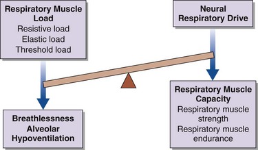
Breathlessness, alveolar hypoventilation and hypercapnic respiratory failure result from an imbalance between the respiratory muscle load, capacity, and neural respiratory drive (Figure 64-1). By using this model the physician can develop a simple clinical tool to assess the breathless patient and the patient in chronic respiratory failure. More commonly, in patients with restrictive lung disease, the reduction in capacity occurs as a consequence of intrinsic muscle weakness. On occasion, however, capacity may be relatively preserved, but in the context of a significant respiratory muscle load or poor orientation of the inspiratory muscles (e.g., kyphoscoliosis), this may limit the pressure-generating capacity, so that dyspnea and alveolar hypoventilation can develop subsequently. In critically ill patients, the respiratory muscle weakness may be transient and reversible, such as occurs in respiratory acidosis, hypokalemia, and hypophosphatemia (Box 64-1).

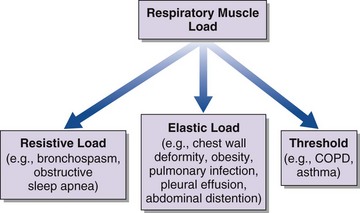
An increase in respiratory muscle load occurs in patients with increased airway resistance, chest wall deformity, or obesity. Any of these conditions will accelerate the onset of dyspnea and alveolar hypoventilation in patients with respiratory muscle weakness (Figure 64-2).

In healthy subjects, a significant reduction in neural respiratory drive to skeletal muscle, including the respiratory muscles, occurs during sleep. Despite the complete loss of skeletal muscle activity during rapid eye movement (REM) sleep, in normal subjects significant nocturnal hypoxia and hypercapnia are avoided because the activation of the diaphragm to maintain ventilation is preserved. By contrast, in patients with respiratory muscle weakness, especially diaphragmatic weakness and paralysis, hypoventilation during REM sleep often is the first sign of declining respiratory function. Of interest, in patients with idiopathic diaphragmatic paralysis, hypoventilation is prevented by a neuroadaptive mechanism with cyclic activation of the sternocleidomastoid muscle during REM sleep, which maintains ventilation. Also, there is substantial reserve in the respiratory muscle pump such that inspiratory muscle strength must fall to one third of normal before the onset of respiratory failure. Furthermore, clinical deterioration is observed at an earlier stage if an increased load is applied to the system, such as in pneumonia, or if the neural respiratory drive is modified with drugs such as benzodiazepines, opiates, and other anesthetic agents used during routine anesthesia.

Assessment of the Respiratory Muscle Pump

The respiratory muscle pump is essential for effective ventilation and to maintain gas exchange. In addition to a directed clinical history and examination, the physician can use simple bedside tests as well as more advanced measurements of respiratory muscle function to perform a detailed assessment of the patient with restrictive respiratory muscle and chest wall disease.

Clinical Features

As always, a detailed history will facilitate making a correct diagnosis (Table 64-1). Of particular importance is the rate of onset of symptoms. Acute respiratory muscle weakness tends to progress rapidly, culminating in a medical emergency requiring intubation and mechanical ventilation. Some causes of acute respiratory muscle weakness, such as an acute myasthenic crisis or Guillain-Barré disease, may be reversible, and it is important to facilitate invasive respiratory support in the acute setting until a diagnosis can be made and appropriate therapies initiated. In chronic respiratory failure, the onset may not be clear, but clues to the course of events can be gained from the history. Often patients will describe dyspnea on exertion, but if peripheral muscle weakness precedes respiratory muscle weakness, the consequent loss of significant locomotor function will not permit exertion of sufficient degree to produce dyspnea. Classic features of diaphragm weakness are not always present in patients with generalized neuromuscular disease but may include dyspnea on lying supine, on bending forward, or on immersion in water above the midchest level. If respiratory weakness is severe, patients also may describe symptoms of nocturnal hypoventilation, such as extensive daytime somnolence, reduced concentration, and morning headaches that typically resolve within 30 minutes of waking. Specifically, in the pediatric and adolescent populations, clinicians should be alerted to more subtle clinical features such as failing school performance, recurrent episodes of chest sepsis, reduction in appetite, and weight loss.

Table 64-1 Important Clinical Features in Diseases of the Thoracic Cage and Respiratory Muscles

Disorder Clinical Manifestations
Sleep-disordered breathing Morning headache, daytime sleepiness, disrupted sleep pattern, impaired intellectual function, generalized fatigue, loss of appetite and weight
Respiratory muscle weakness Orthopnea, breathlessness on immersion in water, breathlessness on leaning forward, breathlessness on exertion, poor cough, poor chest expansion, paradoxical abdominal motion during inspiration (inward motion of the anterior abdominal wall due to diaphragm weakness), abdominal muscle recruitment in expiration
Bulbar dysfunction Low volume voice, difficulty swallowing, drooling, difficulty clearing secretions, poor cough, staccato/slurred speech, coughing on swallowing

In all cases of respiratory muscle weakness, an important aspect of the evaluation is to identify any symptoms that may suggest generalized neuromuscular weakness—in particular, decrements in the patient’s speech and swallowing function, as well as weakness of the arms and legs. Weakness of the abdominal muscles may result in difficulty achieving an effective cough to clear secretions and debris from the airways. This impairment leads to issues with sputum retention and increases the risk of chest infection. In the case of chest wall disease, the common causes kyphoscoliosis and obesity usually are self-evident. As highlighted earlier, certain drugs have effects that can accelerate respiratory decline, so attention should be given to the common offenders, such as benzodiazepines, opiates, and corticosteroids. More recently, in particular, in the Duchenne muscular dystrophy (DMD) population, corticosteroids are increasingly being used to maintain locomotor muscle function, and these agents are associated with substantial weight gain and consequent upper airway obstruction. Clinicians must be aware of this potential problem in this younger patient population.

Physical examination requires a careful and considered approach, which must include a thorough neurologic examination to observe for signs of tongue and peripheral muscle fasciculation, peripheral muscle wasting and weakness, and peripheral sensory loss. Other signs, such as pseudohypertrophy of the calf muscles, are observed in patients with muscular dystrophies. Scars of previous operations may indicate possible trauma to underlying neuromuscular structures or an imposed restrictive chest wall deformity, such as from a phrenic nerve crush, thoracoplasty, coronary artery bypass grafting, thyroidectomy, or thymoma resection. With severe diaphragm weakness, abdominal inspiratory paradox is observed: The anterior abdominal wall moves inward as a result of the failure of a weak or paralyzed diaphragm to descend against the force exerted by the abdominal contents. With more generalized weakness, global loss of thoracic expansion on inspiration is observed.

Pulmonary Function Tests

Patients with respiratory neuromuscular weakness and those with chest wall disease both are characterized by a reduced ability to expand the thoracic rib cage. In turn, this failure to expand the rib cage results in a lack of generation of sufficient negative intrathoracic pressure to facilitate adequate inspiratory airflow. As a result, these patients present with clinical features of a restrictive lung function pattern characterized by a reduced forced vital capacity (FVC) and an elevated ratio of forced expiratory volume in 1 second (FEV1) to FVC (FEV1/FVC greater than 80%). Although vital capacity (VC) is in widespread use and is relatively simple to determine, its utility is limited because it has low sensitivity. In particular, significant inspiratory muscle weakness is required before a significant fall in VC is observed. A fall in VC on adoption of the supine position is specific for respiratory muscle weakness, and a fall in VC of more than 20% suggests bilateral diaphragmatic weakness. Of importance, the VC can be preserved in persons with a moderate degree of global respiratory muscle weakness or hemidiaphragm weakness.

The pattern of respiratory muscle weakness must be considered in interpreting results of pulmonary function tests in patients with chest wall disease and respiratory muscle weakness. Although total lung capacity (TLC) and VC are reduced in patients with predominantly inspiratory muscle weakness and combined inspiratory and expiratory muscle weakness, the pattern of weakness influences functional residual capacity (FRC), residual volume (RV), overall gas transfer (DLCO), and gas transfer corrected for alveolar volume (Table 64-2).

Table 64-2 Pulmonary Function Tests for Patients with Predominantly Inspiratory Muscle Weakness and Combined Inspiratory/Expiratory Muscle Weakness

Inspiratory Muscle Weakness Combined Inspiratory/Expiratory Muscle Weakness
Reduced VC Reduced VC
Fall in supine VC >20% N/A
FEV1/FVC ratio >80% FEV1/FVC ratio >80%
Reduced TLC Reduced TLC
Normal RV Increased RV
Reduced FRC Increased FRC
Reduced TLCO Reduced TLCO
“Supranormal” KCO Reduced KCO

FEV1, forced expiratory volume in 1 second FRC, functional residual capacity; FVC, forced vital capacity; KCO, gas transfer coefficient corrected for alveolar volume; N/A, not available; RV, residual volume; TLC, total lung capacity; TLCO, overall gas transfer; VC, vital capacity.

Tests of Respiratory Muscle Function

Because respiratory muscle contraction generates tension, the consequent pressure change that occurs can serve as an in vivo measure for the quantification of respiratory muscle strength. In addition, recent developments have expanded the current understanding of the mechanical actions and interactions of the respiratory muscles. A greater emphasis has been placed on assessing and quantifying respiratory muscle strength and pulmonary mechanics in a variety of patient groups, including children, adolescents, and adults (see Chapter 6 for a more detailed discussion).

Maximal Inspiratory and Expiratory Pressures

Maximal inspiratory and expiratory pressure measurements are clinically useful noninvasive tests with established reference ranges. As with any volitional test, however, results will be dependent on subject motivation and maximal effort, which explains in part the wide range of normal values. Furthermore, the observed pressure depends on mouthpiece design and patient posture, and it often is difficult to distinguish between mild weakness and normal strength on an individual basis. Nevertheless, maximum inspiratory mouth pressure (PImax) can provide a simple rapid estimation of global inspiratory muscle strength (in men, below −80 cm H2O; in women, below −70 cm H2O), but it does not allow specific conclusions to be drawn about the function of the diaphragm. If the maneuver is performed from FRC, PImax reflects the strength of the inspiratory muscles, whereas with performance from RV, the test will be influenced by the elastic recoil of the chest wall. In clinical practice, patients find it easier to perform the test from FRC rather than RV, and previous data have shown little difference between the peak or plateau values measured from either RV or FRC.

The strength of the expiratory muscles, principally the abdominal muscles, is assessed by measuring the static expiratory pressure generated at the mouth (PEmax). As with the PImax, the range of the normal values is wide (in men, above +130 cm H2O; in women, above +100 cm H2O) and some patients can find this maneuver difficult to perform, particularly those patients with weakness of the orofacial muscles. Thus, as with PImax, although a high value of PEmax excludes expiratory muscle weakness, a low value can be difficult to interpret. PEmax are can be measured from either TLC or FRC. PImax and PEmax measurements are reduced in females and decline with age.

Sniff Inspiratory Pressure

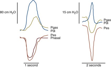
The rapid inspiratory effort of a sniff maneuver (see Chapter 6) is accompanied by momentary equilibration of intrathoracic and upper airway pressures. This equilibration occurs above a pressure of 10 to 12 cm H2O, so employing sniff nasal pressure (Pnsn) allows noninvasive measurement of inspiratory muscle strength. Pnsn is a particularly useful additional investigation in patients with a low or equivocal PImax to confirm or exclude the presence of inspiratory muscle weakness.

A study of 241 patients with moderate to severe neuromuscular disorders showed a positive correlation between Pnsn and PImax, but with a relatively poor agreement observed between Pnsn and PImax. However, the findings of this study differ from those in earlier studies in that the value of PImax was at least the same as or even greater than Pnsn, particularly in those patients with severe ventilatory restriction, which highlights the potential limitation of Pnsn in this particular patient population. These findings add support to the idea that Pnsn can underestimate inspiratory muscle strength in patients with moderate to severe neuromuscular disease (VC less than 40% of predicted), as demonstrated previously in smaller studies of patients with chronic stable inspiratory muscle weakness and acute respiratory failure. Specifically, as VC falls, a greater decrease occurs in Pnsn than in PImax. Strictly speaking, PImax and Pnsn are not interchangeable measurements but are complementary tests, and they should be used in combination with VC for a complete sequential assessment of inspiratory muscle strength in patients with neuromuscular and chest wall disease. In clinical practice, Pnsn is usually measured through occlusion of one of the nasal passages with a nasal bung fitted with a small piece of tubing that connects to a handheld pressure transducer. Normal Pnsn values are below −70 cm H2O in men and below −60 cm H2O in women with the measurement made from FRC.

Twitch Transdiaphragmatic Pressure

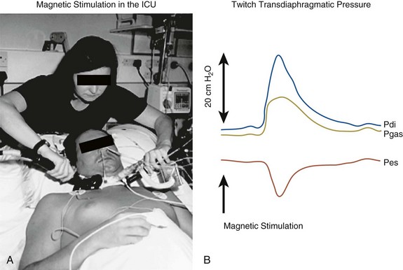
Faraday’s law states that in a conducting material, a changing magnetic field will induce an electric current. Magnetic stimulation stores electrical energy in a capacitor and then discharges this energy by way of a coil to generate a rapidly changing magnetic field. This magnetic field has advantages over the electrical stimulus in that it is less attenuated by distance and therefore can more easily penetrate the deep structures in the neck and stimulate the phrenic nerve. This stimulation produces a single “twitch” of the diaphragm that is well tolerated by patients.

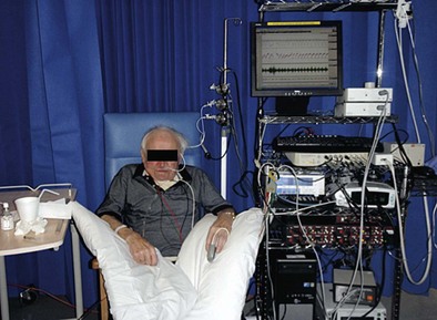
Clinical magnetic stimulation to allow bilateral stimulation of the roots supplying the phrenic nerves was first reported in 1989. Subsequent work has demonstrated its clinical usefulness, and this technique provides a nonvolitional assessment of diaphragm strength in a number of patient groups, including children and adolescents, as well as critically ill patients in intensive care in whom motivation and cooperation to perform maximal volitional maneuvers are difficult to achieve. In addition to improved tolerability, magnetic stimulation has advantages over electrical stimulation in that it is easier to stimulate the phrenic nerves and reduces the technical difficulties involved in isolation of the nerve (Figure 64-5). Further details can be found in the recommended reading section.

Role of Imaging in Respiratory Muscle Weakness and Chest Wall Disease

An elevated hemidiaphragm on a plain chest radiograph often is considered to indicate diaphragmatic weakness or paralysis, but in fact this finding is confirmed by diaphragmatic testing in only approximately 24% of cases. The most recent data indicate that hemidiaphragm elevation on the chest radiograph has a 93% sensitivity but only a 44% specificity for predicting diaphragmatic weakness. Thus, elevation of the hemidiaphragm on the chest radiograph is likely to represent hemidiaphragmatic weakness; however, a normally positioned hemidiaphragm does not exclude weakness on that side.

The contraction of the diaphragm can be directly visualized by other techniques such as fluoroscopic screening, ultrasonography, and dynamic magnetic resonance imaging. Of these, ultrasonography has the advantage of being cheap and radiation-free; however, false positives may occur in approximately 6% of subjects, and even when correctly identifying weakness, this technique gives no functional information with regard to diaphragm strength. Accordingly, ultrasound imaging should not routinely be used in the assessment of patients with suspected hemidiaphragm or bilateral diaphragm weakness. For these purposes, unilateral and bilateral magnetic phrenic nerve stimulation should be used. Although computed tomography (CT) cross-sectional imaging may have particular value in the assessment of pleural causes of lung restriction, it is relatively unhelpful in obese patients, except to exclude coexisting thromboembolic disease and parenchymal lung disease.

Sleep Studies

Overnight oximetry and capnometry constitute an essential part of the assessment for the early detection of nocturnal hypoventilation. These are recommended investigations in patients with neuromuscular and chest wall disease who report symptoms of sleep-disordered breathing (see Table 64-1). Although some investigators advocate the use of polysomnography, including an electroencephalogram, to confirm and quantify arousals during respiratory events, this study is not necessary in the routine clinical management of these patients. Nocturnal hypoventilation can be managed simply with overnight titration of ventilatory support to a defined level of hypercapnia, irrespective of the sleep stage of the patient.

Respiratory Presentations of Neurologic and Chest Wall Disease

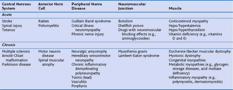
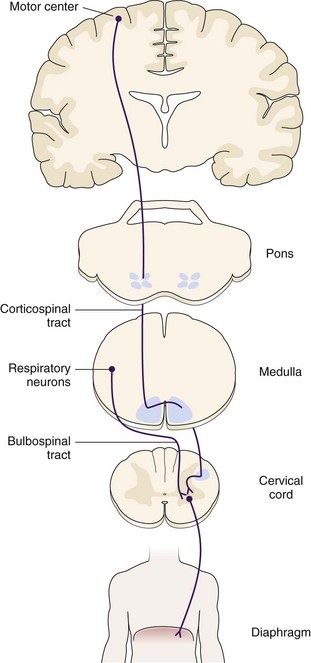
The remainder of this chapter describes specific neurologic/neuromuscular disorders and chest wall diseases, as well as obesity, recognized to be associated with restrictive lung disease. These conditions are divided into acute and chronic presentations (Table 64-3), and the neuromuscular conditions are discussed in the context of the disease and grouped according to their impact on the relevant portion of the anatomic pathway from the central cortex to the peripheral muscle (Figure 64-6).

Acute Disorders Affecting the Central Nervous System

Stroke

Stroke (cerebrovascular accident) is one of the most common causes of acute neurologic deterioration. It is the third leading cause of death in both the United Kingdom and the United States affecting 150,000 and 795,000 patients, respectively, each year. The diaphragm has bilateral cortical representation, whereas the expiratory muscles have contralateral representation, similar to that in the limb muscles. Although hypercapnic respiratory failure is uncommon in stroke patients, chest infection is relatively frequent, and it is likely that impaired voluntary cough coupled with poor glottic coordination as well as swallowing difficulties contribute to the episodes of chest sepsis.

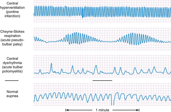
Cheyne-Stokes respiration breathing pattern often is observed as a feature associated with many different types of stroke. This pattern consists of a regular crescendo and decrescendo in breathing interspersed with periods of total apnea that cycles approximately every 30 seconds to 2 minutes. It commonly is observed in chronic heart failure and as a preterminal breathing pattern but also may be seen in healthy subjects at altitude.

Certain classic breathing patterns (Figure 64-7) are considered to reflect aspects of central nervous system pathology, though these are relatively rare in mainstream respiratory clinical practice. These include (1) central neurogenic hyperventilation resulting from a pontine lesion with hyperventilation present throughout both sleep and wakefulness; (2) cluster breathing caused by a mid–brain stem lesion, with periods of hyperventilation alternating with distinct periods of apnea; (3) apneustic breathing as a consequence of damage to the caudal pons with prolonged pauses after each inspiratory breath resulting in hypoventilation; and (4) ataxic breathing observed in patients with cortical lesions of the medulla, which is characterized by an erratic breathing pattern, which differs from the other breathing patterns in having no clear-cut cyclic nature.

image

Figure 64-7 Breathing patterns in central nervous system diseases.

(Modified from Plum F, Swanson AG: Central neurogenic hyperventilation in man, AMA Arch Neurol Psychiatry 81:535–549, 1959.)

Spinal Cord Injury

Spinal cord injury (SCI) commonly is a complication of trauma occurring predominantly in young adults, but it also may result from spinal artery infarction or tumor compression. The average age at insult is 34 years, and males are four times more likely to be affected. The extent of any neuromuscular weakness will be dependent on the site of injury, with higher cervical injuries resulting in more profound impairment. SCI at the level of C1 to C3 results in profound respiratory failure secondary to respiratory muscle denervation. Both voluntary and involuntary muscle control are lost. Unless neurologic recovery occurs, the patient will remain tracheostomy ventilator–dependent. SCI at the level of C3 to C6 results in a variable degree of diaphragmatic weakness, with more caudal lesions conferring a better prognosis. Patients may not have long-term ventilator dependence, and some improvement in respiratory muscle function often occurs over time as a consequence of reduction in inflammation and recruitment of other accessory muscles. SCI affecting the lower cervical and upper thoracic spinal cord carries a significantly better prognosis. The intercostal muscles and the abdominal muscles are affected, but the diaphragm is spared. Patients therefore rarely require long-term mechanical ventilation.

Custom-made corsets that are designed to provide both truncal stability and abdominal support are helpful in such patients. These garments reduce the sensation of respiratory effort by optimizing the operating lung volumes and decreasing abdominal compliance, which in turn enhances diaphragm performance. These patients are susceptible to respiratory tract infections, however, as with other neuromuscular disease, due to the impairment of cough function.

Bilateral diaphragmatic pacing can be considered as an alternative method of ventilatory support in a select group of patients who have high SCI and preserved function of the phrenic nerve–diaphragm unit. The most suitable candidates for pacing are considered to be those patients with a preserved response to peripheral phrenic nerve stimulation but lacking a response to transcranial stimulation of the diaphragm motor area.

Chronic Disorders Affecting the Central Nervous System

Parkinson Disease

Parkinson disease is the most common movement disorder observed in developed countries and frequently is associated with fatal respiratory complications related to infection. Inspiratory muscle weakness occurs late in the progression of the disease, when maximal inspiratory pressures can fall to approximately 30% predicted. Expiratory muscle weakness also is seen; patients are unable to generate a rapid rise in peak expiratory flow. This impairment is akin to the generalized hypokinesia that is a common feature in Parkinson disease and has a deleterious effect on cough function.

Patients with this disease have significant difficulty in coordinating succinct respiratory muscle recruitment, which may partly explain the breathlessness associated with many of the dystonias. Of interest, early in the course of the disease, when no respiratory muscle weakness is evident from objective lung function parameters, the patients are still less able to perform repetitive respiratory muscle contractions than healthy age-matched control subjects. L-Dopa and apomorphine therapy have been shown to reverse these phenomena, suggesting that the defect is one of a central control and coordination of the respiratory muscles.

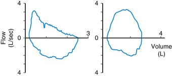
Patients with Parkinson disease commonly exhibit involuntary movements affecting the upper airways. A saw-tooth flutter wave often is present on the flow-volume loop trace (Figure 64-8); this aberration results from repetitive adduction of the vocal cords, occurring at the same frequency as a tremor of 4 to 8 Hz. It can cause significant difficulties if tracheal intubation is required. Obstructive sleep apnea is observed more frequently in patients with Parkinson disease, although patients also may experience sleep fragmentation because of pain or dystonias. Finally, the related condition of multisystem atrophy is characterized by features of Parkinson disease and autonomic failure. It has been associated with irregular breathing patterns and life-threatening upper airway obstruction related to glottis and vocal cord dysfunction narrowing during sleep.

image

Figure 64-8 Characteristic flow-volume loop in Parkinson disease (left) compared with a normal flow-volume loop (right).

(From Laghi F, Tobin MJ: Disorders of the respiratory muscles, Am J Respir Crit Care Med 168:10–48, 2003.)

Acute Disorders Affecting the Anterior Horn Cells

Chronic Disorders Affecting the Anterior Horn Cells

Motor Neuron Disease

Motor neuron disease (MND), also known as amyotrophic lateral sclerosis (ALS), has a European annual incidence of approximately 2 to 3 cases per 100,000 population. Only 1% to 3% of MND cases manifest with respiratory muscle weakness and acute respiratory failure. It is uncommon for patients to describe the classical signs of respiratory muscle weakness (see Table 64-1) as a presenting complaint, but respiratory physicians should consider this diagnosis in patients with coexisting muscle wasting and weakness. A more common presentation is one of exertional and positional dyspnea, ineffective cough, and low speech volume in a patient who has already received the diagnosis. Bulbar features, notably aspiration, are common in advanced disease. When muscle weakness is severe, the patient may report disrupted sleep and excessive fatigue, morning headaches and confusion. Physical findings may include tachypnea at rest, inability to complete full sentences, use of accessory muscles, and abdominal inspiratory paradox.

Respiratory muscle weakness may be present in the context of other generalized muscle involvement without chronic respiratory failure. Early detection of respiratory muscle involvement may facilitate appropriate timing of ventilatory support to prevent respiratory crises. Studies have shown that Pnsn is easier to obtain in patients with MND and can be used to predict prognosis. Routine monitoring of Pnsn has been recommended; if the Pnsn is less than 40 cm H2O, referral for home noninvasive ventilation should be considered. The median survival is 3 to 5 years from onset of symptoms; home noninvasive ventilation has been shown to prolong survival in non-bulbar MND and to enhance health-related quality of life in bulbar MND.

Acute Disorders Affecting the Peripheral Nerves

Guillain-Barré Syndrome

Guillain-Barré syndrome is an acute inflammatory demyelinating motor polyneuropathy. Patients characteristically present with a symmetric ascending motor weakness with associated depressed reflexes. Symptoms usually progress over a 2-week period, with 90% of patients reaching the peak of the disease course within 4 weeks. Severe respiratory weakness is a common associated feature, and ventilatory support is required at some point in approximately 10% to 30% of cases. Despite many effective immunomodulatory treatments, the mortality rate for Guillain-Barré syndrome requiring intubation is around 5%.

Inflammatory demyelination starts at the nerve root and progresses in a patchy fashion along the nerve sheath, resulting in weakness and paralysis of the distal muscles. This is confirmed by nerve conduction tests whereby conduction velocity of the signal is slowed or absent. A reduction in the amplitude of the action potential obtained through diaphragm needle electromyelography in response to phrenic nerve stimulation has been correlated with the requirement for invasive ventilation. In view of the risk of respiratory failure, it is essential that respiratory muscle strength is monitored in patients with confirmed or suspected Guillain-Barré syndrome. Predictive indicators for requirement of invasive ventilation are cranial nerve involvement, infection in the preceding 8 days, time from onset of weakness to admission less than 7 days, inability to stand, inability to lift elbow above head, inability to lift head off the pillow, and ineffective cough. Respiratory muscle strength commonly is assessed by serial VC and mouth pressure measurements, for which a declining trend is indicative of impending ventilator failure.

The “20-30-40” rule often is applied in clinical practice, with the combination of a VC less than 20 mL/kg, a maximum inspiratory pressure above −30 cm H2O, and maximum expiratory pressure below +40 cm H2O indicating the onset of respiratory failure. A clinical challenge is in maintaining an adequate mouth seal in patients with bulbar involvement, and a full face mask can be attached to the spirometer or pressure monitor to ensure optimal delivery of ventilatory support. A minority of patients, mainly those in their seventh and eighth decade of life with significant bulbar involvement, fail to wean from invasive mechanical ventilation, and these patients will require long-term tracheostomy ventilation.

Iatrogenic Disease

Chronic Disorders Affecting the Peripheral Nerves

Chronic Neuromuscular Junction Disorders

Myasthenia Gravis

Myasthenia gravis is the most common condition associated with interruption of neuromuscular signaling, but the disorder remains rare, with a prevalence of approximate 15 to 20 cases per 100,000 population. It is caused by antibodies against the acetylcholine receptor in the post synaptic nerve terminal. Patients typically present with a fluctuating muscular weakness that progresses with fatigue of the muscles, which classically is described as most pronounced toward the end of the day. Ocular and bulbar symptoms are the most common manifestations, but respiratory weakness also can occur and occasionally may be a presenting symptom of the condition. Acute respiratory failure can occur in a myasthenic crisis that may be precipitated by factors such as infection and surgery.

The mainstay of treatment is immunosuppression and use of cholinesterase inhibitors, although care must be taken to achieve an optimal balance, because excessive cholinergic blockade can result in issues with increased bronchial secretions or frank cholinergic block of the respiratory muscles. Despite appropriate treatment, a proportion of patients exhibit nocturnal hypoventilation, which will be exacerbated by steroid-induced weight gain.

Thymoma commonly is associated with the myasthenic syndrome, and clinicians must be aware that after thymectomy, acute respiratory failure may occur. Diagnostic considerations in this setting include both postoperative myasthenic syndrome, although risk for this syndrome should be much less after surgery, and phrenic nerve damage incurred during the procedure, because the nerve can be embedded in the tumor, with consequent intentional or unintentional resection.

Chronic Muscle Disorders

Duchenne Muscular Dystrophy

DMD is an X-linked inherited condition arising from a mutation in the dystrophin gene. It affects males from early childhood, with an incidence of 1 in 3000 births. The decline in function in DMD manifests as a proximal to distal limb muscle weakness, leading to wheelchair dependency by the early teenage years and to respiratory muscle weakness with progression. Respiratory failure is the major cause of death in this group of patients, although mortality also may be associated with the dilated cardiomyopathy that can occur in the absence of respiratory muscle weakness.

As with other progressive neuromuscular conditions, respiratory muscle weakness progresses gradually. The deterioration in respiratory function within these groups manifests initially as nocturnal hypoventilation in REM sleep and progresses to hypoventilation in REM and non-REM sleep and finally to daytime hypercapnia and chronic respiratory failure. This progression in chronic respiratory failure is associated with increasing sleep disturbance, frequency of chest infections, hospital admissions, and deteriorating quality of life. The monitoring of respiratory muscle strength and VC in these patients is a key clinical assessment. VC decreases from the age of 10 years, and a VC of less than 1 L confers a poor prognosis, with mean survival of approximately 2 years without ventilatory support. The presence of scoliosis in these patients may accelerate this process. Mean survival with the onset of chronic respiratory failure is 9.7 months if home nocturnal noninvasive ventilation is not initiated. Although the prophylactic use of home nocturnal noninvasive ventilation before the onset of chronic respiratory failure is controversial, most authorities support the view that noninvasive ventilation brings improvements in gas exchange, normalization of sleep patterns, enhanced quality of life, and prolonged survival. A more recent study indicates that nocturnal ventilatory support should be considered when patients develop nocturnal hypoventilation, and without home noninvasive ventilation, progression to chronic diurnal hypercapnic respiratory failure occurs within 18 months.

Respiratory support also will include secretion clearance, specifically during any episode of pneumonia, with the introduction of a mechanical insufflation-exsufflation device to enhance cough function and secretion management (see earlier under “Cough Peak Flow”). As bulbar function declines, in addition to a reduction in effective cough, patients will be at increased risk for aspiration, and these swallowing problems reduce nutritional intake, resulting in weight loss. Early gastrostomy feeding tube insertion, accomplished with endoscopic or radiologic guidance, is recommended, although evidence supporting this practice is limited. It is possible, with appropriate expertise, to do such procedures with sedation using noninvasive ventilation alone in patients with established respiratory failure.

Other Rarer Myopathies

Congenital myopathies are a group of inherited disorders of muscle that typically manifest with generalized muscle weakness in infancy. A muscle biopsy is required to characterize the type of myopathy. All patients present with hypotonia and a predominant proximal muscular weakness. The severity of the muscle impairment depends on the individual genetic mutation. A majority of the patients require mechanical ventilation from early life and do not survive childhood.

Metabolic myopathies constitute a heterogenous group of disorders that are related to a deficit in the metabolic pathways of the muscle. Among such disorders, the most common are the glycogen storage diseases. From a respiratory perspective, the most important metabolic myopathy is acid maltase deficiency, because respiratory muscle weakness can be the primary presentation of the condition in the adult-onset form. Treatment is supportive, as with the other myopathic conditions, although enzyme replacement therapy may have a role in slowing the onset of chronic respiratory failure.

Mitochondrial myopathies result from dysfunction of mitochondrial ability to release energy form adenosine triphosphate (ATP). The clinical manifestations are highly variable but can include respiratory failure.

Idiopathic inflammatory myopathies, including dermatomyositis and polymyositis, manifest with muscular weakness due to inflammatory destructive processes directly affecting the muscle fibers and their blood supply. Dermatomyositis is characterized by typical skin manifestations such as Gottron’s papules on the hands and feet and a heliotropic rash over the eyelids and can be associated with malignancy. The muscular weakness may rarely result in respiratory failure. These inflammatory myopathies are associated with interstitial parenchymal lung disease in approximately 20% of cases. Such disease can be rapidly progressive and frequently is fatal.

Thoracic Disorders

Chest wall deformities, such as kyphosis, scoliosis, and kyphoscoliosis, and obesity hypoventilation syndrome are described in detail in Chapters 63 and 62, respectively. Obesity itself also has clinical consequences for pulmonary function, and an overview of this issue is presented next, with specific emphasis on pulmonary function tests, pulmonary mechanics, hypercapnic ventilatory response, and respiratory muscle strength.

Obesity

Respiratory Muscle Pump in Obese Patients

It is hypothesized that breathlessness and alveolar hypoventilation in obese patients result from an imbalance between respiratory muscle load and/or capacity and neural respiratory drive, although the exact pathophysiologic details are yet to be determined. Respiratory muscle capacity, albeit estimated using volitional PImax and PEmax measurements, was found to be reduced in hypercapnic obese patients compared with eucapnic obese patients. However, direct measurement of diaphragm strength, using Pdimax, demonstrated no difference between eucapnic and hypercapnic obese patients, indicating that diaphragm weakness does not contribute to the development of ventilatory failure in obese patients. By contrast, a difference is seen in respiratory muscle load between hypercapnic obese patients and eucapnic obese subjects with greater upper airway resistance in both sitting and supine positions and reduced respiratory system compliance. This increasing load on the respiratory muscles results in a reduction in lung volumes, with a reduction in FEV1 and FVC and an elevated FEV1/FVC ratio confirming a restrictive defect. In addition, TLC, expiratory reserve volume (ERV), and FRC are all reduced. Of interest, these reductions are more marked in obese patients with hypercapnic respiratory failure than in eucapnic obese subjects with a matched BMI. It is appreciated that in addition to absolute fat load, the distribution of the fat is important in determining the severity of lung restriction.

The obese patient breathes at a lower lung volume, which reduces chest wall and lung compliance. Breathing at a lower lung volume also results in closure of the small airways during early expiration, causing expiratory airflow limitation and development of intrinsic positive end-expiratory pressure (PEEPi). This phenomenon is exacerbated by adoption of a supine position, such as occurs during sleep. It has been shown that in obese patients, moving from a sitting to supine posture is associated with an increase in neural respiratory drive, as measured by the diaphragm electromyogram, with a corresponding increase in diaphragm pressure generation, as measured from transdiaphragmatic pressure swings, but without a corresponding increase in tidal volume. Furthermore, these patients develop PEEPi on change in position, indicating expiratory flow limitation, although it is difficult to separate whether this is a consequence of early airway closure during expiration or upper airway obstruction, or a combination of both mechanisms. More studies in this area are required to confirm the previous data demonstrating changes in hypercapnic ventilatory response in obese hypercapnic patients. These previous studies have shown that obese hypercapnic patients, compared with eucapnic obese subjects, have reduced hypercapnic ventilatory response as a consequence of an inadequate ability to increase tidal volume, resulting in dead space ventilation. Such research will allow investigators to define the pathophysiologic cause of alveolar hypoventilation in obese patients.

Controversies and Pitfalls

Respiratory muscle weakness is a common complication in many neuromuscular and chest wall conditions, and although it may occasionally be a presenting feature of the underlying disorder, it generally is a complication of an already established diagnosis. In view of the impact of chronic respiratory failure on health-related quality of life and the potential life-threatening consequences of respiratory muscle weakness, respiratory muscle function should be monitored at regular intervals with noninvasive respiratory muscle tests in patients with progressive neuromuscular disease. Furthermore, patients with neuromuscular disease or chest wall disease, or both, who present with unexplained dyspnea should be referred for formal respiratory assessment. This evaluation will include a detailed history and clinical examination with a focus on symptoms suggestive of significant muscle weakness, sleep-disordered breathing, and associated complications such as bulbar symptoms. Other components of the evaluation will include a basic lung volume measurement, noninvasive respiratory muscle testing, and, in special circumstances, invasive respiratory muscle testing. It is well to be mindful, however, of the limitations of performing a single measure of respiratory muscle strength in attempting to diagnose respiratory muscle weakness. Previous data have demonstrated that isolated sniff inspiratory nasal pressure often overdiagnosed the severity of weakness in patients with severe restrictive ventilator deficits from neuromuscular disease. Accordingly, for an accurate assessment of respiratory muscle strength, performing an array of noninvasive tests of respiratory muscle function, such as Pnsn, PImax, and PEmax, is recommended.

With these clinical data, the timing of initiation of noninvasive ventilation can be planned. In patients with neuromuscular and chest wall disease, recent data suggest that chronic respiratory failure will occur 18 to 24 months after the onset of nocturnal hypoventilation. This will be the time to consider the introduction of noninvasive ventilation, and for those patients who are relatively asymptomatic at this stage and are reluctant to pursue noninvasive ventilation, close monitoring of clinical status is indicated. In addition to ventilatory strategies, clinicians must consider other respiratory adjunctive measures, such as physiotherapy techniques and the use of devices including mechanical insufflation-exsufflation devices for patients with neuromuscular disease in whom poor cough function has been documented.

Finally, clinicians need to be cognizant of the rising trend in obesity and of its negative effects on respiratory function. Thus far, no prospective randomized controlled trials have been conducted to determine an optimal ventilation strategy for treating obesity hypoventilation syndrome. A single-center randomized controlled trial of selected patients with obesity hypoventilation showed equivalence between continuous positive airway pressure and noninvasive ventilation, leading consensus opinion to advocate noninvasive ventilation in patients for whom continuous positive airway pressure has not achieved symptom control. With the increasing incidence of superobesity, it is clear that further research in this area is urgently required.

Suggested Readings

American Thoracic Society/European Respiratory Society. ATS/ERS statement on respiratory muscle testing. Am J Respir Crit Care Med. 2002;166:518–624.

Bassetti CL, Gugger M. Sleep disordered breathing in neurologic diseases. Swiss Med Wkly. 2002;132:109–115.

Hart N. Respiratory failure. Clin Med. 2008;36:242–245.

Hart N, Polkey MI. Investigation of respiratory muscle function. Clin Pulm Med. 2001;8:180–187.

Laghi F, Tobin MJ. Disorders of the respiratory muscles. Am J Respir Crit Care Med. 2003;168:10–48.

Luo YM, Moxham J, Polkey MI. Diaphragm electromyelography using an oesophageal catheter: current concepts. Clin Sci (Lond). 2008;115:233–244.

Murphy P, Polkey MI, Hart N. Diagnostic tests in the assessment of patients for home mechanical ventilation. In: Elliott MW, Nava S, Schönhofer B. Principles and practices of non-invasive ventilation and weaning. London: Hodder Arnold; 2010:151–166.

Mustfa N, Moxham J. Respiratory muscle assessment in motor neurone disease. Q J Med. 2001;94:497–502.

Polkey MI, Lyall RA, Davidson AC, et al. Ethical and clinical issues in the use of home non-invasive mechanical ventilation for the palliation of breathlessness in motor neurone disease. Thorax. 1999;54:367–371.

Polkey MI, Lyall RA, Moxham J, Leigh PN. Respiratory aspects of neurological disease. J Neurol Neurosurg Psychiatry. 1999;66:5–15.

Polkey MI, Moxham J. Clinical aspects of respiratory muscle dysfunction in the critically ill. Chest. 2001;119:926–939.

Tobin M. Principles and practices of mechanical ventilation, ed 2. New York: McGraw-Hill; 2006.

Share this: