Diphtheria (Corynebacterium diphtheriae)

Published on 27/03/2015 by admin

Filed under Pediatrics

Last modified 22/04/2025

Print this page

rate 1 star rate 2 star rate 3 star rate 4 star rate 5 star
Your rating: none, Average: 0 (0 votes)

This article have been viewed 3771 times

Chapter 180 Diphtheria (Corynebacterium diphtheriae)

Diphtheria is an acute toxic infection caused by Corynebacterium species, typically Corynebacterium diphtheriae and rarely toxigenic strains of Corynebacterium ulcerans. Although diphtheria was reduced from a major cause of childhood death to a medical rarity in the Western hemisphere in the early 20th century, current reminders of the fragility of this success emphasize the necessity to continue vigorous promotion of those same principles across the global community.

Etiology

Corynebacteria are aerobic, nonencapsulated, non–spore-forming, mostly nonmotile, pleomorphic, gram-positive bacilli. C. diphtheriae is by far the most commonly isolated agent of diphtheria. C. ulcerans is more commonly isolated from cattle and can cause similar disease. As corynebacteria are not fastidious in growth requirements, their isolation is enhanced by use of a selective medium (i.e., cystine-tellurite blood agar or Tinsdale agar) that inhibits growth of competing organisms and, when reduced by C. diphtheriae, renders colonies gray-black. Differentiation of C. diphtheriae from C. ulcerans is based on urease activity, because C. ulcerans is urease-positive.

Four C. diphtheriae biotypes (mitis, intermedius, belfanti, gravis) are capable of causing diphtheria and are differentiated by colonial morphology, hemolysis, and fermentation reactions. The ability to produce diphtheritic toxin results from acquisition of a lysogenic Corynebacteriophage by either C. diphtheriae or C. ulcerans, which encodes the diphtheritic toxin gene and confers diphtheria-producing potential on these strains. Thus, indigenous nontoxigenic C. diphtheriae can be rendered toxigenic and disease-producing after importation of a toxigenic C. diphtheriae and transmission of the bacteriophage. Demonstration of diphtheritic toxin production or potential for toxin production by an isolate is necessary to confirm disease. The former is done in vitro using the agar immunoprecipitin technique (Elek test) or in vivo with the toxin neutralization test in guinea pigs, the latter by polymerase chain reaction testing for carriage of the toxin gene. Toxigenic and nontoxigenic strains are indistinguishable by colony type, microscopic features, or biochemical test results.

Epidemiology

Unlike other diphtheroids (coryneform bacteria), which are ubiquitous in nature, C. diphtheriae is an exclusive inhabitant of human mucous membranes and skin. Spread is primarily by airborne respiratory droplets, direct contact with respiratory secretions of symptomatic individuals, or exudate from infected skin lesions. Asymptomatic respiratory tract carriage is important in transmission. Where diphtheria is endemic, 3-5% of healthy individuals can carry toxigenic organisms, but carriage is exceedingly rare if diphtheria is rare. Skin infection and skin carriage are silent reservoirs of C. diphtheriae, and organisms can remain viable in dust or on fomites for up to 6 mo. Transmission through contaminated milk and an infected food handler has been proven or suspected.

In the 1920s, >125,000 diphtheria cases, with 10,000 deaths, were reported annually in the USA, with the highest fatality rates among the very young and the elderly. The incidence then began to decrease and, with widespread use of diphtheria toxoid in the USA after World War II, declined steadily through the late 1970s. Since then, ≤5 cases have occurred annually in the USA, with no epidemics of respiratory tract diphtheria. Similar decreases occurred in Europe. Despite the worldwide decrease in disease incidence, diphtheria remains endemic in many developing countries with poor immunization rates against diphtheria.

When diphtheria was endemic, it primarily affected children <15 yr of age. Since the introduction of toxoid immunization, the disease has shifted to adults who lack natural exposure to toxigenic C. diphtheriae in the vaccine era and have low rates of booster immunization. In the 27 sporadic cases of respiratory tract diphtheria reported in the USA in the 1980s, 70% occurred among persons >25 yr of age. The largest outbreak of diphtheria in the developed world since the 1960s occurred from 1990 to 1996 in the newly independent countries of the former Soviet Union, involving >150,000 cases in 14 of 15 countries. Of these, >60% of cases occurred in individuals >14 yr of age. Case fatality rates ranged from 3% to 23% by country. Factors contributing to the epidemic included a large population of underimmunized adults, decreased childhood immunization rates, population migration, crowding, and failure to respond aggressively during early phases of the epidemic. Cases of diphtheria among travelers from these endemic areas were transported to many countries in Europe.

Most proven cases of respiratory tract diphtheria in the USA in the 1990s were associated with importation of toxigenic C. diphtheriae, although clonally related toxigenic C. diphtheriae has persisted in this country and Canada for at least 25 yr.

Cutaneous diphtheria, a curiosity when diphtheria was common, accounted for >50% of reported C. diphtheriae isolates in the USA by 1975. This indolent local infection, compared with mucosal infection, is associated with more prolonged bacterial shedding, greater contamination of the environment, and increased transmission to the pharynx and skin of close contacts. Outbreaks are associated with homelessness, crowding, poverty, alcoholism, poor hygiene, contaminated fomites, underlying dermatosis, and introduction of new strains from exogenous sources. It is no longer a tropical or subtropical disease; 1,100 C. diphtheriae infections were documented in a neighborhood in Seattle (site of the last major U.S. outbreak), from 1971 to 1982; 86% were cutaneous, and 40% involved toxigenic strains. Cutaneous diphtheria is an important source for toxigenic C. diphtheriae in the USA, and its importation is frequently the source for subsequent sporadic cases of respiratory tract diphtheria. To focus attention on respiratory tract diphtheria, the condition more likely to cause acute respiratory complications and toxic manifestations, C. diphtheria isolates from cutaneous disease were removed from annual diphtheria statistics reported by the Centers for Disease Control and Prevention (CDC) after 1979.

Clinical Manifestations

The manifestations of C. diphtheriae infection are influenced by the anatomic site of infection, the immune status of the host, and the production and systemic distribution of toxin.

Respiratory Tract Diphtheria

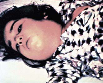
In a classic description of 1,400 cases of diphtheria in California (1954), the primary focus of infection was the tonsils or pharynx (94%), with the nose and larynx the next 2 most common sites. After an average incubation period of 2-4 days, local signs and symptoms of inflammation develop. Infection of the anterior nares is more common among infants and causes serosanguineous, purulent, erosive rhinitis with membrane formation. Shallow ulceration of the external nares and upper lip is characteristic. In tonsillar and pharyngeal diphtheria, sore throat is the universal early symptom: Only half of patients have fever, and fewer have dysphagia, hoarseness, malaise, or headache. Mild pharyngeal injection is followed by unilateral or bilateral tonsillar membrane formation, which can extend to involve the uvula (which may cause toxin-mediated paralysis), soft palate, posterior oropharynx, hypopharynx, or glottic areas (Fig. 180-1). Underlying soft tissue edema and enlarged lymph nodes can cause a bull-neck appearance. The degree of local extension correlates directly with profound prostration, bull-neck appearance, and fatality due to airway compromise or toxin-mediated complications (Fig. 180-2).

image

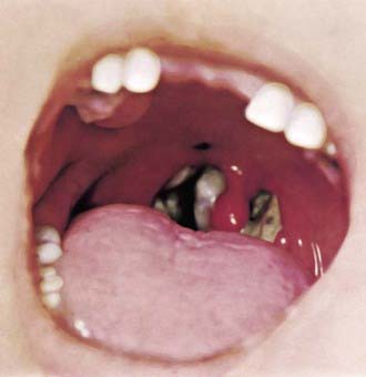
Figure 180-1 Tonsillar diphtheria.

(Courtesy Franklin H. Top, MD, Professor and Head of the Department of Hygiene and Preventive Medicine, State University of Iowa, College of Medicine, Iowa City, IA; and Parke, Davis & Company’s Therapeutic Notes.)

image

Figure 180-2 Diphtheria. Bull-neck appearance of diphtheritic cervical lymphadenopathy.

(Courtesy of the Centers for Disease Control and Prevention.)

The characteristic adherent membrane, extension beyond the faucial area, dysphagia, and relative lack of fever help differentiate diphtheria from exudative pharyngitis caused by Streptococcus pyogenes or Epstein-Barr virus. Vincent angina, infective phlebitis with thrombosis of the jugular veins (Lemierre disease), and mucositis in patients undergoing cancer chemotherapy are usually differentiated by the clinical setting. Infection of the larynx, trachea, and bronchi can be primary or a secondary extension from the pharyngeal infection. Hoarseness, stridor, dyspnea, and croupy cough are clues. Differentiation from bacterial epiglottitis, severe viral laryngotracheobronchitis, and staphylococcal or streptococcal tracheitis hinges partially on the relative paucity of other signs and symptoms in patients with diphtheria and primarily on visualization of the adherent pseudomembrane at the time of laryngoscopy and intubation.

Patients with laryngeal diphtheria are at significant risk for suffocation because of local soft tissue edema and airway obstruction by the diphtheritic membrane, a dense cast of respiratory epithelium, and necrotic coagulum. Establishment of an artificial airway and resection of the pseudomembrane can be lifesaving, but further obstructive complications are common, and systemic toxic complications are inevitable.

Complications

Respiratory tract obstruction by pseudomembranes may require bronchoscopy or intubation and mechanical ventilation. Two other tissues usually remote from sites of C. diphtheriae infection can be significantly affected by diphtheritic toxin: the heart and the nervous system.

Toxic Cardiomyopathy

Toxic cardiomyopathy occurs in 10-25% of patients with respiratory diphtheria and is responsible for 50-60% of deaths. Subtle signs of myocarditis can be detected in most patients, especially the elderly, but the risk for significant complications correlates directly with the extent and severity of exudative local oropharyngeal disease as well as delay in administration of antitoxin. The 1st evidence of cardiac toxicity characteristically occurs during the 2nd and 3rd weeks of illness as the pharyngeal disease improves but can appear acutely as early as the 1st wk of illness, a poor prognostic sign, or insidiously as late as the 6th wk. Tachycardia out of proportion to fever is common and may be evidence of cardiac toxicity or autonomic nervous system dysfunction. A prolonged PR interval and changes in the ST-T wave on an electrocardiographic tracing are relatively frequent findings; dilated and hypertrophic cardiomyopathy detected by echocardiogram has been described. Single or progressive cardiac dysrhythmias can occur, including 1st-, 2nd-, and 3rd-degree heart block. Temporary transvenous pacing may improve outcomes. Atrioventricular dissociation and ventricular tachycardia are also described, the latter having a high associated mortality. Heart failure may appear insidiously or acutely. Elevation of the serum aspartate aminotransferase concentration closely parallels the severity of myonecrosis. Severe dysrhythmia portends death. Histologic postmortem findings are variable: little or diffuse myonecrosis with acute inflammatory response. Recovery from toxic myocardiopathy is usually complete, although survivors of more severe dysrhythmias can have permanent conduction defects.

Treatment

Specific antitoxin is the mainstay of therapy and should be administered on the basis of clinical diagnosis. Because it neutralizes only free toxin, antitoxin efficacy diminishes with elapsed time after the onset of mucocutaneous symptoms. Equine diphtheria antitoxin is available in the USA only from the CDC. Physicians treating a case of suspected diphtheria should contact the CDC diphtheria duty officer (770-488-7100 at all times). Antitoxin is administered as a single empirical dose of 20,000-120,000 U based on the degree of toxicity, site and size of the membrane, and duration of illness. Antitoxin is probably of no value for local manifestations of cutaneous diphtheria, but its use is prudent because toxic sequelae can occur. Commercially available intravenous immunoglobulin preparations contain low titers of antibodies to diphtheria toxin; their use for therapy of diphtheria is not proven or approved. Antitoxin is not recommended for asymptomatic carriers.

The role of antimicrobial therapy is to halt toxin production, treat localized infection, and prevent transmission of the organism to contacts. C. diphtheriae is usually susceptible to various agents in vitro, including penicillins, erythromycin, clindamycin, rifampin, and tetracycline. Resistance to erythromycin is common in populations if the drug has been used broadly. Only erythromycin or penicillin is recommended; erythromycin is marginally superior to penicillin for eradication of nasopharyngeal carriage. Appropriate therapy is erythromycin (40-50 mg/kg/day divided every 6 hr by mouth [PO] or intravenously [IV]; maximum 2 g/day), aqueous crystalline penicillin G (100,000-150,000 U/kg/day divided every 6 hr IV or intramuscularly [IM]), or procaine penicillin (25,000-50,000 U/kg/day divided every 12 hr IM) for 14 days. Antibiotic therapy is not a substitute for antitoxin therapy. Some patients with cutaneous diphtheria have been treated for 7-10 days. Elimination of the organism should be documented by negative results of at least 2 successive cultures of specimens from the nose and throat (or skin) obtained 24 hr apart after completion of therapy. Treatment with erythromycin is repeated if either culture yields C. diphtheriae.

Prevention

Protection against serious disease caused by imported or indigenously acquired C. diphtheriae depends on immunization. In the absence of a precisely determined minimum protective level for diphtheria antitoxin, the presumed minimum is 0.01-0.10 IU/mL. In outbreaks, 90% of individuals with clinical disease have had antibody values <0.01 IU/mL, and 92% of asymptomatic carriers have had values >0.1 IU/mL. In serosurveys in the USA and Western Europe, where almost universal immunization during childhood has been achieved, 25% to >60% of adults lack protective antitoxin levels, with very low levels common in the elderly.

All suspected diphtheria cases should be reported to local and state health departments. Investigation is aimed at preventing secondary cases in exposed individuals and at determining the source and carriers to halt spread to unexposed individuals. Reported rates of carriage in household contacts of case patients are 0-25%. The risk for development of diphtheria after household exposure to a case is approximately 2%, and the risk is 0.3% after similar exposure to a carrier.

Vaccine

Universal immunization with diphtheria toxoid throughout life, to provide constant protective antitoxin levels and to reduce severity of C. diphtheriae disease, is the only effective control measure. Although immunization does not preclude subsequent respiratory or cutaneous carriage of toxigenic C. diphtheriae, it decreases local tissue spread, prevents toxic complications, diminishes transmission of the organism, and provides herd immunity when at least 70-80% of a population is immunized.

Diphtheria toxoid is prepared by formaldehyde treatment of toxin, standardized for potency, and adsorbed to aluminum salts, enhancing immunogenicity. Two preparations of diphtheria toxoids are formulated according to the limit of flocculation (Lf) content, a measure of the quantity of toxoid. The pediatric preparation (i.e., DTaP [diphtheria and tetanus toxoids with acellular pertussis vaccine], DT [diphtheria and tetanus toxoids vaccine]) contains 6.7-25.0 Lf units of diphtheria toxoid per 0.5-mL dose; the adult preparation (dT; 10% of pediatric diphtheria toxoid dose) contains no more than 2 Lf units of toxoid per 0.5-mL dose. The higher-potency (D) formulation of toxoid is used for primary series and booster doses for children through 6 yr of age because of superior immunogenicity and minimal reactogenicity. For individuals 7 years of age or older, dT is recommended for the primary series and booster doses because the lower concentration of diphtheria toxoid is adequately immunogenic and because increasing the content of diphtheria toxoid heightens reactogenicity with increasing age.

For children from 6 wk up to 7 yr of age, five 0.5-mL doses of diphtheria-containing (D) vaccine are given in a primary series, including 3 doses at 2, 4, and 6 mo of age, with a 4th dose, an integral part of the primary series, 9-12 mo after the third dose. A booster dose is given at 4-6 yr of age (unless the 4th primary dose was administered after the 4th birthday). For persons 7 yr of age and older, three 0.5-mL doses of lower-level diphtheria-containing (d) vaccine are given in a primary series of 2 doses 4-8 wk apart and a 3rd dose 6-12 mo after the 2nd dose. The only contraindication to tetanus and diphtheria toxoid is a history of neurologic or severe hypersensitivity reaction after a previous dose. For children <7 yr of age in whom pertussis immunization is contraindicated, DT is used. Those whose immunization is begun with DTaP or DT before 1 yr of age should have a total of five 0.5-mL doses of diphtheria-containing (D) vaccines by 6 yr of age. For those whose immunization is begun at around 1 yr of age, the primary series is three 0.5-mL doses of diphtheria-containing (D) vaccine, with a booster given at 4-6 yr, unless the 3rd dose was given after the 4th birthday.

A booster dose, consisting of an adult preparation of Tdap, is recommended at 11-12 yr of age. Adolescents 13-18 yr of age who missed the Td or Tdap booster dose at 11-12 yr or in whom it has been ≥5 yr since the Td booster dose also should receive a single dose of Tdap if they have completed the DTP/DTaP series.

There is no association of DT or dT with convulsions. Local adverse effects alone do not preclude continued use. The patient who experiences an Arthus-type hypersensitivity reaction or a temperature >103°F (39.4°C) after a dose of dT, which is rare, usually has high serum tetanus antitoxin levels and should not be given dT more frequently than every 10 yr, even if he or she sustains a significant tetanus-prone injury. The DT or dT preparation can be given concurrently with other vaccines. Haemophilus influenzae conjugate vaccines containing diphtheria toxoid (PRP-D) or the variant of diphtheria toxin, CRM197 protein (HbOC), are not substitutes for diphtheria toxoid immunization and do not affect reactogenicity.