Chapter 26
Dilated Cardiomyopathy
1. What is the definition of dilated cardiomyopathy?
4. What kind of diagnostic studies should be carried out for DCM?
Multigated radionuclide angiocardiography (MUGA) may be used to assess LV systolic function in those with poor echocardiographic windows. Compared with echo assessment of LVEF, MUGA may have less interobserver and intertest variability. Thallium-201 myocardial scintigraphy is not a reliable technique for differentiating patients with ischemic cardiomyopathy (ICM) from those with DCM, as patients with DCM may have both reversible and fixed perfusion abnormalities related to the presence of myocardial fibrosis, although a completely normal scan (without reversible or fixed defects) would favor the diagnosis of NICM.
Endomyocardial biopsy may be performed if and only if a specific diagnosis is suspected in which specific therapy may be efficacious upon establishment of diagnosis (see Chapter 23 on endomyocardial biopsy).
5. What is the natural history of DCM?
6. What are the prognostic features of DCM?
7. What are the common causes of DCM?
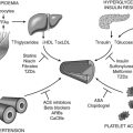
8. What are the features of alcohol-induced cardiomyopathy?
9. What are the features of cocaine-induced cardiomyopathy?
10. What are the features of chemotherapy-induced cardiomyopathy?
Anthracyclines and trastuzumab are among the more prominent chemotherapeutic agents associated with cardiomyopathy. Three major types of anthracycline cardiotoxicity are distinguished: acute, chronic, and late onset, which differ considerably in clinical picture and prognosis. Cardiac failure is rare with acute toxicity. Chronic cardiotoxicity is observed in 0.4% to 23%, several weeks or months after chemotherapy. Such anthracycline-induced cardiomyopathy carries a 27% to 61% mortality rate despite aggressive medical treatment. Late cardiotoxicity occurs years after chemotherapy, at 5% at 10 years, and presents as heart failure, arrhythmia, or conduction abnormalities. The primary risk factor for cardiomyopathy is cumulative dose administered. Toxicity is rare (less than 3%) with cumulative doses below 400 mg/m2. Other risk factors include extremes of age and coexisting cardiac disease. Elevated cardiac troponin and BNP after chemotherapy may allow identification of patients who may develop cardiac toxicity. Anthracyclines should not be administered to patients with a baseline LVEF of 30% or less. LV function should be assessed repeatedly before each subsequent dose (or if the initial ejection fraction [EF] is more than 50%, after a cumulative dose of 350 to 500 mg/m2), and treatment stopped if EF declines by 10% or more, or to a value less than 30% (or less than 50% if EF was normal at baseline). Reversibility has been documented anecdotally. The most effective protection is dexrazoxane (an iron chelator), with a two- to threefold decrease in the risk of cardiomyopathy.
11. What are the features of HIV-related cardiomyopathy?
HIV-related cardiomyopathy accounts for up to 4% of DCM. The incidence is higher among patients with a CD4 count less than 400 cells/mm3. HIV-related cardiomyopathy is discussed in detail in Chapter 46 on cardiac manifestations of HIV and acquired immunodeficiency syndrome (AIDS).
12. What are the associations between collagen vascular disease and DCMs?
In systemic lupus erythematosus (SLE), global LV dysfunction has been reported in 5% of patients, segmental LV wall motion abnormalities in 4% of patients, and right ventricular enlargement in 4% of patients, but DCM is rare. In rheumatoid arthritis, symptomatic cardiac disease, including myocarditis, develops in 8% of patients. Progressive systemic sclerosis can rarely lead to heart failure through myocardial fibrosis, arrhythmia, or intermittent vascular spasm. Dermatomyositis may be associated with cardiomyopathy in some patients. For further discussion, see Chapter 47 on cardiac manifestations of connective tissue disorders and vasculitides.
13. What is peripartum cardiomyopathy?
14. What are the features of tachycardia-induced cardiomyopathy?
Sustained elevated ventricular rates have been shown to cause changes in LV geometry and dilation. This entity should be considered in patients with no other explanation for LV dysfunction and a concomitant tachyarrhythmia. Hyperthyroid-induced sinus tachycardia or atrial fibrillation should be excluded in such patients. Treatment involves restoration of normal sinus rhythm or control of ventricular rate, with which there is typically resolution as quickly as 4 to 6 weeks.
15. What are the features of nutritional causes of DCM?
16. What are the features of cardiomyopathy caused by iron overload?
17. What are the pharmacologic treatments to be used in DCM?
18. What are the device treatments to be used in DCM?
19. Should DCM patients be anticoagulated?
Patients with DCM have multiple risk factors that predispose to thromboembolic events—with the incidence ranging from 0.8 to 2.5 per 100 patient-years. Pooled data from small, randomized, controlled clinical trials and current guidelines do not support routine anticoagulation in heart failure with sinus rhythm. The results of the Warfarin versus Aspirin in Reduced Cardiac Ejection Fraction (WARCEF) trial showed that in a large randomized comparison of aspirin versus warfarin in patients with heart failure and reduced ejection fraction (not in atrial fibrillation) there was no overall difference in the combined primary outcome of death, ischemic stroke, or intracranial hemorrhage between treatment groups. Available data do support the use of anticoagulants in the presence of atrial fibrillation, previous stroke or other thromboembolic events, or visible protruding or mobile thrombus on echocardiography.
Bibliography, Suggested Readings, and Websites
1. Arnold, J.M.O. Heart Failure. Available at: http://www.merckmanuals.com/professional/cardiovascular_disorders/heart_failure/heart_failure_hf.html. Last full review/revision January 2010; last modified April 2012. Accessed March 19, 2013
2. Cooper, L.T., Baughman, K.L., Feldman, A.M., et al. The role of endomyocardial biopsy in the management of cardiovascular disease: a scientific statement from the American Heart Association, the American College of Cardiology, and the European Society of Cardiology. Endorsed by the Heart Failure Society of America and the Heart Failure Association of the European Society of Cardiology. J Am Coll Cardiol. 2007;50:1914–1931.
3. Desai, A.S., Fang, J.C., Maisel, W.H., Baughman, K.L. Implantable defibrillators for the prevention of mortality in patients with nonischemic cardiomyopathy: a meta-analysis of randomized controlled trials. JAMA. 2004;292:2874–2879.
4. Dries, D., Exner, D., Gersh, B., et al. Racial differences in the outcome of left ventricular dysfunction. N Engl J Med. 1999;340:609–616.
5. Epstein, A.E., DiMarco, J.P., Ellenbogen, K.A., et al. ACC/AHA/HRS 2008 Guidelines for Device-Based Therapy of Cardiac Rhythm Abnormalities: a report of the American College of Cardiology/American Heart Association Task Force on Practice Guidelines (Writing Committee to Revise the ACC/AHA/NASPE 2002 Guideline Update for Implantation of Cardiac Pacemakers and Antiarrhythmia Devices) developed in collaboration with the American Association for Thoracic Surgery and Society of Thoracic Surgeons. J Am Coll Cardiol. 2008;51(21):e1–e62.
6. Heart Failure Society of America. Heart failure in patients with left ventricular systolic dysfunction. J Card Fail. 2006;12:e38–e57.
7. Heart Failure Society of America. HFSA website. Available at: http://www.hfsa.org. Accessed March 19, 2013
8. Homma, S., Thompson, J.L.P. Results of the Warfarin versus Aspirin in Reduced Cardiac Ejection Fraction (WARCEF) trial. International Stroke Conference 2012. New Orleans: Abstract LB; February 3, 2012. 12–4372
9. Hunt, S.A. American College of Cardiology; American Heart Association Task Force on Practice Guidelines (Writing Committee to Update the 2001 Guidelines for the Evaluation and Management of Heart Failure): ACC/AHA 2005 guideline update for the diagnosis and management of chronic heart failure in the adult. J Am Coll Cardiol. 2005;46:1–82.
10. Koniaris, L., Goldhaber, S. Anticoagulation in dilated cardiomyopathy. J Am Coll Cardiol. 1998;31:745–748.
11. Mann, D.L. Management of heart failure patients with reduced ejection fraction. In Libby P., Bonow R., Mann D., eds.: Braunwald’s heart disease: a textbook of cardiovascular medicine, ed 8, Philadelphia: Saunders, 2008.
12. Piano, M. Alcoholic cardiomyopathy: incidence, clinical characteristics, and pathophysiology. Chest. 2002;121:1638–1650.
13. Richardson, P., McKenna, W., Bristow, M., et al. Report of the 1995 World Health Organization/International Society and Federation of Cardiology Task Force on the Definition and Classification of Cardiomyopathies. Circulation. 1996;93:841–842.
14. Saxon, L., De Marco, T. Arrhythmias associated with dilated cardiomyopathy. Card Electrophysiol Rev. 2002;6:18–25.
15. Sliwa, K., Fett, J., Elkayam, U. Peripartum cardiomyopathy. Lancet. 2006;368:687–693.
16. Swedberg, K., Cleland, J., Dargie, H., et al. Guidelines for the diagnosis and treatment of chronic heart failure: executive summary (update 2005): The Task Force for the Diagnosis and Treatment of Chronic Heart Failure of the European Society of Cardiology. Eur Heart J. 2005;26:1115–1140.
17. Wigner, M., Morgan, J.P., Causes of dilated cardiomyopathy. Basow D.S., ed. UpToDate, Waltham, MA 2013 UpToDate. Available at: http://www.uptodate.com/contents/causes-of-dilated-cardiomyopathy Accessed March 26, 2013
18. Dumitru, I. Heart Failure. Available at: http://www.emedicine.com. Accessed March 19, 2013