Development and Function of the Gonads

Published on 25/03/2015 by admin

Filed under Pediatrics

Last modified 22/04/2025

Print this page

rate 1 star rate 2 star rate 3 star rate 4 star rate 5 star
Your rating: none, Average: 0 (0 votes)

This article have been viewed 1937 times

Chapter 576 Development and Function of the Gonads

Genetic Control of Embryonic Gonadal Differentiation

Gonadal differentiation is a complex, multistep process that requires the sequential action and interaction of multiple gene products.

Early in the 1st trimester, the undifferentiated, bipotential fetal gonad begins as a thickening of the urogenital ridge, near the developing kidney and adrenal cortex. At 6 wk of gestation, the gonad contains germ cells, stromal cells that will become Leydig cells in testes, or theca, interstitial or hilar cells in the ovary; and supporting cells that will develop into Sertoli cells in testes or granulosa cells in ovaries. In the absence of a testis-determining factor, thought to be the SRY (Sex-determining Region on the Y chromosome), the gonad develops into an ovary. SRY may suppress a putative factor 2 that functions as repressor of male development.

A chromosome complement of 46,XX is necessary for the development of normal ovaries. Both the long and short arms of the X chromosome contain genes for normal ovarian development. The DSS (Dosage Sensitive/Sex reversal) locus associated with the DAX1 (DSS Adrenal hypoplasia on the X chromosome) gene, which is defective in patients with X-linked congenital adrenal hypoplasia and hypogonadotropic hypogonadism, is a member of the nuclear receptor superfamily and acts as a repressor of male gene expression. DAX1 acts by binding to a related nuclear receptor SF-1 (Steroidogenic Factor-1). In vitro, the signaling gene WNT4 stimulates expression of DAX1, resulting in the suppression of androgen synthesis in XX females. The WNTs are ligands that activate receptor-mediated signal transduction pathways and are involved in modulating gene expression as well as cell behavior, adhesion, and polarity. A key to its role in humans was elucidated by loss-of-function mutation of the WNT4 gene that was found in an 18 yr old 46,XX woman. She had absence of Müllerian-derived structures (uterus and fallopian tubes), unilateral renal agenesis, and clinical signs of androgen excess.

Mutations of the Wilms tumor 1 (WT1) gene, including alternative splicing, may also impact sex differentiation. WT1 mutations are associated with the Denys-Drash syndrome (early-onset renal failure with abnormal external genitalia and Wilms tumor). Haploinsufficiency of a 3-amino-acid (KTS) form of WT1 has been implicated in the gonadal dysgenesis of patients with Fraser syndrome (late-onset progressive glomerulopathy and 46,XY gonadal dysgenesis). Mutations in the FOXL2 and SF-1 gene are associated with ovarian failure. Other autosomal genes also play a role in normal ovarian organogenesis and testicular development. Several conditions of gonadal dysgenesis are associated with gross abnormalities of both autosomes and sex chromosomes. A deletion affecting the short arm of the X chromosome produces the typical somatic anomalies of Turner syndrome.

Development of the testis requires the short arm of the Y chromosome; a testis-determining factor at this site has been identified, and the gene for it has been cloned and designated SRY. During male meiosis, the Y chromosome must segregate from the X chromosome so that both X and Y chromosomes do not occur in the same spermatozoa. The major portion of the Y chromosome is composed of Y-specific sequences that do not pair with the X chromosome. However, a minor portion of the Y chromosome shares sequences with the X chromosome and pairing does occur in this region. The genes and sequences in this area recombine between the sex chromosomes, behaving like autosomal genes. Therefore, the term pseudoautosomal region is used to describe this portion of the chromosome, and the term indicates genetic behavior of these genes. The SRY gene is localized to the 35-kb portion proximal to the pseudoautosomal region of the Y chromosome. It contains a high-mobility group nonhistone protein (HMG box), suggesting that SRY may be a transcriptional regulator of other genes involved in sex differentiation. The gonadal ridge forms at around 33 days of gestation. SRY is detected at 41 days, peaks at 44 days when testis cords are 1st visible, and persists into adulthood.

Other genes that are found on autosomes are important in this process. SOX9, a SRY-related gene containing a region homologous with the high-mobility group box 9 (HMG box 9) of SRY, is located on chromosome 17. Mutations of this gene result in XY sex reversal and camptomelic dysplasia. Steroidogenic factor 1 (SF-1) on chromosome 9q33, is important in adrenal and gonadal development, as well as the development of gonadotropin-releasing hormone (GnRH)-secreting neurons in the hypothalamus. The Wilms tumor gene (WTI), especially the -KSTiso form on chromosome 11p13, is needed for early gonadal, adrenal, and renal development. Fibroblast growth factor-9 (FGF-9), GATA-4, XH-2, and SOY9 are also important.

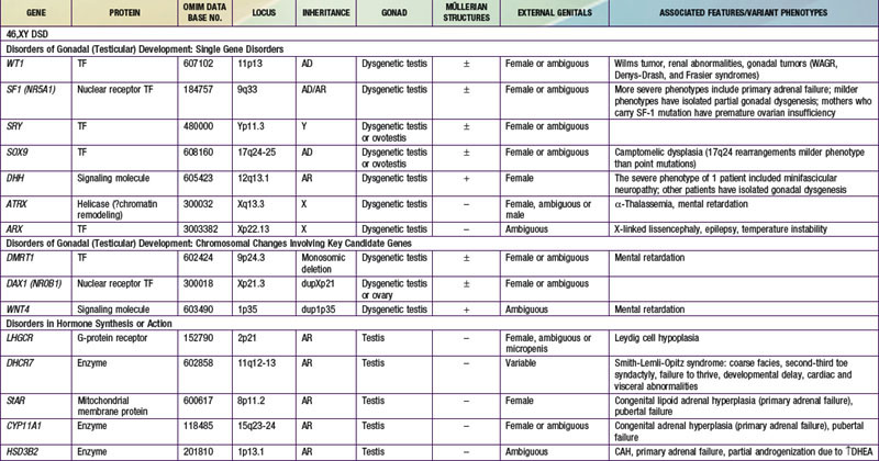
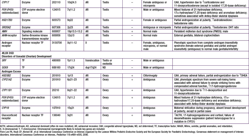
When genetic recombination events on sex chromosomes extend beyond the pseudoautosomal region, X- and Y-specific DNA may be transferred between the chromosomes. Such aberrant recombinations result in X chromosomes carrying SRY, resulting in XX males, or Y chromosomes that have lost SRY, resulting in XY females. SRY acts as a transcriptional regulator to increase cellular proliferation, attract interstitial cells from adjacent mesonephros into the genital ridge, and stimulate testicular Sertoli cell differentiation. Sertoli cells act as an organizer of steroidogenic and germ cell lines and produce antimüllerian hormone (AMH) that causes the female duct system to regress. These cells express low levels of SRY. For additional genes involved in sex development, see Table 576-1.

Function of the Testes

Levels of placental chorionic gonadotropin peak at 8-12 wk of gestation and stimulate the fetal Leydig cells to secrete testosterone, the main hormonal product of the testis. Testosterone is then converted by the enzyme 5α-reductase to its more potent metabolite, dihydrotestoterone. This early period is critical for normal and complete virilization of the XY fetus. Defects in this process lead to different forms of atypical male development (Chapter 582.2). After virilization occurs, fetal levels of testosterone decrease but are maintained at lower levels in the latter half of pregnancy by luteinizing hormone (LH) secreted by the fetal pituitary; this LH-mediated testosterone secretion is required for continued penile growth, and to some degree also for testicular descent.

As part of the normal transition from intrauterine to extrauterine life, perhaps as a result of the sudden withdrawal of maternal and placental hormones, the newborn experiences a transient postnatal surge of gonadotropins and sex steroids. This is the so-called minipuberty.

In males, LH and testosterone peak at 1-2 mo of age and reach prepubertal levels by 4-6 mo of age. Follicle-stimulating hormone (FSH), along with inhibin B, peak at 3 mo and decline to prepubertal levels by 9 and 15 mo, respectively. The LH rise is, however, dominant. By contrast, in females the FSH surge predominates. FSH peaks around 3-6 mo of age, declines by 12 mo, but remains detectable for 24 mo. Under LH influence, estradiol peaks at 2-6 mo of age. The inhibin B response is variable, peaking between 2-12 mo and remaining above prepubertal levels until 24 mo.

The neonatal surge may be important for postnatal maturation of the gonads, stabilization of male external genitals, and perhaps also for gender identity and sexual behaviors. The postnatal surge in LH and testosterone is absent or blunted in infants with hypopituitarism, cryptorchidism, and complete androgen insensitivity syndrome (CAIS). The development of nocturnal pulsatile secretion of LH marks the advent of puberty.

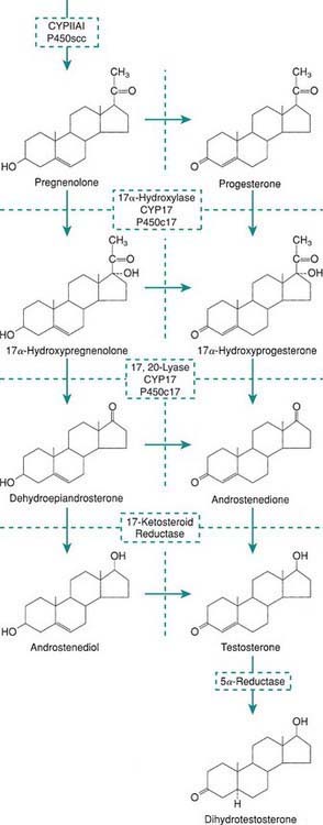
Within specific target cells, 6-8% of testosterone is converted by 5α-reductase to dihydrotestosterone, a more potent androgen (Fig. 576-1), and about 0.3% is acted on by aromatase to produce estradiol. Approximately half of circulating testosterone is bound to sex hormone–binding globulin (SHBG) and half to albumin; only 2% circulates in the free form. Plasma levels of SHBG are low at birth, rise rapidly during the 1st 10 days of life, and then remain stable until the onset of puberty. Thyroid hormone may play a role in this physiologic increase because neonates with athyreosis (absence of the thyroid gland) have very low levels of SHBG.

Antimüllerian hormone (AMH; previously referred to as müllerian inhibitory substance [MIS]), inhibin, and activin are members of the transforming growth factor-β (TGF-β) superfamily of growth factors. This group, which has over 45 members, also includes bone morphogenic proteins (BMPs). Members of the TGF-β superfamily are involved in the regulation of developmental processes and multiple human disease states, including chondrodysplasias and cancer.

AMH, a homodimeric glycoprotein hormone encoded by a gene on chromosome 19, is the earliest secreted product of the Sertoli cells of the fetal testis. Produced as a prohormone, its carboxyterminal fragment needs to be removed before it is active. AMH transcription is initiated by SOX9 acting through the HMG box, while its expression is upregulated by SF-1 binding to its promoter and further interacting with SOX9, WTI, and GATA4. AMH binds to 2 distinct serine/threonine receptors, each having a single transmembrane domain. The activated type 1 receptor signals to the SMAD family of intracellular mediators.

The gene for the AMH receptor (on chromosome 12) is expressed in Sertoli cells. In the female it is expressed in fetal müllerian duct cells, and in fetal and postnatal granulosa cells. During sex differentiation in males, AMH causes involution of the müllerian ducts: embryologic precursors of the cervix, uterus. It works in concert with SF-1 to cause involution of the fallopian tubes.

AMH is secreted in males by Sertoli cells during both fetal and postnatal life. In females, it is secreted by granulosa cells from 36 wk of gestation to menopause but at lower levels. The serum concentration of AMH in males is highest at birth, whereas in females it is highest at puberty. After puberty, both sexes have similar serum concentrations of AMH.

Inhibin is another glycoprotein hormone secreted by the Sertoli cells of the testes and granulosa and theca cells of the ovary. Inhibin A consists of an α-subunit disulfide linked to the β-A subunit, whereas inhibin B consists of the same α subunit linked to the β-B subunit.

Activins are dimers of the B subunits, either homodimers (BA/BA, BB/BB) or heterodimers (BA/BB). Inhibins selectively inhibit whereas activins stimulate pituitary FSH secretion. By means of immunoassays specific for inhibin A or B, it has been shown that inhibin A is absent in males and is present mostly in the luteal phase in women. Inhibin B is the principal form of inhibin in males, and in females during the follicular phase. Inhibin B may be used as a marker of Sertoli cell function in males. FSH stimulates inhibin B secretion in females and males, but only in males is there also evidence for gonadotropin-independent regulation. Levels of inhibin B are currently being studied in children with various forms of gonadal and pubertal disorders.

Like inhibin and activin, follistatin (a single-chain glycosylated protein) is produced by gonads and other tissues such as the hypothalamus, kidney, adrenal gland, and placenta. Follistatin inhibits FSH secretion principally by binding activins, thereby blocking the effects of activins at the level of both ovary and pituitary.

Many additional peptides act as mediators of the development and function of the testis. They include neurohormones such as growth hormone–releasing hormone, GnRH, corticotropin-releasing hormone, oxytocin, arginine vasopressin, somatostatin, substance P, and neuropeptide Y; growth factors such as insulin-like growth factors (IGFs) and IGF-binding proteins, TGF-β, and fibroblast, platelet-derived, and nerve growth factors; vasoactive peptides; and immune-derived cytokines such as tumor necrosis factor and interleukins IL-1, IL-2, IL-4, and IL-6.

Clinical patterns of pubertal changes vary widely (Chapters 12 and 555 covering pubertal maturation). In 95% of boys, enlargement of the genitals begins between 9.5 and 13.5 yr, reaching maturity at 13-17 yr. In a minority of normal boys, puberty begins after 15 yr of age. In some boys, pubertal development is completed in less than 2 yr, but in others it may take longer than 4.5 yr. The adolescent growth spurt occurs later in boys than in girls.

The median age of sperm production (spermarche) is 14 yr. This event occurs in mid-puberty as judged by pubic hair, testis size, evidence of growth spurt, and testosterone levels. Nighttime levels of FSH are in the adult male range at the time of spermarche; the 1st conscious ejaculation occurs at about the same time.

Function of the Ovaries

Without the presence of the SRY gene product, the undifferentiated gonad can be identified histologically as an ovary by 10-11 wk of gestation. Oocytes are present from the 4th mo of gestation and reach a peak of 7 million by 5 mo of gestation. For normal maintenance, oocytes need granulosa cells to form primordial follicles. Functional FSH (but not LH) receptors are present in oocytes of primary follicles during follicular development. Normal X chromosomes are needed for maintenance of oocytes. In contrast to somatic cells, in which only 1 X chromosome is active, both Xs are active in germ cells. At birth, the ovaries contain about 1 million active follicles, which decrease to 0.5 million by menarche. Thereafter, they decrease at a rate of 1000/mo, and at an even higher rate after the age of 35 yr.

The hormones of the fetal ovary are provided in most part by the fetoplacental unit. As in males, peak gonadotropin secretion occurs in fetal life and then again at 2-3 mo of life, with the lowest levels at about 6 yr of age. In both infancy and childhood, gonadotropin levels are higher in females than in males.

The most important estrogens produced by the ovary are estradiol-17 (E2) and estrone (E1); estriol is a metabolic product of these 2, and all 3 estrogens may be found in the urine of mature females. Estrogens also arise from androgens produced by the adrenal gland and the gonads (see Fig. 568-1). This conversion explains why in certain types of disorders of sex differentiation in males, feminization occurs at puberty. In 17-ketosteroid reductase deficiency, for example, the enzymatic block results in markedly increased secretion of androstenedione, which is converted in the peripheral tissues to estradiol and estrone. These estrogens, in addition to those directly secreted by the testis, result in gynecomastia. Estradiol produced from testosterone in the complete androgen insensitivity syndrome causes complete feminization in XY individuals.

Estrogen regulates a host of functionally different activities in multiple tissues. There are 2 distinct estrogen receptors with different expression patterns. The ovary also synthesizes progesterone, a progestational steroid; the adrenal cortex and testis synthesize progesterone as a precursor for other adrenal and testicular hormones.

A host of other hormones with autocrine, paracrine, and intracrine effects have been identified in the ovary. They include inhibins, activins, relaxin, and growth factors IGF-1, TGF-α and TGF-β, and cytokines.

Plasma levels of estradiol increase slowly but steadily with advancing sexual maturation and correlate well with clinical evaluation of pubertal development, skeletal age, and rising levels of FSH. Levels of LH do not rise until secondary sexual characteristics are well developed. Estrogens, like androgens, inhibit secretion of both LH and FSH (negative feedback). In females, estrogens also provoke the surge of LH secretion that occurs in the mid-menstrual cycle. The capacity for this positive feedback is another maturational milestone of puberty.

The average age at menarche in American girls is 12.5-13 yr, but the range of “normal” is wide, and 1-2% of normal girls have not menstruated by 16 yr of age. The age at onset of pubertal signs varies, with recent studies suggesting earlier ages than previously thought, especially in the U.S. African-American population (Chapter 555). Menarche generally correlates closely with skeletal age. Maturation and closure of the epiphyses is at least partially estrogen dependent, as demonstrated by a very tall 28 yr old, normally masculinized male with continued growth due to incomplete closure of the epiphyses, who proved to have complete estrogen insensitivity owing to an estrogen-receptor defect.

Therapeutic Aids

The estrogenic effects of polyhalogenated aromatic hydrocarbons (PHAHs) may in part be due to inhibition of estradiol sulfation by estrogen sulfotransferase (SULT1E1), an important pathway of estradiol inactivation. Naturally occurring estrogens administered orally are rapidly destroyed by gastrointestinal and liver enzymes; accordingly, they are usually given as conjugates or esters. The most widely used oral preparations are equine conjugated estrogens (Premarin) and ethinyl estradiol. Estrogen-containing skin patches for transdermal absorption are also used. With improvements in the understanding of estrogen and estrogen receptor interactions, a new class of compounds called selective estrogen-receptor modulators (SERMs) has been synthesized. For example, raloxifene, a nonsteroidal benzothiophene derivative, acts as an estrogen agonist in bone and liver and as an estrogen antagonist in breast and uterus.

Androgens such as testosterone are generally injected intramuscularly as long-acting esters (enanthate or cypionate, most commonly) because of their potency and steady response. Transdermal testosterone patches and a cutaneously applied gel have to date been used mostly in adults with hypogonadism because of the difficulty in titrating the doses needed during childhood and adolescence. Oral preparations, such as methyltestosterone or fluoxymesterone, do not produce so potent an androgenic response and may be hepatotoxic. Testosterone undecenoate, another oral preparation, is used in Europe but not in the USA. Sublingual (microspheres or pellets) and buccal (absorption via the buccal mucosa) preparations of testosterone are in development.

Bibliography

Bergada I, Bergada C, Campo S. Role of inhibins in childhood and puberty. J Pediatr Endocrinol Metab. 2001;14:343-353.

Biason-Lauber A, Konrad D, Navratil F, et al. A WNT4 mutation associated with Müllerian-duct regression and virilization in a 46,XX woman. N Engl J Med. 2005;351:792-798.

Bouvattier C, Carel JC, Lecointre C, et al. Postnatal changes of T, LH, and FSH in 46,XY infants with mutations in the AR gene. J Pediatr Endocrinol Metab. 2002;87:29-32.

Habert R, Lejeune H, Saez J. Origin, differentiation and regulation of fetal and adult Leydig cells. Mol Cell Endocrinol. 2001;179:47-74.

Hughes IA. Female development—all by default? N Engl J Med. 2004;351:748-750.

Josso N, diClemente N, Gouedard L. Anti-müllerian hormone and its receptors. Mol Cell Endocrinol. 2001;179:25-32.

Koopman P. The genetics and biology of vertebrate sex determination. Cell. 2001;105:843-847.

Ostrer H. Sex determination: lessons from families and embryos. Clin Genet. 2001;59:207-215.

Pierik FH, Vreeburg JTM, Stijnen T, et al. Serum inhibin B as a marker of spermatogenesis. J Clin Endocrinol Metab. 1998;83:3110-3114.

Quigley CA. The postnatal gonadotropin and sex steroid surge: insights from the androgen insensitivity syndrome (editorial). J Clin Endocrinol Metab. 2002;87:24-28.

Rajpert-De Meyts E. Expression of anti-müllerian hormone during normal and pathological gonadal development: association with differentiation of Sertoli and granulosa cells. J Clin Endocrinol Metab. 1999;84:3836-3844.

Rey RA, Belville C, Nihoul-Fékété C, et al. Evaluation of gonadal function in 107 intersex patients by means of serum anti-müllerian hormone measurements. J Clin Endocrinol Metab. 1999;84:627-631.

Sequera AM, Fideleff HL, Boquete HR, et al. Basal ultra sensitive LH assay: a useful tool in the early diagnosis of male pubertal delay? J Pediatr Endocrinol Metab. 2002;15:589-596.

Stenvers KL, Findlay JK. Inhibins: from reproductive hormones to tumor suppressors. Trends Endocrinol Metab. 2010;21:174-180.

Swerdloff RS, Wang C, Cunningham G, et al. Long-term pharmacokinetics of transdermal testosterone gel in hypogonadal men. J Clin Endocrinol Metab. 2000;85:4500-4510.

Teixeira J, Maheswaran S, Donahue PK. Müllerian inhibiting substance: an instructive development hormone with diagnostic and possible therapeutic applications. Endocr Rev. 2001;22:657-674.