Chapter 659 Cutaneous Viral Infections
Wart (Verruca)
Etiology
Human papillomaviruses (HPVs) cause a spectrum of disease from warts to squamous cell carcinoma of the skin and mucous membranes, including the larynx (Chapter 382.2). The human papillomaviruses are classified by genus, species, and type. More than 200 types are now known, and the entire genomes of about 100 are completely sequenced. The incidence of all types of warts is highest in children and adolescents. HPV is spread by direct contact and autoinoculation; transmission by fomites occurs. The clinical manifestations of infection develop ≥1 mo after inoculation and depend on the HPV type, the size of the inoculum, the immune status of the host, and the anatomic site.
Clinical Manifestations
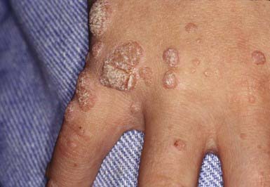
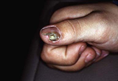
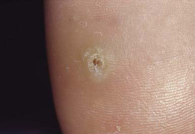
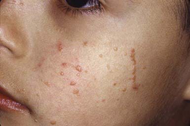
Cutaneous warts develop in 5-10% of children. Common warts (verruca vulgaris), caused most commonly by HPV types 2 and 4, occur most frequently on the fingers, dorsum of the hands (Fig. 659-1), paronychial areas, face, knees, and elbows. They are well-circumscribed papules with an irregular, roughened, keratotic surface. When the surface is pared away, many black dots representing thrombosed dermal capillary loops are often visible. Periungual warts are often painful and may spread beneath the nail plate, separating it from the nail bed (Fig. 659-2). Plantar warts, although similar to the common wart, are caused by HPV type 1 and are usually flush with the surface of the sole because of the constant pressure from weight bearing. When plantar warts become hyperkeratotic (Fig. 659-3), they may be painful. Similar lesions (palmar) can also occur on the palms. They are sharply demarcated, often with a ring of thick callus. The surface keratotic material must sometimes be removed before the boundaries of the wart can be appreciated. Several contiguous warts (HPV type 4) may fuse to form a large plaque, the so-called mosaic wart. Flat warts (verruca plana), caused by HPV types 3 and 10, are slightly elevated, minimally hyperkeratotic papules that usually remain <3 mm in diameter and vary in color from pink to brown. They may occur in profusion on the face, arms, dorsum of the hands, and knees. The distribution of several lesions along a line of cutaneous trauma is a helpful diagnostic feature (Fig. 659-4). Lesions may be disseminated in the beard area and on the legs by shaving and from the hairline onto the scalp by combing the hair. Epidermodysplasia verruciformis (EVER1, EVER2 genes), caused primarily by HPV types 5 and 8 (β-papillomaviruses, species 1), manifests as many diffuse verrucous papules. Wart types 9, 12, 14, 15, 17, 25, 36, 38, 47, and 50 may also be involved. Inheritance is thought to be primarily autosomal recessive, but an X-linked recessive form also has been postulated. Warts progress to squamous cell carcinoma in 10% of patients with epidermodysplasia verruciformis.
Genital HPV infection occurs in sexually active adolescents, most commonly as a result of infection with HPV types 6 and 11. Condylomata acuminata (mucous membrane warts) are moist, fleshy, papillomatous lesions that occur on the perianal mucosa (Fig. 659-5), labia, vaginal introitus, and perineal raphe and on the shaft, corona, and glans penis. Occasionally, they obstruct the urethral meatus or the vaginal introitus. Because they are located in intertriginous areas, they may become moist and friable. When untreated, condylomata proliferate and become confluent, at times forming large cauliflower-like masses. Lesions can also occur on the lips, gingivae, tongue, and conjunctivae. Genital warts in children may occur after inoculation during birth through an infected birth canal, as a consequence of sexual abuse, or from incidental spread from cutaneous warts. A significant proportion of genital warts in children contain HPV types that are usually isolated from cutaneous warts. HPV infection of the cervix is a major risk factor for development of carcinoma, particularly if the infection is due to HPV type 16, 18, 31, 33, 35, 39, 45, 52, 59, 67, 68, or 70. Immunization against types 6, 11, 16, and 18 is now available. Laryngeal (respiratory) papillomas contain the same HPV types as in anogenital papillomas. Transmission is believed to occur from mothers with genital HPV infection to neonates who aspirate infectious virus during birth.
Molluscum Contagiosum
Clinical Manifestations
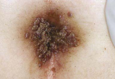
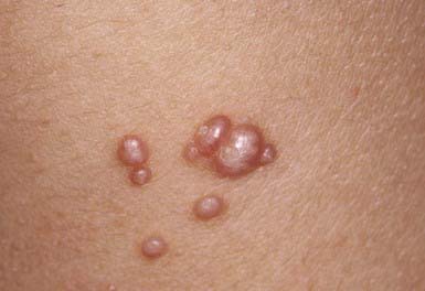
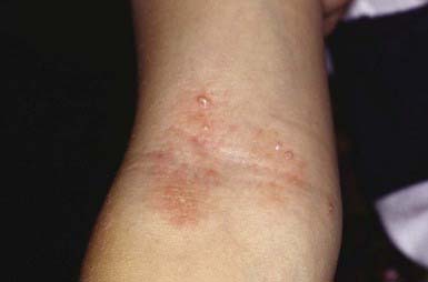
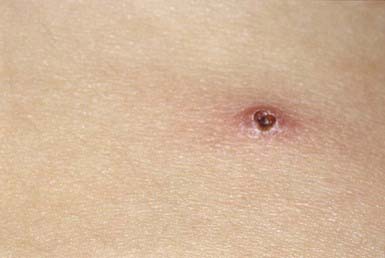
Discrete, pearly, skin-colored, smooth, dome-shaped, papules vary in size from 1 to 5 mm. They typically have a central umbilication from which a plug of cheesy material can be expressed. The papules may occur anywhere on the body, but the face, eyelids, neck, axillae, and thighs are sites of predilection (Fig. 659-6). They may be found in clusters on the genitals or in the groin of adolescents and may be associated with other venereal diseases in sexually active individuals. Lesions commonly involve the genital area in children but in most cases are not acquired by sexual transmission. Mild surrounding erythema or an eczematous dermatitis may accompany the papules (Fig. 659-7). Lesions on patients with AIDS tend to be large and numerous, particularly on the face. Exuberant lesions may also be found in children with leukemia and other immunodeficiencies. Children with atopic dermatitis are susceptible to widespread involvement in areas of dermatitis. A pustular eruption at the site of individual molluscum lesions is seen (Fig. 659-8). It is not a secondary bacterial infection, but an immunologic reaction to the molluscum virus and it should not be treated with antibiotics. Atrophic scars are often seen after this type of reaction.
Gewirtzman A, Bartlett B, Tyring S. Epidermodysplasia verruciformis and human papilloma virus. Curr Opin Infect Dis. 2008;21:141-146.
Gibbs S, Harvey I: Topical treatments for warts, Cochrane Database Syst Rev (19):CD001781, 2006.
Lio P. Warts, molluscum and things that go bump on the skin: a practical guide. Arch Dis Child Educ Pract Ed. 2007;92:119-124.


