100
Cutaneous Metastases
• Relatively rare with the exception of breast cancer and melanoma metastases.
• May be the presenting sign of a malignancy.
• Often associated with a poor prognosis.
• Overall, breast carcinoma most commonly metastasizes to the skin and prostate carcinoma least commonly (Table 100.1).
Table 100.1
Percentages of patients with metastatic cancer who had cutaneous metastases.
| Primary Malignancy | Patients with Metastatic Disease (n = 4020) | Patients with Cutaneous Metastases (n = 420; 10%) |
| Melanoma | 172 | 77 (45%) |
| Breast | 707 | 212 (30%) |
| Squamous cell carcinoma of the head and neck (e.g. laryngeal, oral) | 221 | 29 (13%) |
| Colon/rectum | 413 | 18 (4.5%) |
| Lung | 802 | 21 (2.5%) |
| Prostate | 207 | 0 |
Adapted from Lookingbill DP, Spangler N, Helm KF. Cutaneous metastases in patients with metastatic carcinoma: A retrospective study of 4020 patients. J. Am. Acad. Dermatol. 1993;29(2 Pt 1):228–236.
• Most common skin metastases: women – breast carcinoma, melanoma; men – melanoma, squamous cell carcinoma of the head and neck and lung, and colon carcinoma (Table 100.2).
Table 100.2
Ranking of underlying primary malignancies in patients with cutaneous metastases – men versus women.
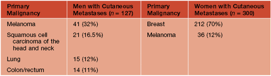
Adapted from Lookingbill DP, Spangler N, Helm KF. Cutaneous metastases in patients with metastatic carcinoma: A retrospective study of 4020 patients. J. Am. Acad. Dermatol. 1993;29(2 Pt 1):228–236.
• In general, nonspecific morphology: firm, mobile, painless papulonodule(s), often skin-colored to pink or red-brown and occasionally ulcerated (Fig. 100.1).
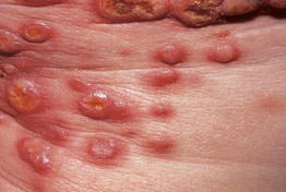
Fig. 100.1 Metastases of adenocarcinoma of the lung. Ulcerated erythematous nodules on the anterior trunk.
• Other clinical morphologies may be characteristic of a particular primary malignancy (Table 100.3).
Table 100.3
Clinical presentations of cutaneous metastases and histologic correlates.
An individual patient can have an admixture of the various types. Occasionally, cutaneous metastases have a zosteriform distribution pattern and clinically they can resemble dermatoses, including eczema, vasculitis, and erythema annulare centrifugum. Obviously, they can also mimic epidermoid or pilar cysts as well as cutaneous tumors, including non-melanoma skin cancers, lipomas, granular cell tumors, or angiosarcoma.
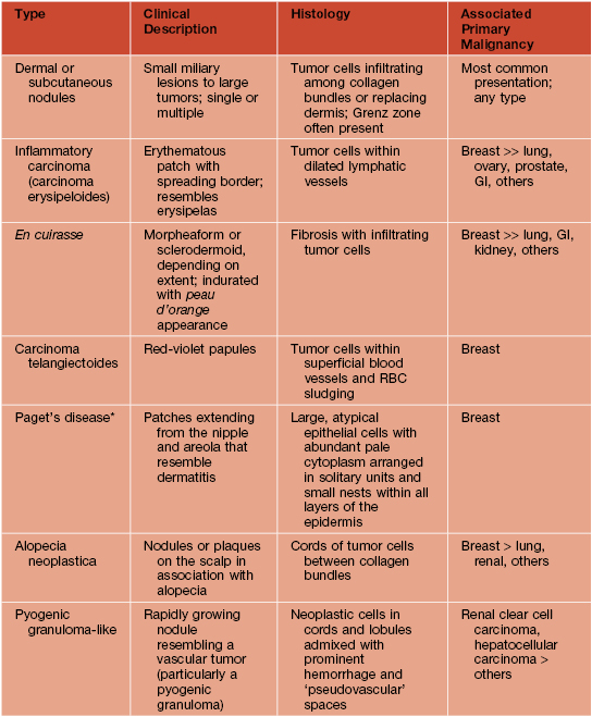
* Lesions of extramammary Paget’s disease share similar histopathologic features with those of Paget’s disease of the breast, but they usually represent (>75% of patients) primary cutaneous adenocarcinomas (see Table 60.6); an underlying visceral malignancy is most likely when there is perianal involvement.
GI, gastrointestinal; RBC, red blood cell.
• Melanoma metastases vary from pink to blue to black and they may develop along the lymphatic drainage between the primary site and regional lymph nodes (referred to as in-transit metastases; AJCC Stage III) or at distant sites (AJCC Stage IV) (Fig. 100.2).
Metastatic Breast Carcinoma
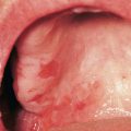
• May have distinctive clinical morphologies (Fig. 100.3): (1) carcinoma erysipeloides – well-demarcated elevated erythema that resembles erysipelas or cellulitis; (2) carcinoma telangiectoides – telangiectasias and red papules; and (3) carcinoma en cuirasse – begins on the chest as induration with a peau d’orange appearance reminiscent of scleredema.
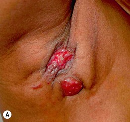
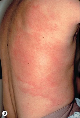
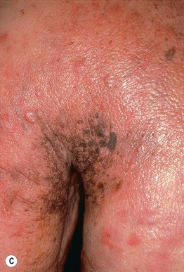
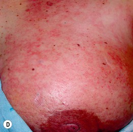
Fig. 100.3 Various presentations of cutaneous metastases of breast carcinoma. A Eroded erythematous nodules in the axilla. B Inflammatory form (carcinoma erysipeloides) with patches of erythema that may initially be misdiagnosed as infectious cellulitis. C Primarily en cuirasse form with obvious induration and peau d’orange appearance in addition to papulonodules. D Mixed pattern – reticulated erythema of carcinoma erysipeloides as well as peau d’orange appearance near the areola. A, D, Courtesy, Matthew B. Zook, MD, and Stuart R. Lessin, MD.
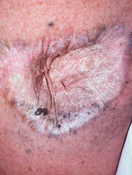
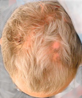
Alopecia Neoplastica
Sister Mary Joseph Nodule
• Pink to red-brown papule, umbilical or periumbilical (Fig. 100.5).
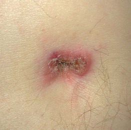
Fig. 100.5 Cutaneous metastasis of colon carcinoma. A Sister Mary Joseph nodule presenting as a pink plaque with scale-crust. Courtesy, Matthew B. Zook, MD, and Stuart R. Lessin, MD.
• Originally described in association with gastric carcinoma.
For further information see Ch. 122. From Dermatology, Third Edition.