[level-membership-for-radiology-category] Faster to perform, less operator dependent, more sensitive and specific


 , transmural inflammation with deep ulcers
  , mesenteric vessel engorgement, and fibrofatty proliferation
  .
.
 .
.
 , as well as local mesenteric fibrofatty proliferation and engorged vasa recta
  .
.
 are opacified. Traditional barium studies remain valuable for evaluation of strictures, fistulas, and sinus tracts.
IMAGING
General Features


DIFFERENTIAL DIAGNOSIS
Ulcerative Colitis (“Backwash” Ileitis)


 .
.
 , along with clusters of mildly enlarged mesenteric nodes
  .
.
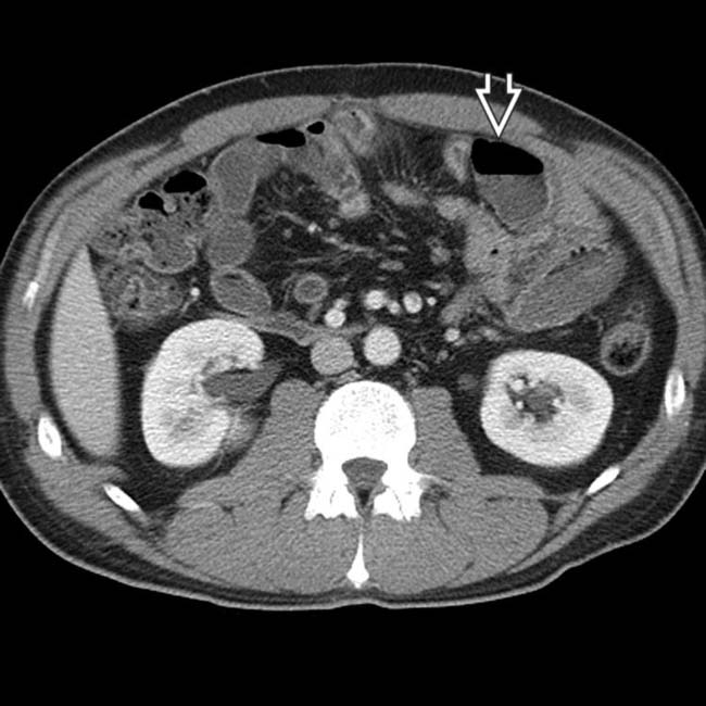
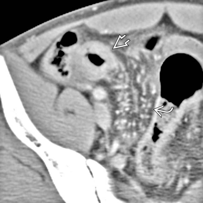
 that were separated by normal segments of bowel, the classic “skip lesions” of Crohn disease. Note the engorged vasa recta
  supplying the more distal segment of inflamed bowel.
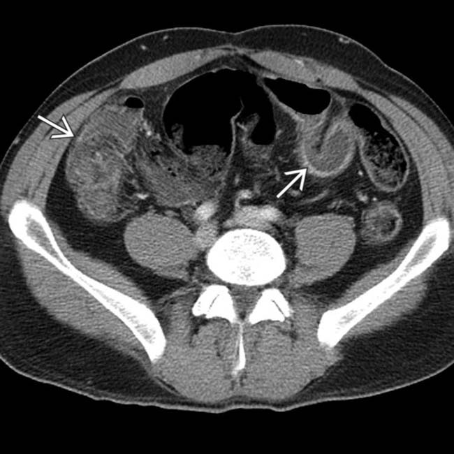
 with mucosal enhancement, wall thickening, and luminal narrowing.
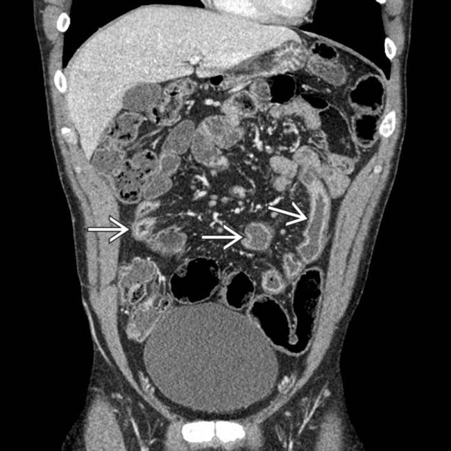
 along with mesenteric lymphadenopathy and prominent vessels.
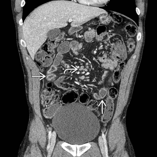
 and mesenteric lymphadenopathy
  . The diagnosis of Crohn disease was confirmed on colonoscopy with biopsy of the terminal ileum.
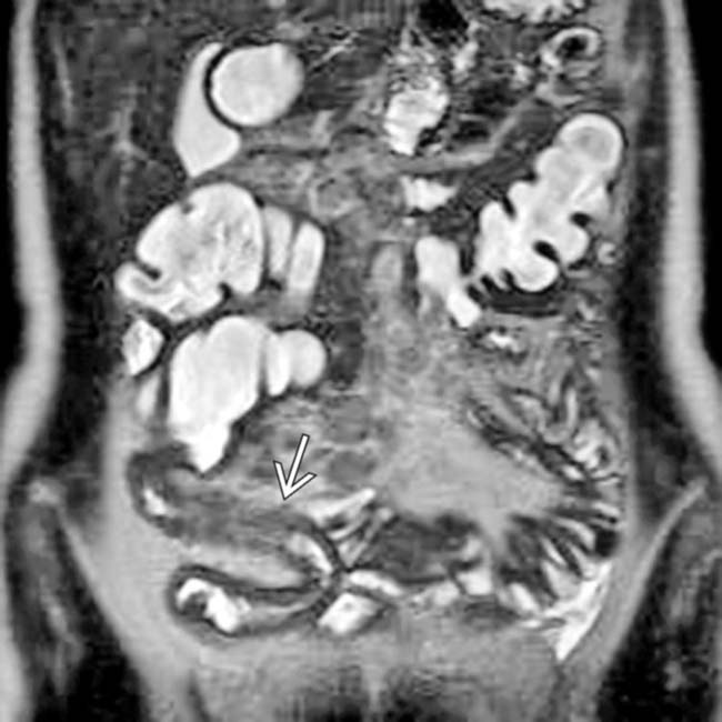
 representing the neoterminal ileum, where recurrent disease often occurs.
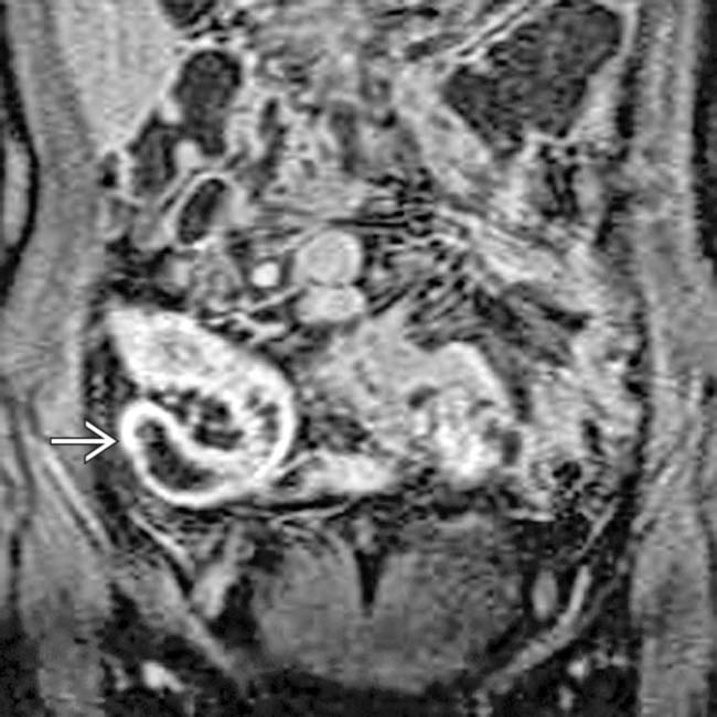
 . The adjacent small bowel and colon do not enhance with the same intensity.
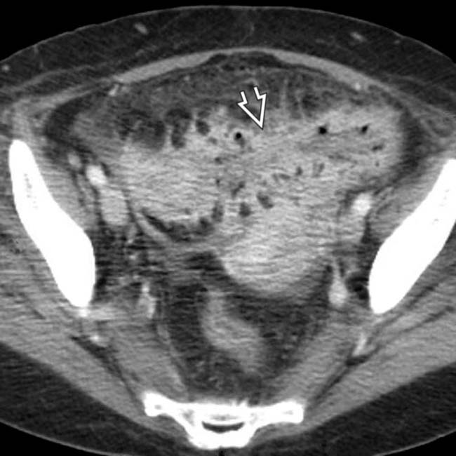
 and mesentery.
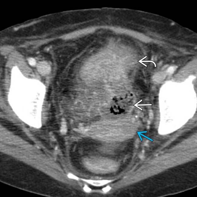
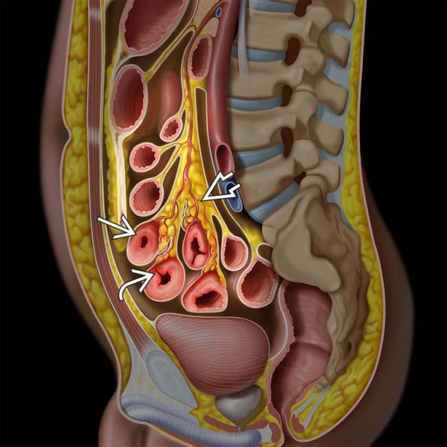
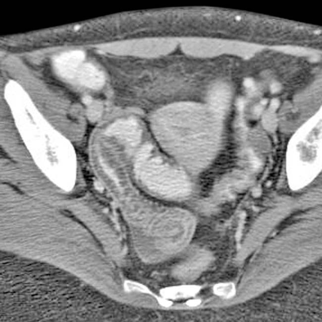
 interposed between the colon, uterus
  , and top of the bladder
  .
.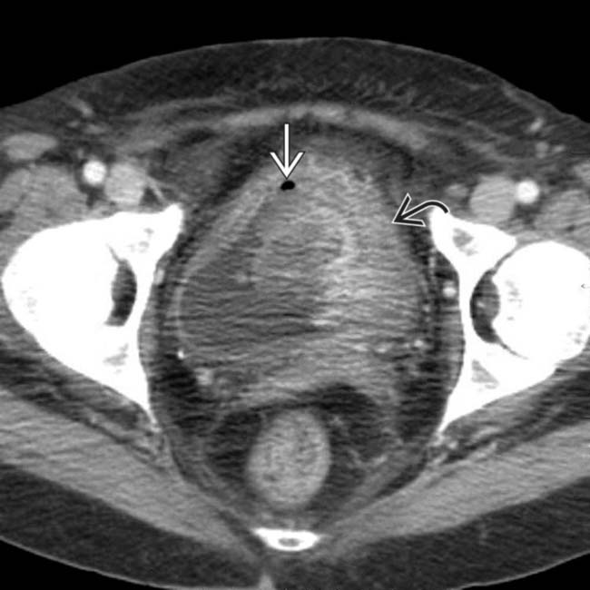
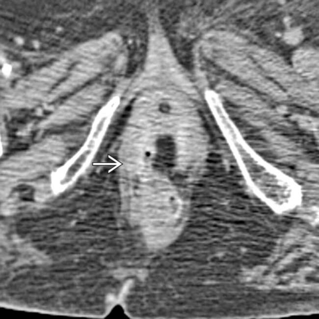
 along with gas
  and debris within the bladder.
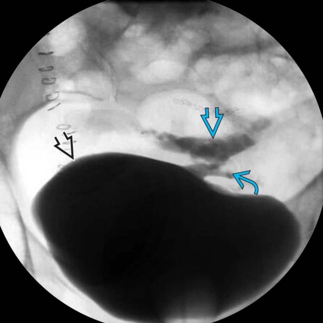
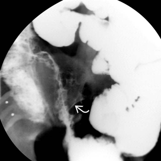
 opacifies the sigmoid colon
  through a fistulous tract
  . Fistulas are a key feature of the transmural inflammation caused by Crohn disease.
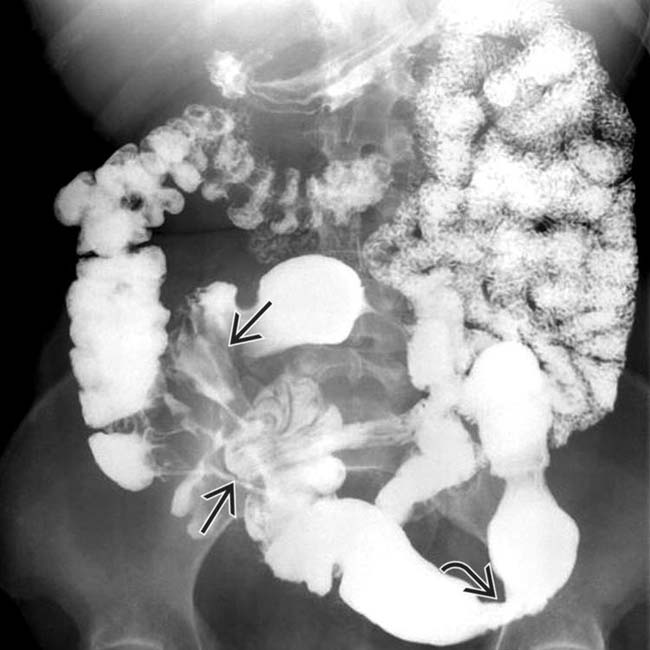
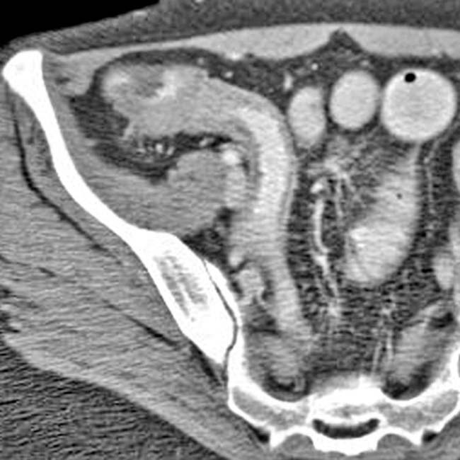
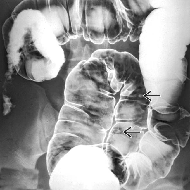
 , and ulceration with skip areas
  .
.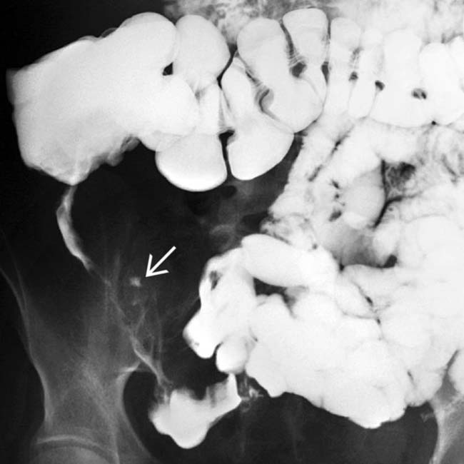
 and indirect evidence of mesenteric fibrofatty proliferation separating the TI from other bowel segments.
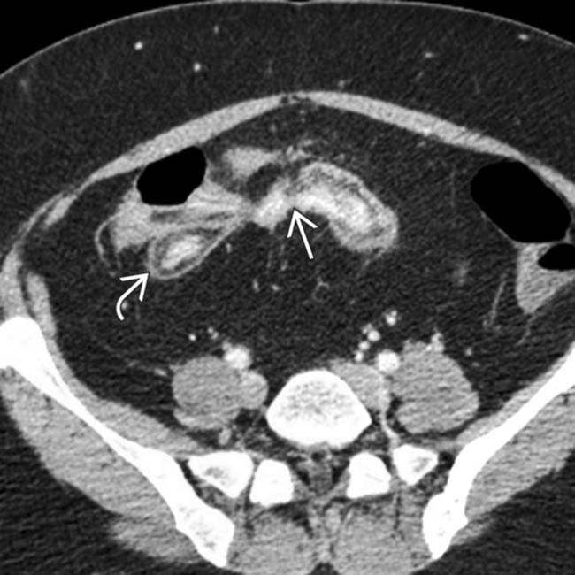
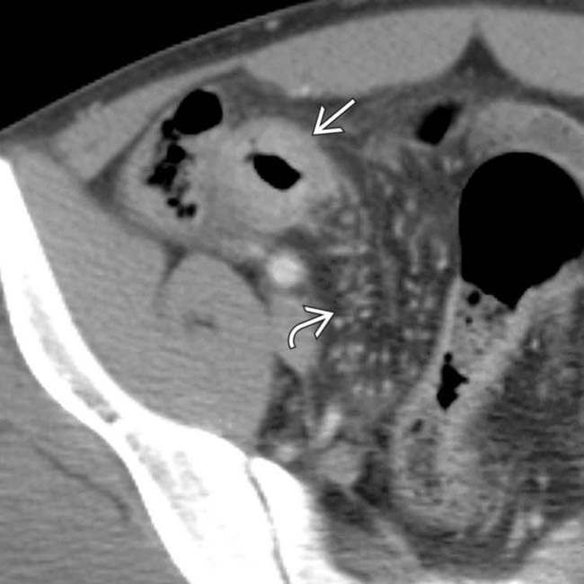
 , indicative of chronic inflammation. The nonepithelialized fistulas
  extend from 1 bowel segment to the others.
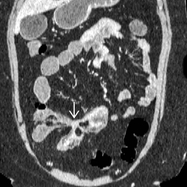
 between the bowel segments.
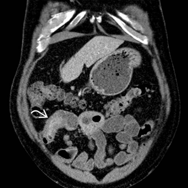
 with enhancing mucosa and submucosal edema.
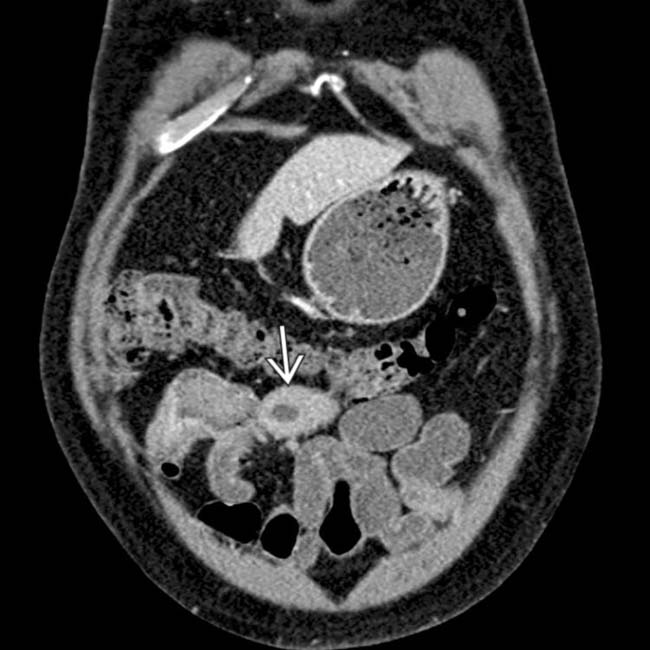
 felt to be concerning for neoplasm. Surgical resection confirmed lymphoma of the SB, a rare but recognized complication of CD or its medical therapy (steroids and immunomodulator drugs).
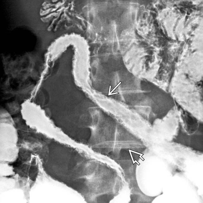
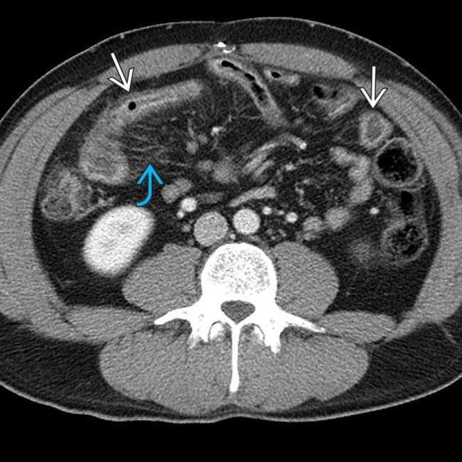
 and wide separation of the loops from creeping fat
  .
.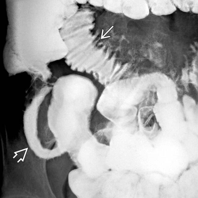
 and the featureless mucosa of terminal ileum in chronic disease
  .
.
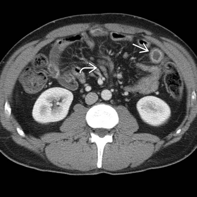
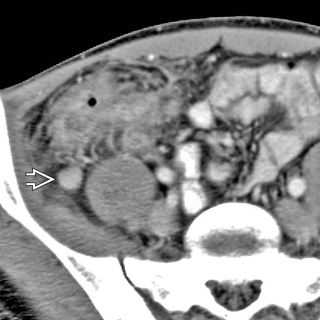
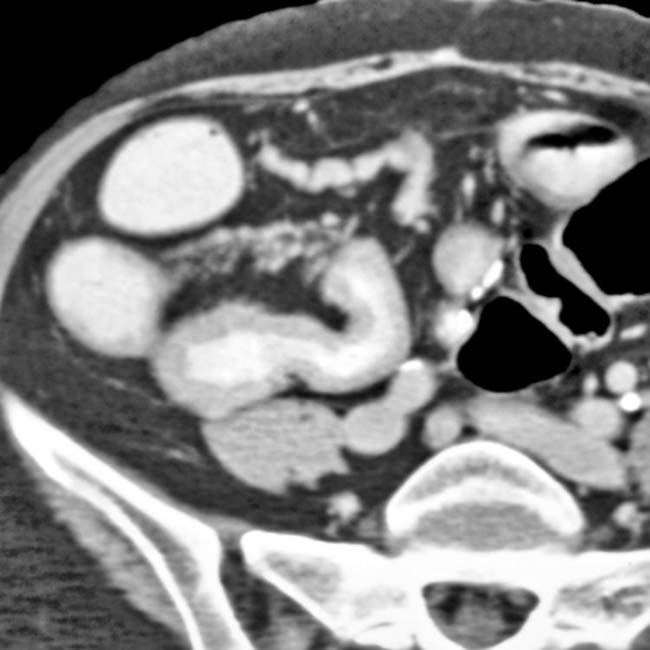
 and luminal narrowing. Note the hyperemia of the mesenteric blood vessels supplying the inflamed bowel
  .
.
 is also seen.
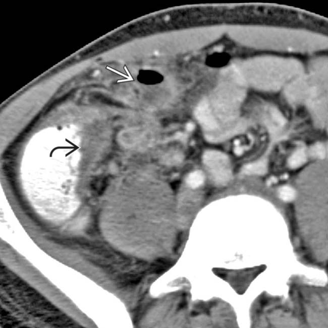
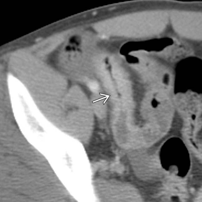
 of the distal ileum and marked inflammatory infiltration of the adjacent mesentery. Note the extraluminal bubbles of gas and the presence of small abscess
  .
.
 .
.
 throughout the colon.
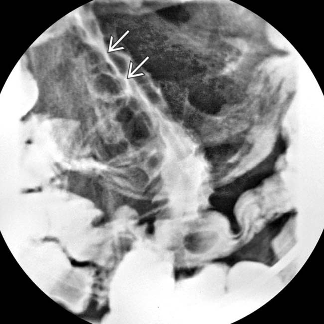
 .
.
 due to Crohn disease.
[/level-membership-for-radiology-category][not-level-membership-for-radiology-category] Faster to perform, less operator dependent, more sensitive and specific
, transmural inflammation with deep ulcers , mesenteric vessel engorgement, and fibrofatty proliferation .., as well as local mesenteric fibrofatty proliferation and engorged vasa recta . are opacified. Traditional barium studies remain valuable for evaluation of strictures, fistulas, and sinus tracts.IMAGING
General Features
Other Modality Findings
• Capsule endoscopy is commonly used to complement imaging studies

Buy Membership for Radiology Category to continue reading. Learn more here
[/not-level-membership-for-radiology-category]
