Chapter 32 Cortical Screw Post Femoral Fixation Using Whipstitches, Fabric Loop, or Endobutton
The Universal Salvage
Background
History
Whipstitch cortical screw post was perhaps the most popular method of soft tissue ACL graft fixation when two-incision methods were commonly used. There are many reports of high stability using this method.1–6 However, the advent of the Endobutton and then the various cross-pins that did not require a formal second incision relegated the two-incision method to a secondary role. More recently it has become apparent that a second incision for outside-in drilling can facilitate the lower femoral tunnel placement that is now recognized as biomechanically preferable, and more two-incision methods are now being described.
Biomechanics
The technique uses rigid cortical bone for anchorage. This has been shown to be the most important factor in producing high-stiffness fixation.7 The stiffness is reduced slightly by the length of the construct, but the rigidity of the cortical bone7 has been shown to more than compensate. A fabric or suture interface has been associated with high-stability ACLR,8–11 as have whipstitches if properly implanted.12–14 Both are described here in conjunction with cortical screw post fixation.
Advantages
The only disadvantage to this technique is that it requires a second incision. However, this disadvantage is usually primarily in the mind of the surgeon. We, and others, have never found the use of a small second incision to be of concern to the patient (see Chapter 49). Furthermore, the incision does not need to be large. Some may dislike the fact that a nonbioabsorbable and nonradiolucent screw remains in the patient. However, we have never seen one of these screws back out, nor have we ever seen one bother the patient2 because the screw sits flush on cortical bone under a thick muscular layer. Plus, because they are metadiaphyseal, they are far enough from the joint to not interfere with subsequent magnetic resonance images (MRIs).
Surgical Technique
Femoral Screw Insertion Technique
The femoral screws should be inserted unicortically as with tibial screw posts for three reasons.
Attaching the Graft to the Femoral Post
1 Endobutton-CL Fabric Loop Passed around the Femoral Post
With a long 2ST/2Gr graft this method is highly satisfactory because there is enough length to have more graft than is needed in each tunnel; usually 3 cm in each tunnel is a reasonable goal. If the measurements are off slightly after the graft is tightened down, the graft does not need to be adjusted. If the end result is too little graft in one of the tunnels by the surgeon’s standards, then the graft can be withdrawn out the tibial tunnel and passed through again using a shorter or longer Endobutton-CL loop. With the shorter 4ST graft this method is still usually satisfactory but requires more precision. A minimal length of 15 mm of graft in the tunnels appears to be adequate.15,16
3 Whipstitch Technique
In this technique the four-strand graft, if it is used, may be made into four single strands of graft of equal length. #2 braided, nonabsorbable whipstitches are then woven into each of the eight ends as described in Chapter 42.
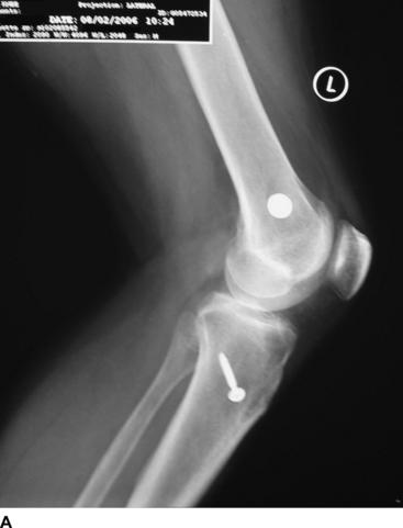
It is of paramount importance that all the sutures are woven in very tightly with strong tension after every pass or every other pass of the suture to maximally tighten the weave so that no tightening of the weave will occur later. Again, the graft should be marked with both a marker and suture, as in method 1. The sutures are then tied, two by two, around a cancellous screw and washer (Fig. 32-1) as follows: Each double suture strand is brought up to the smooth screw shank and crossed around the far side of the screw. These ends are then pulled back and tied two by two, such that the knot is on the graft side of the screw for the first two graft limbs. The process is repeated for the other two graft limbs. This will result in two knots in the femoral post. It is important to keep track of which sutures correspond to which graft segment either by using different color sutures or marking or knotting the suture ends before passing the graft segments.
4 Graft Passed Through the Endobutton-CL Loop and Sutures Used to Tie the Endobutton to the Femoral Post
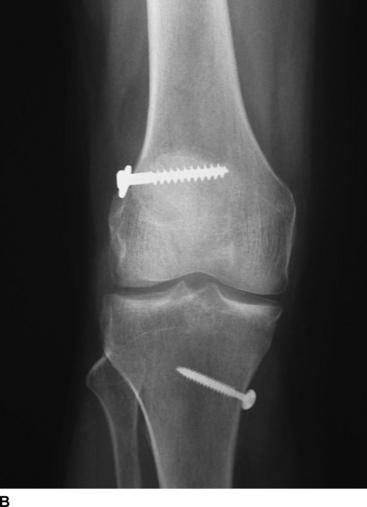
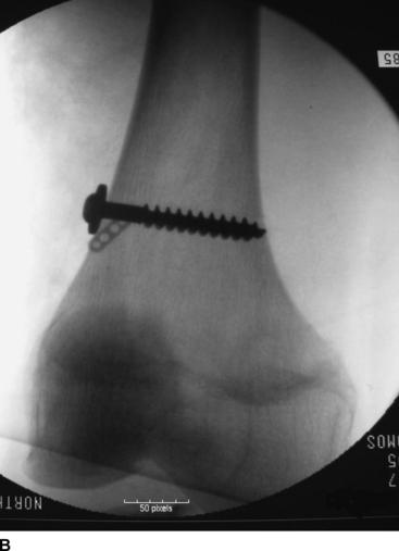
This is our preferred technique. Rather than tying a fabric loop around a cancellous screw, the surgeon can pass the graft through a short Endobutton loop and then pass a #5 braided nonabsorbable suture through each end of the Endobutton. These sutures can then be used to tie the Endobutton (and hence the loop and graft) to the screw as described in the previous paragraph. We prefer this technique if the Endobutton-CL is available because of the known strength of the fabric loop attached to the Endobutton. It also obviates the need to put whipstitches in the femoral end. Generally the surgeon will prefer to use the shortest loop, which is 15 mm. The Endobutton will be closely juxtaposed to the screw (Fig. 32-2) but is sufficiently low in profile to allow secure attachment to the screw without being in the way. A washer should be used on the screw.
Again, the graft should be marked with both a marker and suture as in method 1.
Fixating Single Strands of Graft and Odd Numbers of Strands
Double-length strands that are looped over a femoral fixation device are now the mainstay of autograft and allograft ACL soft tissue femoral fixation. However, single strands may also be used in combination with double strands or other single strands to fashion a variety of grafts. The goal is to have a graft of sufficient strength to provide good stability using the available tissue. Reports of excellent stability now exist for three-, four-, five-, six-, and eight-strand grafts (see Chapter 17). These variegated grafts may be used with any of the four fixation methods described earlier. For an added single strand of graft, whipstitches of braided #2 nonabsorbable suture should be implanted. Then the two ends can be tied one to one either around the Endobutton-CL fabric loop (methods 1 and 4), the Dacron tape (method 2), or the femoral screw itself (method 3).
The Femoral Post Technique Can Salvage the Following Situations
Conclusions
1 Prodromos CC, Joyce BT. Five-strand hamstring ACL reconstruction: a new technique with better long-term stability than four-strand. Presented at the 2006 meeting of the Mid-America Orthopaedic Association, San Antonio, TX, April, 2006. 2006.
2 Prodromos CC, Joyce BT. Five-strand hamstring ACL reconstruction: a new technique with better long-term stability than four-strand. Presented at the 2006 meeting of the Arthroscopy Association of North America, Hollywood, FL, May, 2006. 2006.
3 Goradia VK, Grana WA. A comparison of outcomes at 2 to 6 years after acute and chronic anterior cruciate ligament reconstructions using hamstring tendon grafts. Arthroscopy. 2001;17:383-392.
4 Howell SM, Deutsch ML. Comparison of endoscopic and two-incision techniques for reconstructing a torn anterior cruciate ligament using hamstring tendons. Arthroscopy. 1999;15:594-606.
5 Hamada M, Shino K, Horibe S, et al. Preoperative anterior knee laxity did not influence postoperative stability restored by anterior cruciate ligament reconstruction. Arthroscopy. 2000;16:477-482.
6 Maeda A, Shino K, Horibe S, et al. Anterior cruciate ligament reconstruction with multistranded autogenous semitendinosus tendon. Am J Sports Med. 1996;24:504-509.
7 To JT, Howell SM, Hull ML. Contributions of femoral fixation methods to the stiffness of anterior cruciate ligament replacements at implantation. Arthroscopy. 1999;15:379-387.
8 Prodromos CC, Han YS, Keller BL, et al. Stability of hamstring anterior cruciate ligament reconstruction at two- to eight-year follow-up. Arthroscopy. 2005;21:138-146.
9 Cooley VJ, Deffner KT, Rosenberg TD. Quadrupled semitendinosus anterior cruciate ligament reconstruction: 5-year results in patients without meniscus loss. Arthroscopy. 2001;17:795-800.
10 Gobbi A, Mahajan S, Zanazzo M, et al. Patellar tendon versus quadrupled bone-semitendinosus anterior cruciate ligament reconstruction: a prospective clinical investigation in athletes. Arthroscopy. 2003;19:592-601.
11 Gobbi A, Tuy B, Mahajan S, et al. Quadrupled bone-semitendinosus anterior cruciate ligament reconstruction: a clinical investigation in a group of athletes. Arthroscopy. 2003;19:691-699.
12 Feller JA, Webster KE. A randomized comparison of patellar tendon and hamstring tendon anterior cruciate ligament reconstruction. Am J Sports Med. 2003;31:564-573.
13 Howell SM, Deutsch ML. Comparison of endoscopic and two-incision techniques for reconstructing a torn anterior cruciate ligament using hamstring tendons. Arthroscopy. 1999;15:594-606.
14 Goradia VK, Grana WA. A comparison of outcomes at 2 to 6 years after acute and chronic anterior cruciate ligament reconstructions using hamstring tendon grafts. Arthroscopy. 2001;17:383-392.
15 Zantop T, Brucker P, Bell K, et al. The effect of tunnel-graft length on the primary and secondary stability in ACL reconstruction: a study in a goat model. Presented at the 2006 meeting of the European Society of Sports Traumatology, Knee Surgery, and Arthroscopy,Innsbruck, Australia, May, 2006. 2006.
16 Yamazaki S, Yasuda K, Tomita F, et al. The effect of intraosseous graft length on tendon-bone healing in anterior cruciate ligament reconstruction using flexor tendon. Knee Surg Sports Traumatol Arthrosc. 2006;14:1086-1093.