Coronary circulation and the myocardial conduction system

Published on 07/02/2015 by admin

Filed under Anesthesiology

Last modified 22/04/2025

Print this page

rate 1 star rate 2 star rate 3 star rate 4 star rate 5 star
Your rating: none, Average: 0 (0 votes)

This article have been viewed 1489 times

Coronary circulation and the myocardial conduction system

Harish Ramakrishna, MD, FASE

Coronary circulation

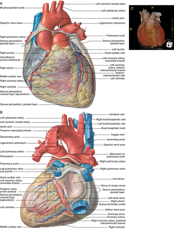
The right and left main coronary arteries arise from ostia (small openings) located behind the right and left aortic valve cusps toward the more cephalad portion of the sinus of Valsalva (Figure 52-1). The third aortic cusp is named the posterior or noncoronary cusp. The left main coronary artery travels anteriorly and leftward from the left coronary sinus and, after a 2-mm to 10-mm course between the pulmonary trunk and the left atrium, divides into the left anterior descending (LAD) and left circumflex arteries. Occasionally, a diagonal branch is also present.

The LAD or left interventricular coronary artery is a direct continuation of the left main coronary artery, traveling anterior and caudad, descending in the anterior interventricular groove. This artery terminates in the inferior aspect of the cardiac apex. Branches of this artery include (1) the first diagonal, (2) the first septal perforator, (3) right ventricular branches (inconstant), (4) three to five additional septal perforators, and (5) two to six additional diagonal branches. The LAD provides blood to most of the ventricular septum (anterior two thirds); the anterior, lateral, and apical walls of the left ventricle; most of the right and left bundle branches; and the anterolateral papillary muscle (double blood supply—see later in the chapter) of the left ventricle. It can provide collateral vessels to the anterior right ventricle via the circle of Vieussens, to the ventricular septum via septal perforators, and to the posterior descending artery via the distal LAD artery or a diagonal branch.

The left circumflex artery (LCA) travels posteriorly around the heart in the left atrioventricular (AV) sulcus. In 85% to 90% of individuals, it terminates near the obtuse margin of the left ventricle; in the remaining 10% to 15%, it continues around to the crux of the heart to become the posterior descending artery. Branches include (1) a branch to the sinoatrial (SA) node in 40% to 50% of individuals, (2) a left atrial circumflex branch, (3) an anterolateral marginal branch, (4) a distal circumflex artery, (5) posterolateral marginal branches, and (6) the posterior descending artery, as noted. This artery provides blood to the left atrium, the posterior and lateral left ventricle, the anterolateral papillary muscle of the left ventricle, and the SA node, as noted earlier. If it continues as the posterior descending artery (in 10% to 15% of hearts), it also supplies blood to the AV node, the proximal bundle branches, the remainder of the inferoposterior left ventricle, the posterior interventricular septum, and the posteromedial papillary muscle of the left ventricle.

The right coronary artery (RCA) passes forward to emerge between the pulmonary trunk and the right atrium and then descends in the right AV sulcus. In most hearts, once it reaches the apex, the RCA continues traveling in the posterior AV sulcus around the posterior of the heart to terminate as a left ventricular branch or to anastomose with the LCA. Branches include (1) the conus artery, (2) the artery to the SA node (in 50% to 60% of hearts), (3) anterior right ventricular branches, (4) right atrial branches, (5) an acute marginal branch, (6) an artery to the AV node and proximal bundle branches, (7) the posterior descending artery (in 85% to 90% of hearts), and (8) terminal branches to the left atrium and left ventricle. The RCA supplies blood to the SA node (as noted earlier), the right ventricle, the crista supraventricularis, and the right atrium. If it provides the posterior descending artery, it also supplies blood to those areas discussed previously. The RCA provides collaterals to the LAD artery via the conus artery and septal perforators.

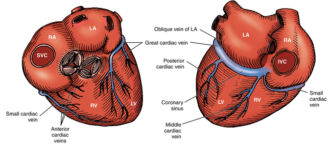
The coronary venous system consists of three primary systems: (1) the coronary sinus, (2) the anterior right ventricular veins, and (3) the thebesian veins (Figure 52-2). The coronary sinus is located in the posterior AV groove and receives blood from the great, middle, and small cardiac veins; the posterior veins of the left ventricle; and the left oblique atrial vein (oblique vein of Marshall). The coronary sinus drains blood primarily from the left ventricle and opens into the right atrium. The two to three anterior right ventricular veins originate in and drain blood from the right ventricular wall. These veins enter the right atrium directly or enter into a small collecting vein at the base of the right atrium. The thebesian veins are tiny venous outlets that drain directly into the cardiac chambers, primarily the right atrium and right ventricle.

Myocardial conduction system

The conducting system of the heart is composed of specially differentiated cardiac muscle fibers that are responsible for initiating and maintaining normal cardiac rhythm as well as ensuring proper coordination between atrial and ventricular contraction. This system comprises the SA node, the AV node, the bundle of His, right and left branch bundles, and Purkinje fibers.

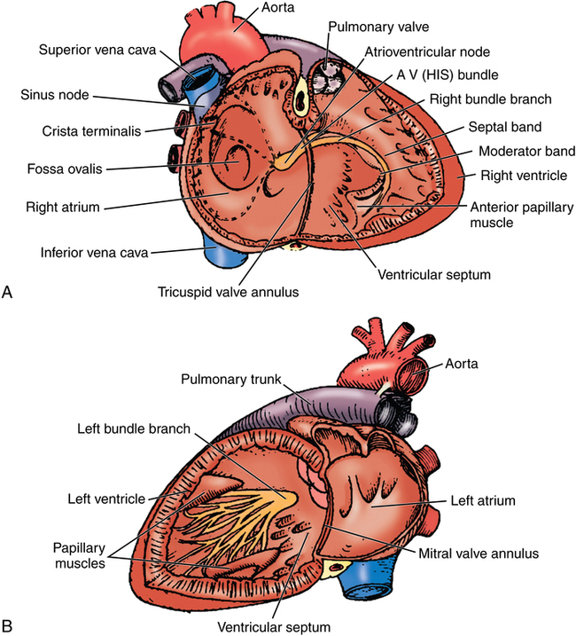
The SA node is a horseshoe-shaped structure located in the upper part of the sulcus terminalis of the right atrium (Figure 52-3). It extends through the atrial wall from epicardium to endocardium. SA nodal fibers have a higher intrinsic rate of depolarization than do any other cardiac muscle fibers and act as the pacemaker of the heart (see Chapter 33). Three internodal pathways facilitate conduction of impulses between the SA and AV nodes: the anterior (Bachmann bundle), middle, and posterior internodal tracts. The AV node lies in the medial floor of the right atrium at the base of the atrial septum above the orifice of the coronary sinus. The bundle of His begins at the anterior aspect of the AV node and penetrates through the central fibrous body. Here, the bundle of His divides into the left and right branch bundles. The division straddles the upper border of the muscular ventricular septum, and the bundles run superficially down either side of the septum. About midway to the apex, the left bundle divides into the anterior superior and posterior inferior fascicles. These fascicles continue to the base of the papillary muscles of the left ventricle, where they form plexuses of Purkinje fibers that distribute to all portions of the left ventricular myocardium. The right branch bundle continues to the anterior papillary muscle of the right ventricle, where it forms a plexus of Purkinje fibers that distribute to all portions of the right ventricular myocardium.