Congenital heart disease: Congestive heart failure

Published on 07/02/2015 by admin

Filed under Anesthesiology

Last modified 22/04/2025

Print this page

rate 1 star rate 2 star rate 3 star rate 4 star rate 5 star
Your rating: none, Average: 0 (0 votes)

This article have been viewed 1179 times

Congenital heart disease: Congestive heart failure

William C. Oliver, Jr., MD

Anesthetic management of a patient with congenital heart disease (CHD) and congestive heart failure (CHF) requires a thorough understanding of human anatomy and physiology. Anesthesia is more frequently employed in the setting of cardiac surgery; however, as life expectancy increases in this patient population, anesthesia is increasingly necessary for the performance of noncardiac procedures.

The fetal circulation is perfectly designed to adapt to the intrauterine environment; the anatomic and physiologic characteristics of the fetal circulation also allow the fetus to tolerate CHD. It is with the transition from fetal to postnatal circulation that characteristic physiologic changes usually appear that point to the presence of a cardiac anomaly. The degree of “fetal” circulation that persists after birth determines the impact of extrauterine life; consequently, the diagnosis of CHD may be made immediately after birth or may be delayed for days to months. Certain CHDs typically result in poor ventricular function and hemodynamics with progression to CHF. Unfortunately, the cause of CHF is not always readily apparent in neonates and infants. In addition to CHF, patients with CHD may have secondary effects, such as pulmonary hypertension.

CHD has been classified using numerous systems that represent the biases and interests of their authors, but none have been universally accepted. For this discussion, CHD will be classified according to the presence or absence of cyanosis. Cyanotic lesions are caused by shunting of blood from the pulmonary circulation to the systemic circulation, which results in poor pulmonary blood flow and progressive arterial desaturation. In contrast, lesions without cyanosis (acyanotic) are characterized by pulmonary overcirculation because of shunt from the systemic to pulmonary circulation that eventually causes CHF. Excessive blood to the lung reduces lung compliance and increases the work of breathing by two mechanisms: (1) increased left atrial pressure resulting in pulmonary venous congestion and pulmonary edema, which decreases the compliance of the lung itself, and (2) increased size of pulmonary vessels, causing greater obstruction to airflow in both large and small airways. A typical example of acyanotic CHD with CHF is the preterm infant with a ductus arteriosus that does not close postnatally (patent ductus arteriosus, or PDA). A large left-to-right (L-to-R) shunt causes systemic circulatory steal, pulmonary overcirculation, and diastolic hypotension. Pharmacologic or surgical closure of the PDA is required to resolve the CHF.

The orifice of the shunt in a PDA may be described as restrictive or nonrestrictive. If the orifice is restrictive, the primary determinant of shunt fraction is the radius of the orifice and pressure gradient. If the orifice is nonrestrictive, the shunt direction and magnitude depend on the relative resistances of the pulmonary and systemic vascular circulations, which can be manipulated as part of the care of individuals until closure of the PDA.

CHF can also occur from obstructive cardiac defects more quickly than from L-to-R shunts and may progress to circulatory collapse without immediate intervention. Obstructive defects are characterized as subvalvular, primary valvular, or supravalvular obstructions causing reduced left ventricular reserve, hypotension, and ventricular hypertrophy. Furthermore, myocardial ischemia is especially common in obstructive lesions of both ventricles, which show signs of failure. Patients with obstructive defects are at increased risk for developing arrhythmias, such as ventricular fibrillation, in part because of the tenuous myocardial O2 supply-to-demand ratio. Isolated obstructive lesions can be seen in the right ventricle and are exacerbated by increased pulmonary vascular resistance (PVR), resulting in right-sided heart failure.

Anesthetic management

The presence of CHF in a patient with CHD should raise concern, especially if the patient has pulmonary hypertension or an obstructive outflow lesion, because these patients are at increased risk for experiencing serious perioperative morbidity and death. In some patients, the stress of surgery may be enough to lead to acute cardiac decompensation, typically reflected by a respiratory, as well as metabolic, acidosis. There is no single anesthetic technique that has been identified as “ideal” for these patients. Anesthetic management must include knowledge of the individual physiologic aspects of the cardiac anatomy (shunt flow) and a plan to minimize myocardial depression and maintain baseline hemodynamic parameters.

Central to the anesthetic management of patients with CHF secondary to L-to-R shunting is to avoid increasing the shunt and pulmonary overcirculation. Repeated cardiopulmonary evaluations are important to identify influences on the patient that would increase systemic vascular resistance or decrease PVR (Table 202-1). One of the foremost responsibilities of the anesthesia team is to consider factors that may adversely affect shunt flow. However, the team must be cautious about the degree to which the L-to R-shunt is manipulated to reduce pulmonary overcirculation. Efforts to aggressively reduce systemic vascular resistance or increase PVR to reduce pulmonary overcirculation and, hence, improve CHF will lessen the L-to-R shunt; however, the ensuing hypotension or pulmonary hypertension, respectively, reduces coronary perfusion and stresses a poorly functioning right ventricle, leading to hemodynamic deterioration. In contrast, cyanotic CHD with R-to-L shunts can be improved dramatically with aggressive measures to increase systemic vascular resistance or decrease PVR, often in association with immediate hemodynamic improvement.

Table 202-1

Manipulations That Alter Pulmonary Vascular Resistance (PVR)

↑ PVR ↓ PVR
Hypoxia
Hypercarbia
Acidosis
Hyperinflation
Atelectasis
Sympathetic stimulation
High hematocrit
Surgical constriction
O2
Hypocarbia
Alkalosis
Normal functional residual capacity (FRC)
Low hematocrit
Blocking sympathetic stimulation
Nitric oxide

Altering ventilation or oxygenation or both are important ways to influence either L-to-R or R-to-L shunts. The pulmonary vasculature is very sensitive to changes in PaCO2. Values of PaCO2 between 28 and 32 mm Hg are associated with pulmonary vasodilation that will worsen CHF in patients with L-to-R shunts. A PaCO2 above 55 mm Hg raises PVR and lessens pulmonary overcirculation in these patients. However, the patient will tolerate hypercarbia only until the associated respiratory acidosis results in worsening myocardial function and compromised hemodynamics, overcoming any benefit from reduced L-to-R shunt. The effect of O2 as a potent pulmonary vasodilator often goes unnoticed in patients with shunts. The patient’s inspired O2 concentration (FIO2) should be lowered incrementally after induction of anesthesia to avoid hyperoxia, which decreases PVR and could worsen pulmonary overcirculation.

Patients with CHF secondary to L-to-R shunts or obstructive lesions will benefit from anesthetic medications that do not change or only minimally decrease myocardial contractility. Administering appropriate doses of synthetic opioids, ketamine, or both (which have minimal to no negative inotropic effects) provides excellent hemodynamic stability for these patients. Ketamine is widely used for neonates and infants with CHF because it maintains cardiac output and perfusion pressures by enhanced sympathetic stimulation. Propofol has been used infrequently in this patient population, but even with careful dose adjustments, the use of these drugs poses risks to hemodynamic stability. The use of propofol has been associated with significant vasodilation and hypotension in adults. Preservation of baseline heart rate is essential in the neonate or infant because neonates and infants, unlike adults, are unable to augment cardiac output with increases in stroke volume. Ketamine preserves the heart rate better than does any other anesthetic agent and prevents the bradycardia often associated with the administration of fentanyl alone. A benefit to the use of synthetic opioids in patients with CHD and CHF is the ability of these drugs to attenuate increases in PVR. Though these patients with CHD and CHF may have hypertrophied pulmonary vasculature, the vasculature can be very reactive. Any insult that increases PVR may cause severe systemic hypotension by decreasing left ventricular preload and hypoxemia by decreasing perfusion of the lungs.

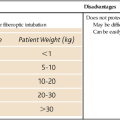
If the intravenous route is not an option for induction of anesthesia, intramuscular or inhalation techniques may be used. For intramuscular induction, ketamine is the drug of choice for use when obtaining venous access by causing dissociative anesthesia. A major advantage of ketamine over other medications for intramuscular induction is the limited respiratory depression and maintenance of airway reflexes caused by ketamine, which increases safety until venous access is obtained. Halogenated inhalation agents have been used for induction for years in pediatric patients with CHD and CHF. In the past, the nonirritating airway effects of halothane made it a popular choice for induction, but the associated significant myocardial depression, bradyarrhythmia, and prolonged time to induction were particularly disadvantageous for neonates and infants with CHD, who responded with a greater degree of hypotension to the use of inhalation agents than do older children and adults. Hypotension occurs with the use of inhalation agents (see Chapter 66) as a result of a combination of reduced myocardial contractility, increased vasodilation, lower heart rate, and inhibition of compensatory reflex mechanisms.

The development of inhalation agents with low solubility and less myocardial depression (desflurane and sevoflurane) since the 1990s has been very beneficial, creating an opportunity to use these inhalation agents in managing the care of neonates and infants with CHF. The lower solubility of these inhalation agents results in more rapid induction and emergence. Unlike desflurane, sevoflurane has been evaluated repeatedly in neonates and infants with either cyanotic or acyanotic CHD and has been found to be acceptable in terms of cardiopulmonary side effects when used appropriately for induction and maintenance of anesthesia. It has replaced halothane as the agent of choice for inhalation induction because it results in more rapid inductions, reasonably well maintained hemodynamics, fewer arrhythmias, better contractility, and more rapid emergence and possesses the nonirritating airway effects of halothane. Furthermore, the use of sevoflurane has been shown to be associated with less breath holding, coughing, and laryngospasm, as compared with halothane.

Anesthesia can be induced by sevoflurane in patients with obstructive lesions, but at the concentration often used for an inhalation induction, the negative inotropic effect and risk of hypotension present a risk of hemodynamic collapse, a phenomenon not seen when ketamine is used to induce anesthesia. However, compared with the hypotension associated with the use of other inhalation agents, hypotension from sevoflurane can be quickly corrected by decreasing the concentration of inhaled drug. When inhalation induction with sevoflurane is used, an intravenously administered induction agent should be substituted to complete induction as soon as intravenous access is obtained because, irrespective of the inhalation agent used, a ketamine-based or an opioid-based anesthetic induction is more likely to provide greater hemodynamic stability in this population of patients.