Chapter 32 Congenital Anomalies of the Coronary Circulation
The word coronary is derived from the Latin coronarius (pertaining to a crown), translated from the Greek stephanos (wreath), which refers to the crown-like or wreath-like arrangement of arteries that encircle the heart.1 This chapter deals with the coronary circulation—arterial and venous—including morphogenesis, the normal coronary arteries, and the congenital anomalies as classified in Table 32-1.
Table 32-1 Classification of Congenital Anomalies of the Coronary Circulation
| Congenital anomalies of coronary arteries unassociated with congenital heart disease |
| Congenital anomalies of coronary arteries associated with congenital heart disease |
| Acquired anomalies of coronary arteries resulting from congenital heart disease |
| Congenital anomalies of the coronary venous circulation |
In 1513, Leonardo da Vinci’s anatomic drawings of a bullock’s heart identified the coronary arteries (see Figure 32-3A) and the great cardiac vein (coronary sinus).2 Leonardo called attention to two vessels that arise from two external openings (aortic sinuses). In 1543, 500 years later, the circulation in the normal heart was characterized,3 and congenital anomalies of the coronary arteries were recognized.4 In 1543, Andreas Vesalius, the celebrated 16th century Flemish anatomist, depicted an anomalous right coronary artery originating from the left aortic sinus and passing anterior to the right ventricular outflow tract (De Humani Corporis Fabrica).

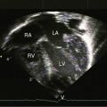
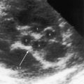
Figure 32-3 A, Drawing of a bullock’s heart by Leonardo da Vinci, circa 1513, with labels by the author. “The heart from the right side. The pulmonary artery has been removed to expose the pulmonary orifice and the semilunar valves guarding it. From the aorta spring the right and left coronary arteries.” B, Echocardiogram (short-axis view) from an asymptomatic 23-year-old man in whom the right coronary artery (RCA) originated from the left aortic sinus and passed between the aorta and right ventricular outflow tract. The left main coronary artery (LCA) originated from the left aortic sinus by a separate ostium and divided into the left anterior descending and circumflex coronary arteries (compare with Figure 32-1D, second illustration). (Ao = aorta; LAD = left anterior descending.) C, Coronary arteriogram from a 29-year-old woman with angina pectoris. The right coronary artery (RCA) originated from the left aortic sinus and was compressed (paired arrows) as it passed between the aorta and right ventricular outflow tract (compare with Figure 32-1D, second illustration). The left coronary artery originated by a separate ostium from the left aortic sinus and divided into the left anterior descending (LAD) and circumflex coronary arteries.
Selective coronary angiography was introduced in 1962 by Mason Sones5 and was a major step forward in imaging coronary artery anatomy in the living beating human heart. Techniques continue to advance and include transesophageal echocardiography, magnetic resonance imaging, computerized axial tomography, electron beam tomography, and three-dimensional reconstruction.6–8 Concurrently, nomenclature has evolved.9
Myocardial morphogenesis begins with the emergence of trabeculations in the luminal ventricular layers that permit an increase mass in the absence of a coronary circulation.10 Cardiac jelly in the embryonic luminal layer is the primitive site of metabolic exchange between cardiac mesenchyma and blood in the ventricular cavity.4,11 The human heart begins to beat as early as the 22nd day after conception, and a few days later, an ebb and flow circulation permits metabolic exchange.
The sequence of development of the coronary vascular bed begins with blood islands and coronary venous connections followed by coronary artery–to–aortic connections. Blood islands are epicardial layers of endothelial cells distended by nucleated erythrocytes. The blood islands proliferate, coalesce, and form rudimentary networks of vascular channels with no discernible connections to other islands or to the cavity of the ventricle. The second stage of development is a venous connection between the network of vascular channels and the coronary sinus, an arrangement that drains the blood islands and reduces their size. The distal coronary bed remains a loose intermingling vascular network until myocardial mass develops. In the third stage of development, the coronary arteries join the aorta through an arterial connection to the plexus of vascular channels. The third stage is established after aortopulmonary septation and after formation of the semilunar valves. Flow commences from the aorta into the proximal coronary arteries through myocardial capillaries, then into coronary veins and into the coronary sinus. It is unknown why the two coronary arteries originate from the right and left aortic sinuses that face the right ventricular outflow tract (Figure 32-1A). The embryonic great arteries contain six sinuses, three in the aorta and three in the pulmonary trunk. The endothelial outgrowths or anlagen (buds, sprouts) in the third aortic sinus and in all three pulmonary sinuses undergo rapid involution or do not develop. Aortic–to–coronary artery connections become evident before the appearance of precursor connections in the aortic sinuses. It has been speculated that the aortic sinus sites of the two coronary ostia are determined by mural tension that is increased by the catenoid configuration of the right and left sinuses, so that endothelium penetrates the aortic wall preferentially in the right and left sinus sites, establishing connections with the epicardial coronary plexus.

The capillary bed interposed between the coronary arterial and coronary venous circulations plays a pivotal role in the transport of oxygen and nutrients to the myocardium. Capillary density, defined as the ratio of the number of capillaries to the number of cardiomyocytes, is basic to the metabolic exchange between blood and tissues. Capillary density depends on angiogenesis—the capacity of capillaries to replicate. Fetal capillaries replicate in response to the hypoxic intrauterine environment. Postnatal angiogenesis is a diminishing continuation of that response.11
The coronary circulation includes three separate systems of veins.12 The largest system terminates in the coronary sinus and drains blood from most of the left ventricle. A second venous system drains most of the blood from the right ventricle. Adam Christian Thebesius, a German anatomist (1686-1732), described a third venous system consisting of tributaries that drain directly into the cardiac chambers.12 Before the discovery of the pulmonary circulation, Thebesian veins were considered pathways of blood flow from right heart to left heart.
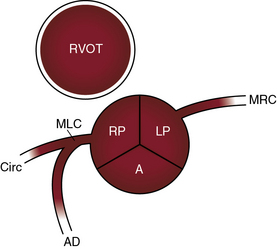
The round or ovoid ostia of the two coronary arteries are located in the right and left anterior aortic sinuses, which are therefore called the coronary sinuses.1 The posterior aortic sinus is devoid of a coronary ostium and is appropriately called the noncoronary sinus.1 Normal coronary artery ostia are located in the middle of an aortic sinus just below the sinotubular junction and just above the upper margin of the aortic cusps. Abnormally located ostia originate above the sinotubular junction, in the posterior aortic sinus, or eccentrically near a commissure.
There are typically two coronary ostia, but three or four ostia are considered normal variants. A third ostium usually results from a separate conus branch in the right coronary sinus (see Figure 32-1A). Less commonly, three coronary ostia are the result of origin of the left anterior descending and circumflex coronary arteries from the left aortic sinus and of origin of the right coronary artery from the right aortic sinus, an arrangement that has been designated absent left main coronary artery (Figure 32-2A).

A relatively large left ventricular mass is served by both the left and the right coronary arteries. Either the right or the left coronary artery can be dominant, with dominance encompassing a wide range. Extreme dominance assumes that the dominant and nondominant coronary arteries arise from separate ostia in separate aortic sinuses, in contrast to dominance that results from a single coronary artery that originates from a single coronary ostium in a single aortic sinus (see subsequent).13
Congenital anomalies of the coronary arteries are the subject of comprehensive clinical and necropsy reviews.13–16 The widespread use of selective coronary angiography has gone far in establishing the types and prevalence of these anomalies (see Table 32-1).13,15,17 The incidence rate was 1.3% in 126,595 coronary arteriograms performed at the Cleveland Clinic.18 Transesophageal echocardiography19,20 and multislice computed tomographic coronary angiography21–25are established procedures for depicting the origin and course of anomalous coronary arteries.
Anomalous aortic origins of coronary arteries unassociated with congenital heart disease
Anomalous origins of coronary arteries that are unassociated with congenital heart disease are shown in Table 32-2.
Table 32-2 Congenital Anomalies of Coronary Arteries Unassociated with Congenital Heart Disease
A normal coronary ostium is located in the middle of the aortic sinus and is considered anomalous when it is located above the sinotubular junction, in the posterior sinus, or in close proximity to an aortic commissure.4 In 30% to 50% of normal human hearts, a small conus artery arises from the right aortic sinus and is of no functional significance (Figure 32-1A).17,26 The circumflex coronary artery may arise from a separate ostium in the right aortic sinus and pass behind the aorta (Figure 32-1B) or may arise from the proximal right coronary artery and enter the left atrioventricular groove as if it were a proximal branch of the left coronary artery.18,27 Anomalous origin of the circumflex coronary artery is regarded as benign, but there is one report of myocardial ischemia in the absence of coronary atherosclerosis. Also regarded as benign is the absence of the circumflex coronary artery with a dominant right coronary artery that perfuses the lateral and posterolateral left ventricular walls.18
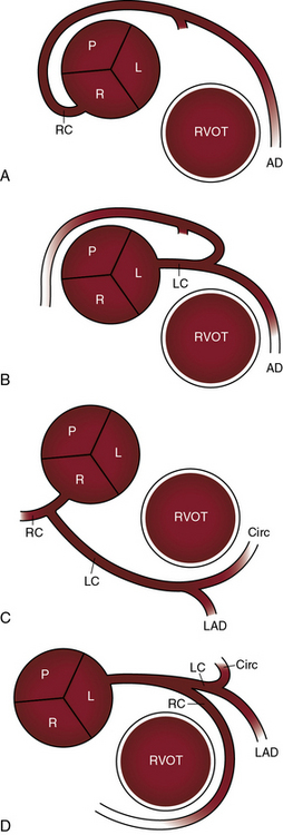
Origin of the left main coronary artery from the right aortic sinus is uncommon but clinically important.13,18,26 The right coronary artery and the left main coronary artery can arise from separate ostia in the right aortic sinus (see Figure 32-1C).28 The left main coronary artery passes either anterior or posterior to the right ventricular outflow tract (see Figure 32-1C) or between the right ventricular outflow tract and the aorta (see Figure 32-1D) or is within the ventricular septum (intramyocardial).13,18,26,29 The risk associated with passage of the left main coronary artery between the aorta and the right ventricular outflow tract has been convincingly established (see subsequent).
Absence of a left main coronary artery refers to a rare anomaly in which the left anterior descending and circumflex coronary arteries originate from separate ostia in the left aortic sinus (see Figure 32-2A).13,18,30 Both arteries are then normally distributed, so the arrangement is functionally benign.18
Atresia of the left main coronary artery includes ostial atresia, which is represented by an imperforate dimple, and atresia of the proximal course of the coronary artery, which is represented by a fibrous strand (see Figure 32-2B). A large conus branch originates from the right coronary artery and supplies the left anterior descending and circumflex coronary arteries (see Figure 32-2B).
Origin of the right coronary artery from the left aortic sinus (see Figures 32-1C and D, and 32-3B) represents about one quarter of ectopic coronary artery origins.13,28 Less common is origin of the right coronary artery from the posterior aortic sinus or from the left coronary artery.18 The functional significance of these arrangements does not depend on the anomalous origin but on whether the ectopic right coronary artery passes between the aorta and right ventricular outflow tract (see Figures 32-1D, and 32-3B and C).13,18,28
Single coronary artery has been known since 1903.31 The single coronary artery originates from a single ostium in the left or right aortic sinus and gives rise to the entire coronary circulation (Figures 32-4 and 32-5).13,18 A second coronary ostium is neither present nor implied. A single coronary artery unassociated with congenital heart disease divides into normally formed and normally distributed branches irrespective of the aortic sinus from which it originates (see Figure 32-4C and D).13,18 A single coronary artery associated with a congenital heart disease is characterized by branching patterns that bear no resemblance to normal.13 A single coronary artery functions normally unless a major branch is congenitally hypoplastic32 or passes between the aorta and right ventricular outflow tract (Figure 32-6).


Anomalous origin of a coronary artery from a systemic artery is relatively rare (see Table 32-2). The anomalous coronary artery can originate from the innominate, subclavian, internal mammary, or carotid or bronchial artery or from the descending aorta.
The Coronary Artery Surgery Study (1989) asked whether a coronary artery that originated anomalously from an aortic sinus was predisposed to atherosclerosis.33 The study concluded that atherosclerosis was more prevalent in anomalous circumflex coronary arteries than in matched controls with nonanomalous circumflex arteries.
The courses taken by anomalous coronary arteries and their branches are more important than their ectopic origins.26,29,34,35 These courses include: (1) passage anterior to the right ventricular outflow tract (see Figure 32-1C); (2) passage posterior to the aorta (see Figure 32-1C); (3) passage between the aorta and right ventricular outflow tract (see Figure 32-1D); and (4) intramyocardial passage (bridging).13,34–38 The clinical significance of bridging is uncertain.39 Angina pectoris, myocardial infarction, and sudden death are, with few exceptions, reserved for anomalous coronary arteries that pass between the aorta and the right ventricular outflow tract (see previous).13,26,29,34,40–42
Ischemic risk is determined by a number of variables in addition to the course of an anomalous coronary artery, namely13,26,28: (1) the coronary artery or branch that is involved; (2) the slit-like ostium and acute angulation of the origin of the ectopic coronary artery34,41,43; (3) coronary dominance28; and (4) the effect of strenuous exercise.26,34,43,44 Risk is greatest when the left main coronary artery arises from the right aortic sinus and courses between the aorta and right ventricular outflow tract (see Figure 32-1D)26,29,34,42,45 or when the left branch of a single coronary artery courses between the aorta and the right ventricular outflow tract (see Figure 32-6B). Risk is less when the right coronary artery originates from the left aortic sinus and passes between the aorta and the right ventricular outflow tract (see Figure 32-1D).28,41,43 Sudden death typically occurs during or immediately after physical exercise.26,29,34,42,44 Fatal impairment to coronary blood flow appears to be sporadic in light of the fact that highly trained competitive athletes perform intense physical exercise repeatedly and for many years before experiencing sudden death.46 It has been proposed that expansion of the aortic root and pulmonary trunk during exercise is responsible for an increase in acute angulation of the ectopic coronary artery and for flap-like closure of its slit-shaped ostium.13,26,46 An analogous risk may confront the fetus during the hemodynamic stress of labor and delivery. A stillborn term fetus that died during labor had at necropsy a right coronary artery that originated from the left aortic sinus.41 Exercise-induced dilation of the aortic root can compress an ectopic coronary artery against the base of the pulmonary trunk to which the infundibular septum is firmly anchored.13,26 This mechanism may account for sudden death when an ectopic coronary artery is not characterized by acute angulation or a slit-like ostium.28
Coronary arteries that originate normally from their respective aortic sinuses can have abnormal distal connections. A small coronary arterial fistula incidentally discovered during routine coronary arteriography is a case in point (see Chapter 22).18 Another example is a functionally benign distal intercoronary communication in which contiguous peripheral coronary artery branches form an open-ended circulation with bidirectional flow.18
Anomalous pulmonary arterial origins of coronary arteries13,18,47,48 are listed in Table 32-2. Anomalous origin of the left coronary artery from the pulmonary trunk is the most important (see Chapter 21).24 Anomalous origin of the right coronary artery from the pulmonary trunk is a rare anomaly that was first reported in 188549 and may be discovered incidentally at necropsy.50,51
Congenital anomalies of coronary arteries unassociated with congenital heart disease
These congenital anomalies are shown in Table 32-2.
Anomalies of coronary arterial size include congenital hypoplasia or atresia, or conversely congenital coronary artery ectasia or aneurysm (Tables 32-1 and 32-3). Atresia of the ostium of the left main coronary artery extends proximally as a fibrous chord from a blind dimple in the left aortic sinus (see Figure 32-2B).13,52–56 Survival depends on adequate connections between a normally arising right coronary artery and the distal nonhypoplastic portion of the left coronary artery (see Figure 32-2B). Myocardial infarction and death occur from infancy54 to the teens56 to 60 years and 71 years of age.55 The clinical presentation of infants with ostial atresia or left main coronary artery atresia is similar to the presentation of infants with anomalous origin of the left coronary artery from the pulmonary trunk (see Chapter 21).54
| Partial or complete absence: |
Modified from Mantini E, Grondin CM, Lillehei CW, Edwards JE: Congenital anomalies involving the coronary sinus. Circulation 1966;33:317-327.
Hypoplasia of coronary arteries exists when the right coronary artery and left circumflex coronary artery do not go beyond the lateral border of the heart.57 Hypoplasia of the left main coronary artery57 is analogous to atresia described previously (see Figure 32-2B).
Coronary artery ectasia accompanies cyanotic congenital heart disease (see Figure 32-14). Hereditary hemorrhagic telangiectasia or Rendu-Osler-Weber disease is another association58 (see Chapter 30). Congenital aneurysms in the proximal right or left coronary artery are believed to result from an occult developmental fault.59

Congenital coronary artery anomalies associated with congenital heart disease
Congenital anomalies of the coronary arteries associated with congenital heart disease are shown in Box 32-1.
Congenital and acquired abnormalities of the coronary arteries coexist with congenital malformations of the heart.60 Routine coronary angiography during catheterization of adults with congenital heart disease has revealed an 11% incidence rate of congenital abnormalities and a 5% incidence rate of acquired lesions.60
Fallot’s tetralogy is associated with a 10% to 36% incidence rate of anomalies of origin, course, and distribution of coronary arteries (see Chapter 18).61–63 Most common are origin of a conus artery from the right coronary artery or from the right aortic sinus (Figure 32-7A), origin of a circumflex coronary artery from the right coronary artery, origin of a left anterior descending artery from the right aortic sinus (Figure 32-7B), and origin of a single coronary artery from the right aortic sinus (Figures 32-7C, and 32-8). Less common anomalies include origin of a coronary artery from the posterior aortic sinus, anastomoses between coronary arteries and bronchial arteries,64,65 fistulas between coronary arteries and pulmonary arteries or right atrium,64 hypoplastic coronary arteries, and pulmonary arterial origin of the left anterior descending coronary artery.66 The physiologic consequences of these coronary anomalies are unimportant, but the surgical risk incurred when anomalous coronary arteries cross the right ventricular outflow tract is important indeed (see Figure 32-7B and C).


In complete transposition of the great arteries (see Chapter 27), the origins of the coronary arteries are relevant to arterial switch operations.67–69 Coronary artery morphology and ventricular morphology are concordant (i.e., a morphologic right coronary artery is assigned to the morphologic subaortic right ventricle and a morphologic left coronary is assigned to the morphologic subpulmonary left ventricle). The left aortic sinus and the posterior aortic sinus face the right ventricular outflow tract (Figure 32-9). The rightward left sinus is designated sinus 1.69,70 The posterior sinus is leftward and is designated sinus 2 (see Figure 32-9).69,70 Dual sinus origin means that a main coronary artery branch arises from each of the two sinuses that face the right ventricular outflow tract, an arrangement that is present in 90% of cases.69 Most dual sinus origins are represented by the circumflex coronary artery, the left anterior descending artery arising from left aortic sinus, and the right coronary artery arising from the posterior sinus (Figure 32-9A).69 Less common is dual sinus origin of circumflex coronary artery and right coronary artery from the posterior sinus and origin of the left anterior descending artery from the left aortic sinus (Figure 32-9B).69 Single sinus origin means that main coronary arterial branches arise from one of the two but not both of the sinuses that face the right ventricular outflow tract.69 In single sinus origin, the anterior descending, circumflex and right coronary arteries arise from the posterior sinus.69 Rarely, an anomalous coronary artery is intramyocardial or passes between the aorta and the right ventricular outflow tract.

Congenitally corrected transposition of the great arteries is characterized by morphologic right and morphologic left coronary arteries that are concordant with morphologic right and morphologic left ventricles (see Chapter 6).67,71 The two aortic sinuses that face the right ventricular outflow tract are designated right posterior and left posterior (Figure 32-10). The nonfacing anterior sinus is noncoronary (see Figure 32-10). The morphologic right coronary artery originates from the left posterior sinus, and the morphologic left coronary artery originates from the right posterior sinus, arrangements that are inverted (see Figure 32-10). The course of the left anterior descending coronary artery establishes the position of the ventricular septum. Coronary anomalies are uncommon, but there are reports of hypoplasia of the circumflex and left anterior descending coronary arteries72 and of a single coronary ostium arising from the right posterior sinus.71,72
In double outlet right ventricle, the origin, course, and distribution of the coronary arteries is the same as in the normal heart (see Chapter 19).56,67 The right coronary artery arises from the right aortic sinus, and the left coronary artery arises from the left aortic sinus (Figure 32-11).56,67

Occasionally, both coronary arteries arise from the same aortic sinus, or a single coronary artery arises from a single coronary artery ostium. Double outlet left ventricle is associated with a relatively high incidence of congenital anomalies of the origin and course of coronary arteries, including single coronary artery, origin of the left anterior descending artery from the right aortic sinus, and origin of both coronary arteries or all three coronary arteries from the right or left aortic sinus.73 In univentricular hearts with left ventricular morphology (see Chapter 26), the aortic sinus from which the right or left coronary artery originates is determined by inversion or noninversion of the outlet chamber. When the outlet chamber is inverted, the aortic sinus from which each coronary artery originates is the same as in congenitally corrected transposition of the great arteries with a biventricular heart (see Figure 32-10), but a major branch of each coronary artery delimits or outlines the surface boundaries of the outlet chamber.74 When the outlet chamber is noninverted, the aortic sinus from which each coronary artery originates is the same as in complete transposition of the great arteries with a biventricular heart (see Figure 32-9). Tricuspid atresia (see Chapter 25) is characterized by delimiting coronary arteries that meet at the apex of the rudimentary morphologic right ventricle.75
Coronary artery origins in truncus arteriosus (see Chapter 28) correspond to the number and spatial relationships of the quadricuspid, tricuspid, or bicuspid truncal valve (Figure 32-12).76–78 Truncal cusps that face the atrioventricular orifices lie posterior, and the cusp or cusps that do not face the atrioventricular orifices lie anterior.76 When the truncal valve is tricuspid, the right coronary artery arises from the right anterior sinus, and the left coronary artery arises from the posterior sinus or the left anterior sinus (see Figure 32-12A). Bicuspid truncal valve cusps are oriented right/left or anterior/posterior (see Figure 32-12B). When the cusps are anterior/posterior, the left coronary artery or a single coronary artery arises from the posterior sinus, and the right coronary artery arises from the anterior sinus (see Figure 32-12B).76,79 When bicuspid truncal valves are oriented right/left, the right coronary artery arises from the right sinus, and the left coronary artery arises from the left sinus.76 Quadricuspid truncal valves are associated with two types of coronary artery arrangements. When two of the four truncal cusps are posterior and two are anterior, the left coronary artery arises from the left posterior sinus, and the right coronary artery arises from the right or left anterior sinus (see Figure 32-12C).76,79 In both of these arrangements, high origins of right and left coronary artery ostia are frequent.79

Isolated quadricuspid valves are rare (see Chapter 7).80,81 In one such patient, a single coronary artery arose from the right anterior sinus.82 Isolated bicuspid aortic valves are associated with two types of cusp relationships and coronary artery origins (Figure 32-13). The two cusps may be oriented anterior/posterior with both coronary arteries arising from the anterior sinus (see Figure 32-13A), or the cusps may be oriented right/left with the right coronary artery arising from the right sinus and the left coronary artery arising from the left sinus (see Figure 32-13B). Bicuspid aortic valves tend to be associated with left coronary artery dominance and have a relatively high incidence of immediate bifurcation of the left coronary artery into circumflex and left anterior descending arteries.83–86

Coronary artery disease as a result of congenital heart disease
Anomalies of coronary arteries that result from congenital heart disease are shown in Box 32-2.
Coarctation of the aorta is associated with extramural and intramural abnormalities of the coronary arteries (see Chapter 8), and systemic hypertension causes intimal proliferation and medial thickening, which are risk factors for premature coronary atherosclerosis.87 Luminal size in coarctation increases in proportion to medial thickness.88 Intramural coronary arteries and arterioles have thick walls, a rich adventitia, and dense collagen and elastic fibers.87
Supravalvular aortic stenosis (see Chapter 7) is associated with extramural coronary artery abnormalities analogous to coarctation of the aorta.89–92 Ostial obstruction results from aortic medial proliferation or from adherence of an aortic cusp.90,93 The extramural coronary arteries are exposed to systolic hypertension proximal to the supravalvular stenosis, so they are thick-walled, dilated, tortuous, and prematurely atherosclerotic.
Severe bicuspid aortic regurgitation is accompanied by large smooth-walled extramural coronary arteries appropriate for the increase in left ventricular mass of a magnified geometrically normal left ventricle. Coronary artery ectasia refers to elongation, tortuosity, and dilation of extramural coronary arteries in adults with cyanotic congenital heart disease (Figure 32-14).94–96
Suprasystemic pressure in the blind hypoplastic right ventricle of pulmonary atresia with intact ventricular septum generates flow through intramural intratrabecular channels with the development of ventriculocoronary artery connections (see Chapter 24).97–99 Myocardial ischemia is caused by the fistulous steal associated with aortic diastolic runoff into the right ventricle through these large intramural channels. Ischemia is also caused by luminal narrowing of the ventriculocoronary arterial channels that extend from their right ventricular origins to the coronary ostia.98,99 Narrowing ranges from mild to obliteration to coronary ostial discontinuity.98,100,101
In aortic atresia with mitral atresia, the coronary circulation is not accompanied by ventriculocoronary arterial channels because the sealed left ventricle cannot generate suprasystemic pressure.102–105 When the mitral valve is hypoplastic but perforate, the small left ventricle receives diastolic flow, contracts isovolumetrically, and generates suprasystemic pressure. Nevertheless, intramyocardial sinusoids with direct ventriculocoronary artery connections do not develop, so suprasystemic left ventricular pressure is not transmitted into epicardial coronary arteries, which are spared the morphologic changes that characterize pulmonary atresia with intact ventricular septum. A sinusoidal/capillary network develops, but coronary arterial abnormalities, if present at all, consist of tortuosity with an increase in wall thickness, but not luminal narrowing.102 Paradoxical emboli to the coronary arteries are rare complications of cyanotic congenital heart disease.106,107 Right-to-left intracardiac shunts deliver paradoxical emboli from peripheral thromboembolic sources.
Congenital anomalies involving the coronary sinus
Congenital anomalies of the coronary sinus are shown in Table 32-3.
The coronary sinus is subject to a variety of congenital abnormalities.108 Partial or complete absence results from loss of the roof, from absence of the entire coronary sinus, or from an atrial septal defect that occupies the site of the ostium of the coronary sinus (see Chapter 15). Hypoplasia of the coronary sinus is a response to diversion of blood from the sinus into dilated Thebesian veins. Stenosis or atresia of the sinus ostium is represented by a coronary sinus that is hypoplastic or that ends blindly. A left superior vena cava carries coronary sinus blood retrograde into the left innominate vein.109 The coronary sinus enlarges when it receives a left superior vena cava or an hepatic vein, when it is joined by a left superior vena cava that receives blood from the inferior vena cava via the hemiazygos vein (see Chapter 29), when it is the drainage site for total anomalous pulmonary venous connection (see Chapter 15), or when there is a coronary artery–to–coronary sinus fistula (see Chapter 22).
1 Anderson R., Becker A.E. Cardiac anatomy. London: Gower Medical Pubishing; 1980.
2 O’Malley C., Saunders J.B., Saunders MC, editors. Translations, text and introduction. Leonardo da Vinci on the Human Body. New York: Greenwich House, 1982.
3 Baroldi G., Scomazzoni G. Coronary circulation in the normal heart and in the pathologic heart. Washington, DC: United States Government Printing Office; 1967.
4 Blake H.A., Manion W.C., Mattingly T.W., Baroldi G. Coronary artery anomalies. Circulation. 1964;30:927-940.
5 Sones F.M.Jr, Shirey E.K. Cine coronary arteriography. Mod Concepts Cardiovasc Dis. 1962;31:735-738.
6 Ropers D., Moshage W., Daniel W.G., Jessl J., Gottwik M., Achenbach S. Visualization of coronary artery anomalies and their anatomic course by contrast-enhanced electron beam tomography and three-dimensional reconstruction. Am J Cardiol. 2001;87:193-197.
7 Taylor A.M., Thorne S.A., Rubens M.B., et al. Coronary artery imaging in grown up congenital heart disease: complementary role of magnetic resonance and x-ray coronary angiography. Circulation. 2000;101:1670-1678.
8 Fernandes F., Alam M., Smith S., Khaja F. The role of transesophageal echocardiography in identifying anomalous coronary arteries. Circulation. 1993;88:2532-2540.
9 Dodge-Khatami A., Mavroudis C., Backer C.L. Congenital Heart Surgery Nomenclature and Database Project: anomalies of the coronary arteries. Ann Thorac Surg. 2000;69:S270-S297.
10 Sedmera D., Pexieder T., Vuillemin M., Thompson R.P., Anderson R.H. Developmental patterning of the myocardium. Anat Rec. 2000;258:319-337.
11 Tomanek R.J. Age as a modulator of coronary capillary angiogenesis. Circulation. 1992;86:320-321.
12 James T. Anatomy of the coronary arteries. New York: Paul B. Hoeber; 1961.
13 Roberts W.C. Major anomalies of coronary arterial origin seen in adulthood. Am Heart J. 1986;111:941-963.
14 Anderson R., Becker A., Lucchesse F., et al. Morphology of congenital heart disease. Baltimore: University Park Press; 1983.
15 Greenberg M.A., Fish B.G., Spindola-Franco H. Congenital anomalies of the coronary arteries. Classification and significance. Radiol Clin North Am. 1989;27:1127-1146.
16 Levin D.C., Fellows K.E., Abrams H.L. Hemodynamically significant primary anomalies of the coronary arteries. Angiographic aspects. Circulation. 1978;58:25-34.
17 Engel H.J., Torres C., Page H.L.Jr. Major variations in anatomical origin of the coronary arteries: angiographic observations in 4,250 patients without associated congenital heart disease. Cathet Cardiovasc Diagn. 1975;1:157-169.
18 Yamanaka O., Hobbs R.E. Coronary artery anomalies in 126,595 patients undergoing coronary arteriography. Cathet Cardiovasc Diagn. 1990;21:28-40.
19 Gaither N.S., Rogan K.M., Stajduhar K., et al. Anomalous origin and course of coronary arteries in adults: identification and improved imaging utilizing transesophageal echocardiography. Am Heart J. 1991;122:69-75.
20 Samdarshi T.E., Hill D.L., Nanda N.C. Transesophageal color Doppler diagnosis of anomalous origin of left circumflex coronary artery. Am Heart J. 1991;122:571-573.
21 Berbarie R.F., Dockery W.D., Johnson K.B., Rosenthal R.L., Stoler R.C., Schussler J.M. Use of multislice computed tomographic coronary angiography for the diagnosis of anomalous coronary arteries. Am J Cardiol. 2006;98:402-406.
22 Datta J., White C.S., Gilkeson R.C., et al. Anomalous coronary arteries in adults: depiction at multi-detector row CT angiography. Radiology. 2005;235:812-818.
23 Duran C., Kantarci M., Durur Subasi I., et al. Remarkable anatomic anomalies of coronary arteries and their clinical importance: a multidetector computed tomography angiographic study. J Comput Assist Tomogr. 2006;30:939-948.
24 Friedman A.H., Fogel M.A., Stephens P.Jr, et al. Identification, imaging, functional assessment and management of congenital coronary arterial abnormalities in children. Cardiol Young. 2007;17(suppl 2):56-67.
25 Girzadas M., Varga P., Dajani K. A single-center experience of detecting coronary anomalies on 64-slice computed tomography. J Cardiovasc Med (Hagerstown). 2009;10:842-847.
26 Barth C.W.3rd, Roberts W.C. Left main coronary artery originating from the right sinus of Valsalva and coursing between the aorta and pulmonary trunk. J Am Coll Cardiol. 1986;7:366-373.
27 Young-Hyman P.J., Tommaso C.L., Singleton R.T. A new double coronary artery anomaly: the right coronary artery originating above the coronary sinus giving off the circumflex artery. J Am Coll Cardiol. 1984;4:1329-1331.
28 Kragel A.H., Roberts W.C. Anomalous origin of either the right or left main coronary artery from the aorta with subsequent coursing between aorta and pulmonary trunk: analysis of 32 necropsy cases. Am J Cardiol. 1988;62:771-777.
29 Roberts W.C., Shirani J. The four subtypes of anomalous origin of the left main coronary artery from the right aortic sinus (or from the right coronary artery). Am J Cardiol. 1992;70:119-121.
30 Dicicco B.S., McManus B.M., Waller B.F., Roberts W.C. Separate aortic ostium of the left anterior descending and left circumflex coronary arteries from the left aortic sinus of Valsalva (absent left main coronary artery). Am Heart J. 1982;104:153-154.
31 Banchi A. Morfologia della arterial coronariae cordiae. Arch Ital Anat Embriol. 1903:3.
32 Newton M.C.Jr, Burwell L.R. Single coronary artery with myocardial infarction and mitral regurgitation. Am Heart J. 1978;95:126-127.
33 Click R.L., Holmes D.R.Jr, Vlietstra R.E., Kosinski A.S., Kronmal R.A. Anomalous coronary arteries: location, degree of atherosclerosis and effect on survival—a report from the Coronary Artery Surgery Study. J Am Coll Cardiol. 1989;13:531-537.
34 Cheitlin M.D., De Castro C.M., McAllister H.A. Sudden death as a complication of anomalous left coronary origin from the anterior sinus of Valsalva, a not-so-minor congenital anomaly. Circulation. 1974;50:780-787.
35 Roberts W.C., Dicicco B.S., Waller B.F., et al. Origin of the left main from the right coronary artery or from the right aortic sinus with intramyocardial tunneling to the left side of the heart via the ventricular septum. The case against clinical significance of myocardial bridge or coronary tunnel. Am Heart J. 1982;104:303-305.
36 Arat N., Altay H., Yildirim N., Ilkay E., Sabah I. Noninvasive assessment of myocardial bridging in the left coronary artery by transthoracic Doppler echocardiography. Eur J Echocardiogr. 2007;8:284-288.
37 Herrmann J., Higano S.T., Lenon R.J., Rihal C.S., Lerman A. Myocardial bridging is associated with alteration in coronary vasoreactivity. Eur Heart J. 2004;25:2134-2142.
38 Konen E., Goitein O., Sternik L., Eshet Y., Shemesh J., Di Segni E. The prevalence and anatomical patterns of intramuscular coronary arteries: a coronary computed tomography angiographic study. J Am Coll Cardiol. 2007;49:587-593.
39 Kuribayashi S. Multidetector-row computed tomography is a powerful tool in detection of myocardial bridges but clinical significance remains uncertain. Journal of Cardiovascular Computed Tomography. 2007;1:84-85.
40 Maron B.J., Roberts W.C., McAllister H.A., Rosing D.R., Epstein S.E. Sudden death in young athletes. Circulation. 1980;62:218-229.
41 Muus C.J., McManus B.M. Common origin of right and left coronary arteries from the region of left sinus of Valsalva: association with unexpected intrauterine fetal death. Am Heart J. 1984;107:1285-1286.
42 Tsung S.H., Huang T.Y., Chang H.H. Sudden death in young athletes. Arch Pathol Lab Med. 1982;106:168-170.
43 Roberts W.C., Siegel R.J., Zipes D.P. Origin of the right coronary artery from the left sinus of valsalva and its functional consequences: analysis of 10 necropsy patients. Am J Cardiol. 1982;49:863-868.
44 Liberthson R.R. Congenital anomalies of the coronary arteries. Cardiovascular Medicine. 1984;9:857.
45 Pelliccia A. Congenital coronary artery anomalies in young patients: new perspectives for timely identification. J Am Coll Cardiol. 2001;37:598-600.
46 Basso C., Maron B.J., Corrado D., Thiene G. Clinical profile of congenital coronary artery anomalies with origin from the wrong aortic sinus leading to sudden death in young competitive athletes. J Am Coll Cardiol. 2000;35:1493-1501.
47 Heifetz S.A., Robinowitz M., Mueller K.H., Virmani R. Total anomalous origin of the coronary arteries from the pulmonary artery. Pediatr Cardiol. 1986;7:11-18.
48 Sreenivasan V.V., Jacobstein M.D. Origin of the right coronary artery from the pulmonary trunk. Am J Cardiol. 1992;69:1513-1514.
49 Brooks H.S. Two cases of an abnormal coronary artery of the heart arising from the pulmonary artery: with some remarks upon the effect of this anomaly in producing cirsoid dilatation of the vessels. J Anat Physiol. 1885;20:26-29.
50 Hekmat V., Rao S.M., Chhabra M., Chiavarelli M., Anderson J.E., Nudel D.B. Anomalous origin of the right coronary artery from the main pulmonary artery: diagnosis and management. Clin Cardiol. 1998;21:773-776.
51 Albertal J., Lynch F.G., Vaccarino G., Vrancic M., Pichinini F., Albertal M. Anomalous origin of right coronary artery. Circulation. 2001;103:E73-E75.
52 Bedogni F., Castellani A., La Vecchia L., et al. Atresia of the left main coronary artery: clinical recognition and surgical treatment. Cathet Cardiovasc Diagn. 1992;25:35-41.
53 Beretta L., Lemma M., Santoli C. Isolated atresia of the left main coronary artery in an adult. Eur J Cardiothorac Surg. 1990;4:169-170.
54 Byrum C.J., Blackman M.S., Schneider B., Sondheimer H.M., Kavey R.E. Congenital atresia of the left coronary ostium and hypoplasia of the left main coronary artery. Am Heart J. 1980;99:354-358.
55 Fortuin N.J., Roberts W.C. Congenital atresia of the left main coronary artery. Am J Med. 1971;50:385-389.
56 Van Der Hauwaert L.G., Dumoulin M., Moerman P. Congenital atresia of left coronary ostium. Br Heart J. 1982;48:298-300.
57 Casta A. Hypoplasia of the left coronary artery complicated by reversible myocardial ischemia in a newborn. Am Heart J. 1987;114:1238-1241.
58 Kurnik P.B., Heymann W.R. Coronary artery ectasia associated with hereditary hemorrhagic telangiectasia. Arch Intern Med. 1989;149:2357-2359.
59 Wong C.K., Cheng C.H., Lau C.P., Leung W.H. Asymptomatic congenital coronary artery aneurysm in adulthood. Eur Heart J. 1989;10:947-949.
60 Koifman B., Egdell R., Somerville J. Prevalence of asymptomatic coronary arterial abnormalities detected by angiography in grown-up patients with congenital heart disease. Cardiol Young. 2001;11:614-618.
61 Fellows K.E., Freed M.D., Keane J.F., Praagh R., Bernhard W.F., Castaneda A.C. Results of routine preoperative coronary angiography in tetralogy of Fallot. Circulation. 1975;51:561-566.
62 Weber H.S., Zangwill S.D., Zachary C.H., Cyran S.E. Transvenous approach to coronary angiography in infants with tetralogy of Fallot. Am J Cardiol. 1999;83:630-632. A610–A631
63 Achenbach S., Dittrich S., Kuettner A. Anomalous left anterior descending coronary artery in a pediatric patient with Fallot tetralogy. Journal of Cardiovascular Computed Tomography. 2008;2:55-56.
64 Dabizzi R.P., Caprioli G., Aiazzi L., et al. Distribution and anomalies of coronary arteries in tetralogy of fallot. Circulation. 1980;61:95-102.
65 Zureikat H.Y. Collateral vessels between the coronary and bronchial arteries in patients with cyanotic congenital heart disease. Am J Cardiol. 1980;45:599-603.
66 Yamaguchi M., Tsukube T., Hosokawa Y., Ohashi H., Oshima Y. Pulmonary origin of left anterior descending coronary artery in tetralogy of Fallot. Ann Thorac Surg. 1991;52:310-312.
67 Elliott L.P., Amplatz K., Edwards J.E. Coronary arterial patterns in transposition complexes. Anatomic and angiocardiographic studies. Am J Cardiol. 1966;17:362-378.
68 Shaher R., Puddu G. Coronary arterial anatomy in complete transposition of the great vessels. Am J Cardiol. 1966;17:355-361.
69 Smith A., Arnold R., Wilkinson J.L., Hamilton D.I., McKay R., Anderson R.H. An anatomical study of the patterns of the coronary arteries and sinus nodal artery in complete transposition. Int J Cardiol. 1986;12:295-307.
70 Rossi M.B., Ho S.Y., Anderson R.H., Rossi Filho R.I., Lincoln C. Coronary arteries in complete transposition: the significance of the sinus node artery. Ann Thorac Surg. 1986;42:573-577.
71 Losekoot T., Anderson R., Becker A.E., et al, editors. Congenitally corrected transposition. Edinburgh: Churchill Livingstone, 1983.
72 Dabizzi R.P., Barletta G.A., Caprioli G., Baldrighi G., Baldrighi V. Coronary artery anatomy in corrected transposition of the great arteries. J Am Coll Cardiol. 1988;12:486-491.
73 Coto E., Jimenez M., Anderson R., et al, editors. Double outlet left ventricle. Edinburgh: Churchill Livingstone, 1983.
74 Lev M., Liberthson R.R., Kirkpatrick J.R., Eckner F.A., Arcilla R.A. Single (primitive) ventricle. Circulation. 1969;39:577-591.
75 Deanfield J.E., Tommasini G., Anderson R.H., Macartney F.J. Tricuspid atresia: analysis of coronary artery distribution and ventricular morphology. Br Heart J. 1982;48:485-492.
76 De La Cruz M.V., Cayre R., Angelini P., Noriega-Ramos N., Sadowinski S. Coronary arteries in truncus arteriosus. Am J Cardiol. 1990;66:1482-1486.
77 Shrivastava S., Edwards J.E. Coronary arterial origin in persistent truncus arteriosus. Circulation. 1977;55:551-554.
78 Van Praagh R., Van Praagh S. The anatomy of common aorticopulmonary trunk (truncus arteriosus communis) and its embryologic implications. A study of 57 necropsy cases. Am J Cardiol. 1965;16:406-425.
79 Anderson K.R., McGoon D.C., Lie J.T. Surgical significance of the coronary arterial anatomy in truncus arteriosus communis. Am J Cardiol. 1978;41:76-81.
80 Kurosawa H., Wagenaar S.S., Becker A.E. Sudden death in a youth. A case of quadricuspid aortic valve with isolation of origin of left coronary artery. Br Heart J. 1981;46:211-215.
81 Lanzillo G., Breccia P.A., Intonti F. Congenital quadricuspid aortic valve with displacement of the right coronary orifice. Scand J Thorac Cardiovasc Surg. 1981;15:149-151.
82 Kim H.S., McBride R.A., Titus J.L. Quadricuspid aortic valve and single coronary ostium. Arch Pathol Lab Med. 1988;112:842-844.
83 Johnson A.D., Detwiler J.H., Higgins C.B. Left coronary artery anatomy in patients with bicuspid aortic valves. Br Heart J. 1978;40:489-493.
84 Line D.E., Babb J.D., Pierce W.S. Congenital aortic valve anomaly. Aortic regurgitation with left coronary artery isolation. J Thorac Cardiovasc Surg. 1979;77:533-535.
85 Roberts W.C. The congenitally bicuspid aortic valve. A study of 85 autopsy cases. Am J Cardiol. 1970;26:72-83.
86 Scholz D.G., Lynch J.A., Willerscheidt A.B., Sharma R.K., Edwards J.E. Coronary arterial dominance associated with congenital bicuspid aortic valve. Arch Pathol Lab Med. 1980;104:417-418.
87 Schneeweiss A., Sherf L., Lehrer E., Lieberman Y., Neufeld H.N. Segmental study of the terminal coronary vessels in coarctation of the aorta: a natural model for study of the effect of coronary hypertension on human coronary circulation. Am J Cardiol. 1982;49:1996-2002.
88 MacAlpin R.N., Abbasi A.S., Grollman J.H.Jr, Eber L. Human coronary artery size during life. A cinearteriographic study. Radiology. 1973;108:567-576.
89 Neufeld H.N., Wagenvoort C.A., Ongley P.A., Edwards J.E. Hypoplasia of ascending aorta. An unusual form of supravalvular aortic stenosis with special reference to localized coronary arterial hypertension. Am J Cardiol. 1962;10:746-751.
90 Pansegrau D.G., Kioshos J.M., Durnin R.E., Kroetz F.W. Supravalvular aortic stenosis in adults. Am J Cardiol. 1973;31:635-641.
91 Roberts W.C. Valvular, subvalvular and supravalvular aortic stenosis: morphologic features. Cardiovasc Clin. 1973;5:97-126.
92 Underhill W.L., Tredway J.B., D’Angelo G.J., Baay J.E. Familial supravalvular aortic stenosis. Comments on the mechanisms of angina pectoris. Am J Cardiol. 1971;27:560-565.
93 Price A.C., Lee D.A., Kagan K.E., Baker W.P. Aortic dysplasia in infancy simulating anomalous origin of the left coronary artery. Circulation. 1973;48:434-437.
94 Bjork L. Ectasia of the coronary arteries. Radiology. 1966;87:33-34.
95 Perloff J.K., Urschell C.W., Roberts W.C., Caulfield W.H.Jr. Aneurysmal dilatation of the coronary arteries in cyanotic congenital cardiac disease. Report of a forty year old patient with the Taussig-Bing complex. Am J Med. 1968;45:802-810.
96 Perloff J.K., Rosove M.H., Sietsema K.E., editors. Cyanotic congenital heart disease: a multisystem systemic disorder, 2nd ed, Philadelphia: W.B. Saunders Company, 1998.
97 Bull C., De Leval M.R., Mercanti C., Macartney F.J., Anderson R.H. Pulmonary atresia and intact ventricular septum: a revised classification. Circulation. 1982;66:266-272.
98 Burrows P.E., Freedom R.M., Benson L.N., et al. Coronary angiography of pulmonary atresia, hypoplastic right ventricle, and ventriculocoronary communications. AJR Am J Roentgenol. 1990;154:789-795.
99 Calder A.L., Co E.E., Sage M.D. Coronary arterial abnormalities in pulmonary atresia with intact ventricular septum. Am J Cardiol. 1987;59:436-442.
100 Lenox C.C., Briner J. Absent proximal coronary arteries associated with pulmonic atresia. Am J Cardiol. 1972;30:666-669.
101 Ueda K., Saito A., Nakano H., Hamazaki Y. Absence of proximal coronary arteries associated with pulmonary atresia. Am Heart J. 1983;106:596-598.
102 Baffa J.M., Chen S.L., Guttenberg M.E., Norwood W.I., Weinberg P.M. Coronary artery abnormalities and right ventricular histology in hypoplastic left heart syndrome. J Am Coll Cardiol. 1992;20:350-358.
103 O’Connor W.N., Cash J.B., Cottrill C.M., Johnson G.L., Noonan J.A. Ventriculocoronary connections in hypoplastic left hearts: an autopsy microscopic study. Circulation. 1982;66:1078-1086.
104 Raghib G., Bloemendaal R.D., Kanjuh V.I., Edwards J.E. Aortic atresia and premature closure of foramen ovale. Myocardial sinusoids and coronary arteriovenous fistula serving as outflow channel. Am Heart J. 1965;70:476-480.
105 Roberts W.C., Perry L.W., Chandra R.S., Myers G.E., Shapiro S.R., Scott L.P. Aortic valve atresia: a new classification based on necropsy study of 73 cases. Am J Cardiol. 1976;37:753-756.
106 Gerber R.S., Sherman C.T., Sack J.B., Perloff J.K. Isolated paradoxical embolus to the right coronary artery. Am J Cardiol. 1992;70:1633-1635.
107 Jungbluth A., Erbel R., Darius H., Rumpelt H.J., Meyer J. Paradoxical coronary embolism: case report and review of the literature. Am Heart J. 1988;116:879-885.
108 Mantini E., Grondin C., Lillehei C., Edwards J. Congenital anomalies involving the coronary sinus. Circulation. 1966;33:317.
109 Gerlis L.M., Gibbs J.L., Williams G.J., Thomas G.D. Coronary sinus orifice atresia and persistent left superior vena cava. A report of two cases, one associated with atypical coronary artery thrombosis. Br Heart J. 1984;52:648-653.