Conditions That Mimic Seizures

Published on 27/03/2015 by admin

Filed under Pediatrics

Last modified 22/04/2025

Print this page

rate 1 star rate 2 star rate 3 star rate 4 star rate 5 star
Your rating: none, Average: 0 (0 votes)

This article have been viewed 4736 times

Chapter 587 Conditions That Mimic Seizures

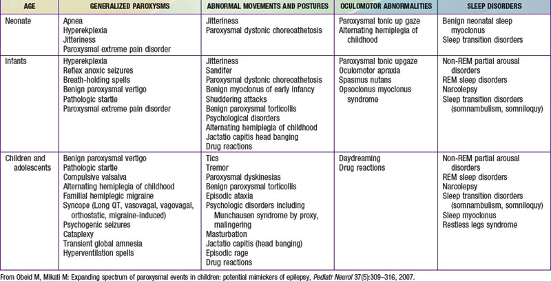
The misdiagnosis of epilepsy has been estimated to be as high as 5-40%, implying that many patients may be subjected to unnecessary therapy and tests. Often all that is needed to differentiate nonepileptic paroxysmal disorders from epilepsy is a careful history and thorough exam; but sometimes, more advanced testing may be necessary. Nonepileptic paroxysmal disorders can be classified according to the age of presentation and the clinical manifestations: (1) generalized paroxysms, (2) abnormal movements and postures, (3) oculomotor abnormalities, and (4) sleep-related disorders (see Table 587-1 on the Nelson Textbook of Pediatrics website at www.expertconsult.com).

Generalized Paroxysms

Breath-Holding Spells

This term has been applied to 2 types of spells. The 1st is the pallid breath-holding spell, which is the vasovagal reflex described below. Often pallid breath-holding spells are made worse by iron deficiency anemia. The 2nd is the cyanotic or, “blue,” breath-holding spell. The term “breath-holding spells” is actually a misnomer, as these are not related to volition or behaviorally-mediated abnormalities. Prolonged expiratory apnea is responsible for the cyanotic episodes, which result from intrapulmonary shunting. On the other hand, reflex vagal-cardiac bradycardia is responsible for the pallid episodes. An episode starts with a cry (often a “silent” cry and marked pallor in the case of the pallid type), and progresses to apnea and cyanosis. Spells usually begin between 6 and 18 mo of age. Syncope, tonic posturing, and even reflex anoxic seizures may follow significant episodes, particularly in breath-holding spells of the pallid type. Injury, anger, and frustration, particularly with surprise, are common triggers. Education and reassurance of the parents is usually all that is needed, as these episodes are, as a rule, self-limited and outgrown within a few years. However, treatment of coexisting iron deficiency is needed if it is present. Education of the parents on how to handle more severe spells by first-aid measures is important. Anticholinergic drugs (e.g., atropine sulfate 0.01 mg/kg/24 hr in divided doses with a maximum daily dose of 0.4 mg), instructing parents in basic cardiopulmonary resuscitation (CPR), or antiepileptic drug therapy for anoxic seizures that are recurrent and not responding to other measures may, rarely, be needed. All parents should be taught not to provide secondary gain when the episodes occur, because this can reinforce the episodes. Also, preparation for unpleasant experiences (such as receiving a shot) rather than surprising the child with them can help limit the number of spells.

Vagal Syncope

Syncope can present with drop attacks and can also lead to generalized convulsions, termed anoxic seizures. These convulsions, triggered by a sudden cutting off of oxygen to the brain, are clinically similar to and can be misdiagnosed as primary generalized seizures. Vasovagal (neurocardiogenic) syncope is one of the most common mimickers of generalized tonic clonic seizures and is usually triggered by dehydration, heat, standing for a long time without movement, hot showers, the sight of blood, pain, or sudden stress. History is usually the clue to distinguishing syncope from epileptic seizures. There is initially pallor and sweating followed by blurring of vision, dizziness, nausea, and then gradual collapse with loss of consciousness. However, these are not invariably present in syncope. Urinary incontinence and a brief period of convulsive jerks are not uncommon in vasovagal syncope. These occur with a frequency of 10% and 50%, respectively. Postictal confusion can also occur, though rarely. Abdominal pain, a common aura in temporal lobe epilepsy, occurs in vasovagal syncope, and can be a trigger or a consequence (intestinal vagal discharge). Most children with vasovagal syncope have an affected first-degree relative. EEG is normal and the tilt test has been used for diagnostic purposes. Although in most cases with typical history, it is not needed. Vagovagal syncope is triggered by swallowing or vomiting, and can progress to convulsive seizure if the asystole is sufficiently prolonged. Sudden cold exposure to the face or to the body can also trigger vagal syncope. Syncope has also been rarely reported to occur in association with cough, tight hair braiding, hair combing, extension of the neck while stretching due to compression of the vertebrals, and with flexion of the neck secondary to an abnormally prolonged stylomastoid process compressing the carotids. The latter 2 conditions require neuroimaging (CT, MRI) for proper diagnosis. Orthostatic hypotension and orthostatic intolerance manifest symptoms that develop during upright standing and can be relieved by recumbence. Postural tachycardia syndrome, the pathophysiology of which remains elusive, is a disease of adolescent females that is characterized by upright tachycardia and hypotension. Primary autonomic failure is rare in children, and familial dysautonomia is the only relatively common form. Familial dysautonomia is a disease common in Ashkenazi Jews, and is characterized by absence of overflow emotional tears, depressed patellar reflexes, and lack of a flare reaction following intradermal histamine. Dopamine beta-hydroxylase deficiency is a very rare cause of primary autonomic failure, and is characterized by impaired ejaculation, ptosis, nocturia, high palate, hyperflexible joints, and a complicated perinatal course (hypotension, hypotonia, hypothermia). Hypotension can also occur in adrenal insufficiency. Tilt test causes a drop in both blood pressure and heart rate in patients with classic vasovagal syncope. It results in a blood pressure drop with minimal change in heart rate in autonomic failure, and in blood pressure drop and an increase in heart rate in postural tachycardia syndrome.

Management of syncope centers on avoidance of precipitating factors (maintenance of hydration, avoidance of standing still, rising slowly from sitting, first aid measures, raise legs, positioning) and treatment of any accompanying or underlying medical conditions (anemia, adrenal insufficiency, cardiac, etc.). In addition, β-blockers (e.g., metoprolol starting dose 1-2 mg/kg once per day up to a maximum of 6 mg/kg/day), or flurohydrocortisone (0.05-0.1 mg/day) therapy may be needed in some selected cases.

Cardiac Syncope

Long QT syndromes (LQT) can cause life-threatening “pallid” or white syncope. Accompanying this are ventricular arrhythmias, usually torsades de pointes or even ventricular fibrillation. There are more than 10 types of prolonged QT syndromes. When accompanied by congenital deafness, it is part of the autosomal recessive Jervell and Lange-Nielson syndrome (type 1, LQT 1, associated with KvLQT1 potassium channel mutation). The Romano-Ward syndrome is an autosomal dominant syndrome with incomplete penetrance that is characterized by episodes of lying still like a dead body for several seconds before the anoxic convulsive episode (LQT 2 associated with an HERG potassium channel mutation). LQT 3 is associated with an SCN1A sodium channel mutation, type 4 ankyrin protein mutation, type 5 (milder form) with the minK KCNE1 mutation, type 6 with KCNE2 potassium gene mutations, type 9 with caveolin sodium channel–related protein mutations, and type 10 with SCN4B sodium channel mutations. Types 7 and 8 are of particular interest due to associated clinical and neurologic manifestations. Type 7 (Andersen-Tawil) syndrome is associated with periodic paralysis, skeletal developmental abnormalities, clinodactyly, low-set ears, and micrognathia (mutations in KCNJ2 gene). Type 8 or the Timothy syndrome (mutations in the calcium channel gene CACNA1c) manifests congenital heart disease, autism, syndactyly, and immune deficiency. All family members of an affected LQT syndrome individual should be investigated. Affected individuals need insertion of cardiac defibrillators, and their families should be taught CPR. As a rule children with new onset seizure disorder should get an electrocardiogram (ECG) to rule out LQT syndrome masquerading as seizure disorder. Cardiac syncope is usually sudden without the gradual onset and the symptoms that accompany vagal syncope. Aortic stenosis can cause sudden syncope at the height of the exercise (usually hypertrophic), or directly at the end (usually valvular) and, if suspected, warrants an echocardiogram.

Psychologic Disorders

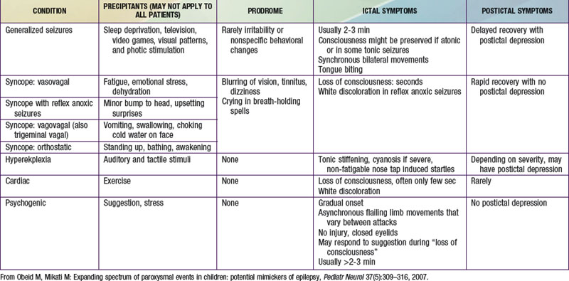
Psychogenic nonepileptic seizures are conversion reactions that are usually suspected clinically based on the characteristics of the spells (Table 587-2). The diagnosis can be confirmed by video EEG with capture of an episode to eliminate any residual doubts about their nature, as they can often occur in patients who also have epileptic seizures. Psychogenic seizures are also less likely than epileptic seizures to be associated with increased serum prolactin levels 15-120 min after the event. They are best managed acutely by reassurance about their relatively benign nature and by a supportive attitude. Psychiatric evaluation and follow-up are needed to uncover potential underlying psychopathology, particularly in adolescents and adults, and to establish continued support as psychogenic seizures can persist over long periods of time. Malingering and Munchausen syndrome by proxy are often difficult to diagnose but an approach similar to that for psychogenic seizures, including video-EEG monitoring, is often helpful.

Abnormal Movements and Postures

Paroxysmal Dyskinesias and Other Movement Disorders

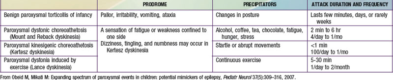
These disorders are characterized by sudden attacks that consist of choreic, dystonic, ballistic, or mixed movements (Table 587-3). A sensation of fatigue or weakness confined to 1 side may herald an attack. Consciousness is preserved and patients may be able perform a motor activity, like walking, despite the attack. The variability in the pattern of severity and localization between different attacks may also help in differentiating them from seizures. The frequency of attacks increases in adolescence, and steadily decreases in the third decade. Neurologic exam between attacks, EEG, laboratory investigations, and imaging studies are normal. These dyskinesias often respond to phenytoin, carbamazepine, clonazepam, or to antidopaminergic drugs such as haloperidol. Drug reactions can result in abnormal movements such as oculogyric crisis with many antiemetics, choreoathetosis with phenytoin, dystonia and facial dyskinesias with antidopaminergic drugs, and tics with carbamazepine. Strokes, focal brain lesions, connective tissue disorders (e.g., systemic lupus erythematosus), vasculitis, or metabolic and genetic disorders can also cause movement disorders. Mutations of glucose transporter 1 (GLUT1/SLC2AI) gene have been described in patients with exercise-induced dyskinesia.

Oculomotor Abnormalities

Sleep Disorders

Paroxysmal nonepileptic sleep events are more common in epileptic patients than in the general population, which makes their diagnosis difficult. Semiology, timing of events, video-EEG and polysomnography help in distinguishing epileptic from non-epileptic events. Parasomnias typically occur less than once or twice a night; more frequent episodes suggest epileptic seizures.