Complications
A Adverse cognitive impairment
2. Postoperative cognitive dysfunction
a) Postoperative cognitive dysfunction (POCD) is characterized by persistent and long-term deterioration of cognitive performance after anesthesia and surgery.
b) POCD is often associated with cardiac and orthopedic surgery, but it can also accompany other surgical procedures. Cognitive dysfunction in both cardiac and noncardiac surgery has largely focused on older adults, who might have a greater vulnerability to neurologic deterioration as a consequence of the aging process.
c) POCD is difficult to diagnose because it requires sophisticated neurophysiologic testing, including preoperative baseline tests.
d) Patient risk factors include age, lower levels of education, and history of stroke even without residual deficit.
e) Increased 1 year mortality is associated with patients who demonstrate cognitive decline at both hospital discharge and 3 months postop.
a) The term confusion is used to describe the general affect and behaviors of patients; however, it is not specific and appears to have a great deal in common with delirium.
b) Confusion (a form of transient cognitive dysfunction) after anesthesia relates to disorders of orientation and is usually a relatively short-lived transient cognitive dysfunction.
c) Confusion after anesthesia is a normal occurrence, and it is suggested that patients refrain from engaging in activities requiring rapid responses for at least 48 hours postanesthesia.
a) Delirium (or acute mental confusion) is transient, often abrupt and fluctuating, typically reversible, and related to increased risk of postoperative adverse reactions (i.e., pulmonary edema, myocardial infarction, respiratory failure, pneumonia, and death), increased length of hospital stay, increased health care cost, and poor functional and cognitive recovery. The onset occurs in hours to days after anesthesia and surgery.
b) Key symptoms include anxiety, incoherent or disorganized thinking and perceiving, reduced ability to sustain and shift attention to new external stimuli, and agitated behavior. There is sensory misperception; a disordered stream of thought; and difficulty in shifting, focusing, and sustaining attention to both external and internal stimuli. Irrelevant stimuli can easily distract the delirious individual.
c) The cause of postoperative delirium is multifactorial. Virtually any drug with central nervous system effects has been implicated, including narcotics (especially meperidine), benzodiazepines, and drugs that possess anticholinergic properties (except glycopyrrolate). Common are perceptual disturbances that result in misinterpretations, illusions, and hallucinations. Disturbances of sleep–wakefulness and psychomotor activity are present.
d) Precipitants are related to physical illness (e.g. cardiovascular disease), infection, hormone disorders, or nutritional deficiencies. Most frequent precipitants are metabolic disturbances; fluid and electrolyte imbalances; drug and alcohol toxicity; and unfamiliar and excessive sensory-environmental stimuli.
e) Delirium may be life threatening and is a medical emergency. Delirium occurs in about 20% of elderly surgical patients and is more common in patients undergoing orthopedic procedures (i.e., femoral fractures) with an incidence rate of 28% to 60% in this surgical population. Thirty-two percent of patients who had coronary artery bypass surgery reported postoperative delirium.
f) Early identification and prompt treatment of the causes of delirium prevent irreversible dementia and death, with interventions targeted at reversing physiologic disturbances and preventing sensory deprivation.
g) The choice of general anesthesia or regional anesthesia does not appear to be a factor, especially when sedation is used in conjunction with regional anesthesia (RA).
h) Management focuses on reversible risk factors such as current meds, pain management, and a better sleep environment. Haloperidol in doses no greater than 1.5 mg can be helpful, especially for agitated delirium. When administered prophylactically, it may reduce the severity of duration but not the incidence of delirium.
a) Awareness, the unambiguous recall of events during general anesthesia, has an incidence of 0.18% in the United States. The term recall, the ensuing retention of an event after it occurs, is a better description of the phenomena; however, episodes of awareness are strong predictors of dissatisfaction with anesthesia care.
b) For every 1000 adult patients who receive a general anesthetic, as many as one or two will express the occurrence of awareness or recall; this figure is higher in children. Inadequate depths of anesthesia may lead to awareness and recall; accounts of awareness rely on patient recollection.
c) Although patient recollection of awareness or recall may be reported, most do not complain of recalling pain during the procedure. Instead, patients report “dreamlike” experiences and auditory remembrance during which they are not in distress.
d) Reports of intraoperative awareness should be addressed immediately and thoroughly evaluated to obtain information for quality assurance.
e) Management of awareness begins when patients are given an opportunity to discuss the causes of the event with the anesthesia provider to gain a clearer understanding of the circumstances surrounding the experience and follow-up consultation.
f) The awareness experience is certainly a distressing event and outside normal operative occurrences. These stressful events may lead to nightmares and sleep disturbances, intrusive memories and avoidance behavior, emotional numbing and forgetting, and other diagnostic criteria for posttraumatic stress disorder.
g) The causes of intraoperative awareness are diverse and are listed in the following box.
h) Awake paralysis, one of the most feared causes, is possibly the most preventable. Awake paralysis is related to lapses in practitioner vigilance and can lead to out-of-sequence neuromuscular blockade administration and medication error.
i) Prevention of awareness is the best treatment for awareness. The American Association of Nurse Anesthetists has an awareness policy that helps identify at-risk patients and measures to address and possibly avoid perioperative awareness. This anesthesia care plan to reduce the incidence and severity of awareness includes the fundamental practices listed in the box on pg. 600.
j) Although awareness and recall of some of the anesthesia experience are impossible to prevent in all patients, vigilance on the part of the anesthesia provider (i.e., close attention to monitoring modalities and to anesthetic levels) should decrease the incidence substantially.
B Eye injury
a) Corneal injury is an infrequent occurrence during anesthesia. The cornea is a tough, transparent, dome-shaped surface covering the eyes that serves as a barrier to infection and trauma in this highly exposed organ.
b) Corneal abrasions are superficial defects of the corneal epithelium. Application of a short-acting topical anesthetic before assessment facilitates an examination.
c) Visual acuity is usually normal unless the abrasion includes the visual axis or if there is considerable edema. There may be miosis caused by ciliary spasm and blepharospasm (marked by an uncontrollable, forcible closure of the eyelids) of the affected eye as a result of photophobia.
d) The cornea may also appear hazy if edema is present.
e) A corneal abrasion, although infrequent, is the foremost injury after general anesthesia and may occur during other types of anesthesia (monitored anesthesia care and major conduction block) when something strikes the eye.
f) Corneal abrasions cause tearing, sensations of a foreign body, photophobia, reduced visual acuity, and eye pain. Postoperative corneal injuries can lead to significant morbidity and lost productivity.
g) Drying of an exposed cornea or patient movement during ophthalmic surgery (e.g., coughing, turning) can lead to abrasion of the cornea with subsequent poor surgical outcomes.
h) Keratoconjunctivitis sicca, also called dry eye disease (DED) and dysfunctional tear syndrome, is a multifactorial disorder of the tear film and ocular surface involving multiple interacting mechanisms.
i) Pressure on the eye (e.g., prone position, leaning of the face) may result from occlusion of choroidal blood flow and reduction of blood flow to the cornea. Because the cornea contains no blood vessels, oxygen delivery is accomplished through diffusion.
j) As a result, reduced blood flow to the cornea leads to a reduction in oxygen availability with subsequent corneal edema. The avascular nature of the cornea, dry environment, pressure, and desquamation of the epithelium of the eye may lead to corneal abrasion.
k) The diagnosis of corneal abrasion is confirmed by green fluorescence in damaged areas of the cornea seen under Wood’s lamp or cobalt blue light on slit lamp examination after the application of fluorescein.
l) The treatment of corneal abrasion varies; however, occlusion of the affected eye is often recommended. Occlusion of the affected eye is also frequently associated with topical application of antibiotic ointment of nonsteroidal medications.
m) Primary goals of therapy are pain control, prevention of infection, and rapid healing of the corneal epithelium. Most corneal abrasions heal within 1 to 3 days; however, defects involving greater than half of the corneal surface may require 4 to 5 days to fully heal.
n) Prevention of corneal abrasion is a primary focus during the course of anesthesia. Although no method is guaranteed to be 100% effective, care and vigilance are required to reduce the associated morbidity of this disturbing complication.
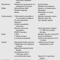
o) Methods to protect the eyes during general anesthesia are listed in the table on pg. 602.
Commonly Used Methods to Protect Eyes During General Anesthesia
| Method | Strengths | Weaknesses |
| Manual closure | Prevents trauma to the globe and eyelids Prevents injury associated with the introduction of agents (ointments, tape) onto the eye |
Inappropriate when surgery involves the head or neck Incompatible with the prone or lateral position; may cause greater harm to the eye |
| Taping the eyelids | Prevents exposure keratopathy and injury associated with the introduction of agents onto the eye | Incorrect placement leads to exposure keratopathy May cause corneal abrasion if tape is placed directly onto the cornea Inappropriate when surgery involves the head or neck Incompatible with the prone or lateral position; may cause greater harm to the eye May cause injury when removing tape at the end of the procedure |
| Ophthalmic ointments, methylcellulose, and hydrogels | Equally effective as taping to prevent corneal abrasion Prevent drying of the eyes for extended periods Permit continuous perioperative monitoring of the eyes during certain procedures |
Postoperative sensation of foreign body in some patients Cause blurred vision and confusion in some patients May cause allergic reactions after the use of ointments containing methylparaben, chlorobutanol, and other preservatives May require reassessment during long procedures |
| Eye shields and pads | Reduce the risk of mechanical injury May be used in prone and lateral positions Equally effective as taping and ointments at preventing corneal abrasion |
Must include taping eyelids or applying ointment |
a) Blindness as a complication of anesthesia during nonocular surgery is a rare but devastating surgical risk and a medicolegal concern for the surgical team (anesthesia provider, surgeon, and nursing staff).
b) The incidence of perioperative blindness ranges from 0.002% of all surgeries to as high as 0.2% of cardiac and spine surgeries.
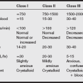
c) Any portion of the visual pathways may be involved, but the most common site of permanent injury is the optic nerves. The most often presumed mechanism is ischemia. With more than 1.2 billion myelinated nerve fibers arising from the cell bodies of cells in the retina, ischemia can be an immense problem, resulting in axonal swelling of these fibers and blindness. Other potential causes of postoperative visual loss include: pressure on the eyes, anemia, prolonged hypotension and increased intraocular pressure.
d) Treatment and prevention of perioperative blindness, especially as a complication of anesthesia, are difficult. Immediate consultation is imperative for patients with perioperative blindness. If an obvious ocular cause is not apparent, urgent neuroimaging should be obtained to rule out intracranial pathology.
C Operating room fire
a) Fires in the operating room are a relatively rare event. The classic fire triangle requires the presence of three elements: fuel, an ignition source, and an oxidizing agent (gas that supports combustion).
b) Of the approximately 100 surgical fires reported each year, approximately 20 cause major thermal injuries; however, one to two of these fires result in death.
c) The most common locations of these fires are in the airway (34%), about the head or face (28%), and several other areas of or in the patient’s body (38%).
d) The most common ignition sources are electrosurgical equipment (68%) and lasers (13%). High-intensity light cords have also been reported to be a source of ignition.
e) Fuels commonly found in the operating room consist of alcohol, solvents, sheets and drapes (cloth and disposable paper), and plastic or rubber materials (including endotracheal tubes [ETTs]).
f) Oxygen is by far the most common oxidizing agent, although nitrous oxide can also support combustion. Materials that are only marginally combustible in air can produce a massive flame in the presence of high oxygen concentration.
2. Fire precaution and prevention
a) The risk of ignition of combustible materials is progressively increased as oxidizing agents build in the operating room.
b) Oxygen levels should be kept as low as can be safely done, and leaks to the ambient air should be kept to a minimum, with special attention to oxygen and nitrous oxide leaks from the anesthesia mask and nasal cannula.
c) Providers should reduce the oxygen concentration when a heat-producing device, such as a cautery unit or fiberoptic light source, is used. Although fiberoptic light is often called “cool light,” heat is generated.
d) When an open oxygen source is used, excessive oxygen can accumulate under the patient drape or about the operative site creating an enriched oxygen environment. If a spark is created in these areas, a flash fire can rapidly spread out of control.
e) Procedures near the head and neck are more likely to be associated with fires because of the frequent presence of enriched oxygen and nitrous oxide. The total oxygen liter per minute flow should be kept as low as possible and should be consistent with the patient’s status and response as monitored by standard physiologic monitors, with special attention to SaO2 values compared with the patient’s baseline values.
f) Avoid petroleum-based ointments in the vicinity of surgery. These include petroleum-based ophthalmic ointments when surgery is in the vicinity of the face.
g) When electrocautery is used, cloth towels are wetted with sterile water to surround a surgical site that is above the xyphoid process when open oxygen is administered by nasal cannula or face mask. Wet surgical towels reduce the likelihood of a flash fire being precipitated by cautery.
h) If possible, administer higher levels of inspired oxygen during the preparation and local anesthetic infiltration if needed and then reduce the inspired oxygen to the lowest possible level consistent with the patient’s condition during the use of electrosurgery, electrocautery, or laser units (drills, defibrillators, static electricity).
i) When surgical drapes are placed over the face (e.g., for eye surgery) during local or local and monitored anesthesia care (MAC) techniques, consider tenting the drapes to allow for O2 escape and active evacuation of the ambient gases given off by nasal cannulas or face masks. This can be achieved by the placement of a funnel-like collection device that evacuates the gases by way of the hospital suction system.
j) Consider giving an oxygen-air blend with an Fio2 of 0.3 or less. This can be done by administering nasal oxygen via oxygen tubing connected with an approximately 5-mm ETT connector placed at the Y-connection outlet from the circle system anesthesia circuit. In this manner, a mixture of oxygen and medical-grade air can be blended from the anesthesia machine flowmeters while using the oxygen analyzer to regulate the O2 level of (ideally) 0.3 or less.
k) The use of an incise drape (clear vinyl drape that is placed over the proposed surgical site so that the incision is made through the clear drape) has been recommended by some sources to keep the oxygen concentrated below the incision. This should not be considered a totally effective method of keeping the oxygen concentration low in the surgical field.
l) Channeling of oxygen can occur through any small opening in the area between the incise drape and the skin.
m) The hot tip of an electrocautery unit laid on a drape can cause ignition of the drapes. The cautery hand unit should be placed in its holster when not in use. Activate heat sources only when the tip is in view and deactivate it before the tip leaves the surgical site.
n) When using an open O2 source: If possible, stop oxygen administration for a minimum of 1 minute before the use of electrosurgical units (cautery) near the head, face, and neck.
o) Arrange drapes to allow ready access to the patient’s face to facilitate observation and maximal exchange of ambient room air, increase communications with the patient, and have ready access for possible airway manipulations.
p) Oxygen and nitrous oxide are heavier than room air, so these gases gravitate down over the patient in a manner similar to that of water poured over a surface. Heavier-than-air gas tends to spread and become concentrated under the drapes. It also contributes to a phenomenon called surface fiber flame propagation (SFFP).
q) When oxygen coats the fine fibers of a surface (e.g., draping fabrics) or even fine facial hair (vellus) and other body hair surfaces, the flames can quickly spread when these fibers ignite rapidly in the presence of oxygen enrichment. Facial hair and any other hair should be coated with a water-soluble gel if it is in the area of surgery and cautery use is anticipated.
a) If a fire starts, immediately cut or tear off any burning drapes or other materials from the patient.
b) Water should be readily available to extinguish fire, especially in the mouth or other open body cavities. Because many drapes (especially disposable ones) are liquid resistant, water will tend to bead up rather than soak the material to reduce flame propagation.
c) If tearing the material away is difficult or ineffective, use a CO2 fire extinguisher. Fire blankets should be available to smother flames if they occur. However, if there is a concern that the fire started under a surgical drape because of an oxygen-enriched environment, a fire blanket may not be able to smother the fire, and the fire could actually continue to thermally injure the patient.
d) Sponges, gauze, pledgets, and strings should be wet and kept moistened when in use in the area of surgery to help prevent airway fires.
D Stroke (brain attack)
a) Stroke has been defined as an acute neurologic deficit that persists for longer than 24 hours.
b) The incidence of acute perioperative ischemic stroke is low and uncommon; however, the overall mortality rate is high, with roughly 80% occurring as a consequence of thromboembolic mechanisms.
c) The major causes include combined cardiac procedures, vascular procedures, lacunar or cerebellar infarction, and hemorrhage. Risk factors include male gender, a history of transient ischemic attacks, and cigarette smoking.
d) Perioperatively, the incidence depends mainly on the type and complexity of the surgical procedure. Slightly fewer than half of all perioperative strokes are recognized within the first day postsurgery, and the remaining cases occur after an uneventful postrecovery period, usually from the second postoperative day forward.
e) The stress of surgery results in hypercoagulability and activation of the hemostatic system, which results in reduced fibrinolysis after surgery.
f) Perioperative withholding of antiplatelet and anticoagulant agents can exacerbate surgery-induced hypercoagulability and add to the risk of perioperative stroke.
g) The neurologic deficit may be mild and transient and may often go unnoticed, depending on the extent of the infarct.
h) The American Heart Association guidelines suggest the use of systemic thrombolysis using alteplase (a recombinant tissue plasminogen activator) within 3 hours of the onset of stroke. Although the mortality rate is unchanged, the use of thrombolytic therapy has been shown to support carotid and vertebrobasilar recanalization in approximately 25% of cases when given within 3 hours of symptoms and to improve 3-month neurologic outcomes.
i) Intraarterial thrombolysis has not been shown to be better than the intravenous (IV) route, and IV streptokinase is not indicated for acute ischemic stroke.
j) Stroke in patients with sickle cell disease is often fatal, particularly in children between the ages of 4 and 15 years. There is a 300-fold increased risk of stroke in these patients, making it the most common cause of childhood stroke.
k) The combination of surgery and anesthesia is an independent risk factor for the development of acute ischemic stroke perioperatively.
l) Acute ischemic stroke has a high mortality rate, and in certain instances of intractable intracranial hypertension, hemicraniectomy or decompressive surgery of the posterior fossa could be lifesaving.
m) Practitioner understanding of the pathologic mechanism of stroke after anesthesia and surgery is vital and requires additional investigation to prevent this overwhelming complication.