Complex regional pain syndrome
Diagnosis
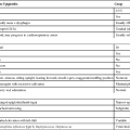
Diagnosis is made by clinical observation, because currently no objective diagnostic criteria exist. In an effort to improve diagnostic predictability, it has been suggested that patients report at least one symptom in each category and demonstrate at minimum one sign in two or more categories (Table 213-1). Using these clinical criteria based on the International Association for the Study of Pain (IASP) guidelines improves diagnostic specificity (94%) at the expense of sensitivity (70%). The differential diagnosis includes small-fiber and diabetic neuropathies, entrapment, degenerative disc disease, thoracic outlet syndrome, cellulitis, vascular insufficiency, thrombophlebitis, lymphedema, angioedema, erythromelalgia, and deep venous thrombosis.
Table 213-1
International Association for the Study of Pain Criteria for the Diagnosis of Complex Regional Pain Syndrome*
| Category | Signs and Symptoms |
| Sensory | Allodynia Hyperalgesia Hyperesthesia Hypoalgesia |
| Vasomotor | Livedo reticularis Skin color changes Temperature variability |
| Sudomotor | Edema Hyperhidrosis Hypohidrosis |
| Motor | Decreased range of motion Neglect Tremor Weakness |
*Diagnostic predictability improves when patients report at least one symptom in each category and one sign in two or more categories.
Laboratory studies may provide objective results to assist in the diagnosis of CRPS. Thermometry, quantitative sudomotor axon reflex test (QSART), thermoregulatory sweat testing, laser Doppler flowmetry, three-phase bone scintigraphy, plain radiographs, and magnetic resonance imaging studies have been shown to be useful in the diagnosis of CRPS. In the past, CRPS types I and II were thought to progress through several stages, which vary significantly in temporal duration (Table 213-2).
Table 213-2
Stages of Complex Regional Pain Syndrome
| Stage | Presentation |
| Acute/warm | Burning or aching pain increasing with physical contact or emotional stress Edema Unstable temperature and color of limb Increased periarticular uptake on scintigraphy Accelerated hair and nail growth Joint stiffness Muscle spasm |
| Dystrophic | Indurated, cool, hyperhidrotic, cyanotic, mottled skin Joint space narrowing Muscle weakness Osteoporotic changes on radiography |
| Atrophic/cold | Ankylosis Hair loss Muscle atrophy Tendon contractures Thickening of fascia Thin shiny skin |


