COMMON PREHOSPITAL COMPLICATIONS AND PITFALLS IN THE TRAUMA PATIENT

Published on 20/03/2015 by admin

Filed under Critical Care Medicine

Last modified 22/04/2025

Print this page

rate 1 star rate 2 star rate 3 star rate 4 star rate 5 star
Your rating: none, Average: 5 (1 votes)

This article have been viewed 1629 times

CHAPTER 16 COMMON PREHOSPITAL COMPLICATIONS AND PITFALLS IN THE TRAUMA PATIENT

The evolution of prehospital care in this country has an interesting and continually evolving record. Although there is recorded history of wagons and carts being used to transport the sick and injured as early as 900 ACE, the term “ambulance” was not used until introduced by Queen Isabella of Spain in the early 15th century. Even at that time, it referred more to military field hospitals and tents for the wounded than to a means of transporting wounded and dead from battlefields. Not until the time of Baron Larrey would the term “ambulance” take up its more current meaning of “a specially equipped motor vehicle, airplane, or ship for carrying sick or injured people, usually to a hospital.”1

Baron Dominique-Jean Larrey was Napoleon Bonaparte’s surgeon and developed what was known as “flying ambulances.” Prior to 1792, there was very little organized transportation of the wounded from the battlefield. As is the case with most medical advances, advances in ambulance transportation occurred as a result of military conflict. Throughout the remainder of the 1800s and the conflicts of the early 20th century, ambulances and other means of transporting individuals from the field of battle were employed.

During the 1950s through the 1970s, helicopters were employed to transport the injured from battlefields to MASH (mobile army surgical hospital) units attaining particular effectiveness in the Korean and Vietnam conflicts. Throughout the first several decades of the 20th century, civilian transport for the injured continued to lag behind advances established in the military.

One of the prime factors identified as contributing to the continued reduction in battlefield casualties from 8% in World War I to less than 2% in the Vietnam War was reducing the time from injury to initiation of medical care. On this backdrop, the mid-1960s and early 1970s sought to improve prehospital care, education, equipment, and processes. The early 1960s called for an extension of basic and advanced first aid training to greater numbers of the lay population, and preparation of nationally accepted texts, training aids, and courses of instruction for rescue squad personnel, policemen, firefighters, and ambulance attendants.2 Ambulance services in the 1960s was very piecemeal and adequate at best. In a few major cities, there were specially equipped ambulances prepared to care for the injured and sick, and trained professional prehospital personnel were available. However, approximately 50% of the country’s ambulance services at that time were provided by over 12,000 morticians mainly because their vehicles were able to accommodate transportation of patients on gurneys or stretchers.

In the mid-1960s, the National Traffic and Motor Safety Act and the Highway Safety Act3 provided for the establishment of national standards for used motor vehicles, motor vehicle inspections, and emergency services. Communications were also problematic. At a time when the United States had just placed a man on the moon, it was easier in most instances to communicate with that extraterrestrial individual than it was for prehospital providers to communicate with the emergency department where they were headed.

Over the next several decades, the education and provision of specifically equipped vehicles progressed until the mid-1980s when Injury in America: a Continuing Public Health Problem was published.4 Although the report found that there had been significant progress in the credentialing and education of prehospital care providers, more than 2.5 million Americans died from injuries in the 1966–1985 period. This prompted the expenditure of more federal dollars to study the continuing public health problem, as the report noted and called for the institution of more systems of communication and transportation of the injured to facilities specially equipped for managing critically injured patients.

In 1992, the Model of Trauma Care Systems Plan, developed by Health Resources and Services Administration under the Authority of the Trauma Systems Planning and Development Act of 1990,5 marked the next major step in the evolution of health policy related to trauma care. This plan emphasized the need for a fully inclusive trauma care system that involved not only trauma centers, but also all health care facilities according to availability of trauma resources, including prehospital providers. As a result, the numbers of dedicated trauma centers and state trauma systems developed, although at a still less-than-adequate pace. Trauma centers were charged with becoming resource facilities for emergency medical response agencies. Educational programs such as Prehospital Trauma Life Support (PHTLS), Basic Trauma Life Support (BTLS), and others were developed with states being empowered to license and credential prehospital providers at various levels.

Today, the initial care of the injured patient continues to reside primarily with trained prehospital providers. Emergency medical technician, basic, intermediate, and paramedic levels of instruction, with police and fire departments also being trained in basic life support, as well as increased communication and education with the lay public with regard to cardiac arrest, seat belt usage, wearing of helmets, and other prevention initiatives are in place to continue to try to combat the unacceptably high level of death and disability in this country from intentional and unintentional injury.

Along with the ever evolving technologies available to the prehospital provider come the unintended risk of complications associated with the implementation of these devices and processes. This chapter addresses some of the more common prehospital complications.

INCIDENCE

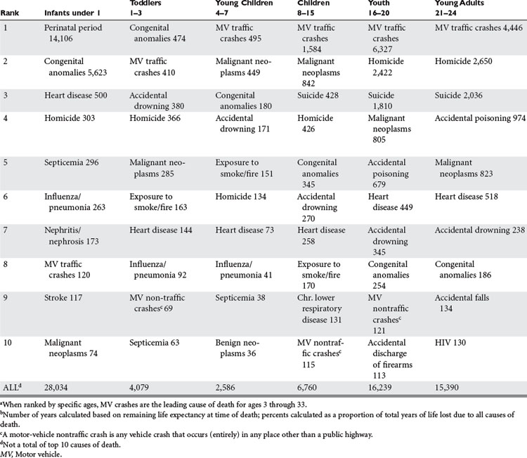
According to the National Highway Traffic Safety Administration, the leading cause of death in the United States in 2002 for people aged 4–34 is overwhelmingly motor vehicle traffic crashes (Table 1). In terms of years of life lost, motor vehicle crashes ranks third, after malignant neoplasms and heart disease, at 5% of total years of life lost for the entire population.

A total of 37% of trauma deaths are caused by motor vehicle crashes and motorcycle crashes. Other important causes of trauma deaths are gunshot wounds, stabbings, and falls. Today’s prehospital provider is in a position to be the first responder to the vast majority of these injuries at or shortly after the time they occur.

The major causes of death in the prehospital period are secondary to severe head injury, respiratory compromise, and exsanguinating hemorrhage. Initial and emergent prehospital treatment focuses on the treatment and prevention of these eventualities.

The foundation of Advanced Trauma Life Support® of the American College of Surgeons6 stresses an ABC (airway, breathing, and circulation) approach. Much of the emphasis on prehospital care and subsequent care involves appropriate management of the airway, providing for ventilation by breathing for the patient, and control of circulation consisting of hemorrhage control and restoration of intravascular volume. Not surprisingly, the most common prehospital complications occur in these three areas.

AIRWAY

Ensuring that the trauma victim has a patent airway is the highest management priority.7 If manual maneuvers (clearing the airway of foreign bodies, jaw thrust, or chin lift) or basic adjuncts (oropharyngeal or nasopharyngeal airways) are not adequate to maintain the airway, then alternate, more invasive methods are required.

Current prehospital techniques used for airway management and ventilation include (1) bag-valve-mask (BVM), (2) laryngeal mask airways (LMA), (3) dual lumen tubes (i.e., Combitubes), (4) endotracheal intubation (with or without the use of paralytics), and (5) emergency cricothyroidotomy.

BVM can be a temporizing method for providing adequate oxygenation and ventilation of the injured patient, but can occasionally be problematic related to obtaining an adequate seal at the mouth, potential for aspiration, problems with bleeding from soft tissue injury, patient cooperation, and the lack of satisfactory ventilation and oxygenation depending on the specific clinical situation. Acute gastric dilatation from overzealous ventilation can also lead to ventilatory impairment from increased intra-abdominal pressure, and, in extreme cases, gastric rupture.

The prehospital use of LMAs and dual lumen tubes has an advantage over conventional endotracheal intubation related to ease of technique and maintenance of insertion skill. LMAs and dual lumen tubes are beneficial in an unconscious patient who cannot be adequately ventilated with a BVM device and/or cannot be successfully intubated. However, because the trachea is not completely protected, the use of an LMA may result in aspiration. The use of dual lumen tubes may also result in aspiration if the gag reflex is intact, and there is also potential for damage to the esophagus, and the possibility for hypoxia if the wrong lumen is used.

The identification of patients requiring definitive airway management may sometimes be problematic based on the patient’s injuries, mental status (secondary to injury, alcohol, and/or drugs), underlying medical conditions, and the experience of the prehospital provider. Delay of intubation until respiratory arrest increases morbidity and mortality and should be avoided if at all possible. Early recognition of the need for intubation is of paramount importance for the prehospital provider.

Late endotracheal intubation may result because of a false sense of security by the provider, the inability to obtain an airway, and lack of recognition of likely deterioration in a patient’s ventilatory status (secondary to airway and chest injuries, traumatic brain injury [TBI], alteration in mental status, or the overall complexity of the injuries). Patients with facial burns and maxillofacial trauma may have progression of their underlying injury, and may deteriorate secondary to edema or hematoma formation, causing airway obstruction. Intubation of these patients can be a difficult challenge with the potential for disastrous results if proactive intubation is not accomplished. This is especially true if paralytic agents have been used and the vocal cords can not be easily visualized. Anticipation of this problem along with early intubation may prevent a catastrophe.

The use of paralytic agents for intubation in the prehospital setting results in a quicker and higher success rate of intubation. However many prehospital providers do not have access to use these agents. Additionally, if paralytic agents are used, it is critical that adequate analgesia and sedation are also administered, so that the injured patient is not chemically paralyzed, while awake and hurting. At the time of hand-off of the trauma patient from the prehospital provider to the trauma team in the emergency department, it is important that all of the medications that have been administered to the patient prior to arrival are reviewed, so that the emergency physician and trauma surgeon will ensure adequate pain management and sedation, even when the patient is chemically paralyzed.

Successful endotracheal intubation is beneficial for the trauma patient whose airway needs to be secured, but there are potential complications and pitfalls that may occur during the process of intubation, regardless of the expertise of the provider. Prehospital personnel should be aware of these potential complications and how to clinically recognize them if they occur.

Esophageal intubation is a known complication of intubation, and should be quickly recognized by the prehospital provider if it occurs. The difficulty of the intubation and in visualizing the vocal cords should increase concerns of an esophageal intubation, and warrants aggressive evaluation to ensure adequate placement of the endotracheal tube. The placement of an esophageal endotracheal tube should be clinically evident by routine chest auscultation immediately after intubation. The routine use of end-tidal CO2 detectors (ETCO2) is beneficial in rapidly detecting the presence of CO2 in the exhaled air. The calorimetric devices have a chemically treated indicator strip that reflects the CO2 level. If there is a question of the exact location, visualization of the ETT location should be repeated and appropriate location confirmed.

Right main-stem intubation is an occasional complication of intubation that occurs up to 30% of the time in pediatric trauma patients, and should be detected by physical exam at the time of intubation, and with frequent routine clinical reassessments, or urgent reassessment if the patient clinically deteriorates. The distance of the tip of the ETT should be evaluated, relative to the size of the patient and the expected appropriate distance of ETT. Repositioning of the ETT while auscultating the chest helps in determining the appropriate location of the ETT. Other possible traumatic injuries that may lead to similar clinical findings must also be considered in severely injured patients, including a pneumothorax, hemothorax, pulmonary contusion, or ruptured hemidiaphragm.

Surgical airways are occasionally needed when endotracheal intubation cannot be successfully achieved secondary to facial trauma, anatomic difficulties, and soft tissue injuries. While the use of surgical airways in the prehospital setting is controversial, local protocols should outline the specific indications and circumstances for their use.

Complications of needle cricothyroidotomy include inadequate ventilation resulting in hypoxia and death, esophageal laceration, hematoma formation, posterior tracheal wall perforation, thyroid laceration, and bleeding.

Complications of surgical cricothyroidotomy include false passage into the tissues, hemorrhage or hematoma formation, esophageal laceration, vocal cord paralysis, and potential subglottic stenosis/edema. If a surgical airway is needed, a surgical cricothyroidotomy should be performed. A formal tracheostomy should not be performed by prehospital providers because of the difficulty and length of time to successfully accomplish the procedure.

BREATHING

A tension pneumothorax is a life-threatening situation as a result of an injury to the lung causing a pneumothorax that results in air leaking into the pleural space, causing increased pressure that results in difficult ventilation and decreased venous return. Typically it is recognized by a variety of signs and symptoms, including tachypnea, dyspnea, decreased breath sounds or unilateral absence of breath sounds, air hunger, respiratory distress, tachycardia, hypotension, tracheal deviation, neck vein distention, and cyanosis (late). Hyperresonant percussion tone and absent breath sounds are typical of a significant pneumothorax. While there are many signs and symptoms that are associated with a tension pneumothorax, some may be difficult to recognize in the prehospital setting, and some may not be present in all situations. In a patient who has required intubation and positive pressure ventilation, a minimal lung injury may develop into a clinically significant tension pneumothorax, and should be anticipated if a patient in this type of setting suddenly deteriorates.

The management of a tension pneumothorax in the prehospital setting includes the recognition of the presence of a clinically significant pneumothorax, and then prompt needle decompression with a large-bore needle, classically inserted into the pleural space in the second intercostal space, midclavicular line. This location minimizes the risk of injuring underlying internal structures; however, the development of a hematoma or lung laceration is possible. There is also the possibility of the needle not being placed deep enough to reach the pleural cavity, thus not relieving the tension pneumothorax. Placement of the needle in the lateral aspect of the affected chest cavity has also been shown to be of benefit in resolving tension pneumothoraces. Once a pleural cavity has been decompressed with a needle, a definitive chest tube should be placed. The lack of recognition of a tension pneumothorax, and a delay in its management may result in a life threatening situation. In a patient who is intubated, the location of the endotracheal tube should be determined to make sure that it is not in a main-stem bronchus as the cause of absent breath sounds, prior to needle decompression of the chest cavity.

CIRCULATION

One of the most common prehospital complications related to circulation is the failure to detect and address ongoing hemorrhage. Control of external hemorrhage is best controlled by applying direct pressure to the bleeding site. Common areas that are missed that are sources of external hemorrhage include the posterior scalp, axillae, perineum, and posterior trunk. Bulky dressings, particularly applied to the scalp, may be dangerous for a number of reasons. First, they can hide posterior scalp hemorrhage from view of the providers while providing a false sense of security that the bleeding has been controlled. Failure to recognize signs and symptoms of intrathoracic, intra-abdominal and pelvic bleeding is another potential prehospital complication related to circulatory insufficiency.

Prehospital intravenous fluid therapy is an area of continued controversy. A thorough discussion of the types and amount of fluid to be administered in the prehospital setting is beyond the scope of this chapter. In general, there are very few complications in the prehospital setting related to fluid excess. There is also evidence that subsets of patients—particularly trauma patients who have suffered penetrating trauma—may do better with no prehospital fluid or limited prehospital fluid than those who receive prehospital fluid.8 There is general agreement among providers of trauma care that extra time spent in the field trying to obtain intravenous access is detrimental to efforts to get the patient to definitive care and attempts to obtain access should be limited in most situations.9 In the case of significant intracavitary bleeding, fluid cannot be administered in adequate amounts in the prehospital setting to restore effective intravascular volume. Therefore, efforts should be directed toward expeditious transfer to definitive care.

Other causes of circulatory insufficiency must be kept in mind. Pericardial tamponade manifested by Beck’s triad of hypotension, jugular venous distension, and muffled heart tones may be difficult to discern in the frenetic prehospital setting. A temporizing measure for this rare prehospital occurrence it pericardiocentesis. Complications related to this procedure are significant and include, but are not limited to, inadvertent ventricular laceration/puncture, laceration of the coronary arteries or vein, injury to the thoracic or upper abdominal great vessels, pneumothorax, or injury to the upper abdominal viscera. Prehospital attempts at pericardiocentesis are discouraged except by the most experienced providers and then in only the most dire of circumstances. Restoration of intravascular volume may temporarily offset the negative circulatory effects of pericardial tamponade, but, again, rapid transport to definitive care is the best approach in this situation.

DISABILITY

The major goal during the resuscitation of patients with traumatic neurologic injury is to avoid or minimize secondary brain injury and spinal cord damage. Avoiding hypoxemia and shock are also major priorities. Management of ventilation and maintenance of cerebral perfusion pressure can and should be addressed, altered, and optimized in the prehospital setting. Maintaining adequate perfusion and oxygenation in order to prevent secondary brain injury makes a positive difference in morbidity and mortality from TBI. The ABCs are important in the management of TBI in order to prevent secondary brain injury. The goals should be to maintain systolic blood pressure greater than 90 mm Hg, oxygenation saturations of at least 95%, and provide ventilation to maintain ETCO2 of 30–35 mm Hg.10

There are multiple reasons for the development of altered mental status in trauma patients other than TBI (hypoxia, hypotension, alcohol, and other mind-altering drugs). However, in the prehospital setting the patient with an altered mental status and a mechanism of injury consistent with a TBI should be assumed to have suffered a significant brain injury until proven otherwise and treated aggressively in order to prevent secondary brain injury. Severity of the TBI may not be readily apparent in the prehospital setting. A high index of suspicion should be maintained based on the mechanism of injury and the patient’s initial neurologic exam.

Historically, prehospital intubation has been the highest priority for patients with TBI with an associated coma (GCS ≤8), but recent evidence has shown that prehospital intubation may worsen outcomes for patients with TBI.11 This has been somewhat controversial and has been refuted based on the variability of the expertise of prehospital providers, and the use of neuromuscular blockade agents (rapid-sequence intubation [RSI]) as an aid to intubation, which varies based on prehospital protocols. These results appear to be related to the expertise of the prehospital provider and the use of neuromuscular blockade agents (RSI). Intubation with pharmacologic agents is thought to lessen the “struggling” and difficulty in intubation. Without adequate sedation or paralytics, the intubation time may be prolonged, resulting in hypoxia, which may be the reason for the worsened outcomes, versus providers that have full pharmacologic agents available. Additionally, some prehospital providers have more expertise in intubation techniques, based on frequency of intubation. Therefore, it likely depends on the expertise and ease of intubation whether intubation is beneficial or harmful in the prehospital setting for patient outcomes with TBI.12 Using the BVM technique and maintaining adequate minute ventilation and oxygenation is preferred compared to a prehospital provider with less proficiency at endotracheal intubation and the potential for the patient to have significant (degree and duration) hypoxemia and/or hypercarbia while attempting to accomplish endotracheal intubation. The use of intravenous lidocaine (1 mg/kg) may blunt an increase in intracranial pressure (ICP) during intubation.

The use of mannitol for severe head injuries should only be used for patients with localizing signs or evidence of elevated intracranial hypertension, when the patient is adequately volume resuscitated. Otherwise, the patient’s volume status may be worsened, producing hypovolemia, and contribute to secondary brain injury and thus further worsening cerebral perfusion. Patients should be maintained in a euvolemic state.

Controlled hyperventilation—mild therapeutic hyperventilation (ETCO2 of 25–30 mm Hg)—may be utilized in situations with acute neurologic deterioration with signs of herniation or obvious increase in ICP. Hyperventilation should be stopped if the signs of intracranial hypertension resolve (i.e., dilated pupil responds). Prophylactic hyperventilation should not be used in the prehospital management of TBI. Overaggressive hyperventilation produces cerebral vasoconstriction that in turn leads to a decrease in cerebral oxygen delivery. Routine prophylactic hyperventilation has been shown to worsen neurologic outcomes and should not be used.10

The use of benzodiazepines for treating seizures should be used with caution (titrated) because of the potential for developing hypotension and ventilatory depression.

Patients with TBI should be taken to the appropriate facility, one that cares for patients with TBI. If the transport time to a facility is prolonged, sedation, analgesia, chemical paralysis, controlled hyperventilation, and treatment with mannitol (osmotherapy) should be utilized as indicated. Prolonged attempts at intubation should be avoided—especially if a short transport time—as an oropharyngeal airway with BVM ventilations is a reasonable alternative.

Patients with suspected TBI should be placed in spinal immobilization, because of the significant incidence of cervical spine fractures. A tight C-collar may impede venous drainage from the head, thereby increasing ICP.

TRANSPORT

Upon arrival at the scene of injury, it is of paramount importance that the prehospital provider have a good understanding of the local resources available in terms of transport times, the transport environment, the transport vehicle, and the receiving facility. It is also very important that information gathered at the time of the initial evaluation be transmitted to the receiving facility in order to facilitate the receiving facility’s ability to properly prepare for the patient’s arrival. At the scene of a crash or injury incident, the paramedic must make a decision based on his or her resources as to which of the patients (if there are multiple patients) requires the first and most resources. Ideally, the most critically injured patient would be attended to first and sent out of the scene toward definitive care in the most expeditious fashion. Toward this end most health providers who care for the injured patient advocate a rapid transport team rather than spending additional time on the scene employing other modalities of advanced access, advanced airway techniques, or any other attempts at what would be definitive control at the scene. Put in another way, there’s more of a desire by health care providers for a “scoop-and-run” than a “stay-and-play” approach. Subsequently, prehospital complications surrounding this include too much time on the scene allowing for the patient with potential intracavitary bleeding to deteriorate beyond the ability to provide definitive control. Second, there is the tendency to perform too many procedures at the scene that may be provided more effectively in the hospital setting.

Finally, there may be a tendency to employ protocol over what may in fact be better for the patient. An example is forcing patients with massive maxillofacial injury, who may otherwise be stable, into a supine position where they may choke on their own secretions. These patients may need to be transported in the decubitus position with cervical spine control or even transported in the sitting position to allow gravity to help secretions fall away from the airway. Similarly, patients who have potential tracheal injuries may actually do worse with repeated attempts at endotracheal intubation, when in fact their airway may be adequate and patent for transfer. Another factor is intravenous access.

COMORBIDITIES

With advances in medicine and medical care, Americans are living longer with chronic medical illnesses. Failure to recognize the underlying chronic medical condition in the victim of trauma is a common prehospital complication.

With advancement of age of the general population, the use of anticoagulants (warfarin, clopidogrel, aspirin) is becoming more common. Failure to recognize the patient who is fully medically anticoagulated may lead to a delay in recognizing significant intracranial, intrathoracic, or intra-abdominal bleeding. Alternatively, recognition of the medically anticoagulated state can lead to a higher index of suspicion on the part of prehospital providers as to the potential for significant injury, despite what might otherwise be considered minor trauma.

Chronic medical conditions that must be considered include chronic cardiopulmonary, renal, hepatic, and endocrine systems.

Patients with chronic congestive heart failure are often on a number of medications that may blunt their ability to mount a response to trauma. Beta-blocking drugs, for instance, may prevent these patients from mounting a tachycardiac response to acute hemorrhage. Chronic diuretic therapy may cause these patients to be chronically intravascularly depleted. These patients are often severely dyspneic, precluding their ability to lay supine on a transport gurney and, finally, their chronic congestive failure may predispose them to easy fluid overload with relatively minimal amounts of intravenous fluid therapy.

Patients with chronic obstructive pulmonary disease (COPD) may be difficult to assess, as they have chronically diminished breath sounds to auscultation. This makes it difficult, if not impossible, to clinically detect a condition such as pneumothorax, hemothorax, and pericardial tamponade. In addition, patients with severe COPD may be dependent on relative hypoxemia to promote respiratory efforts, and too much supplemental oxygen administration may acutely blunt this drive resulting in acute respiratory arrest.

Patients with chronic renal and hepatic insufficiency may manifest diminished clearing of administered intravenous medications used in the prehospital setting. Choice and dosage of these medications such as benzodiazepines and opioids must be made carefully with the patient’s estimated hepatic and renal clearances in mind.

Patients with diabetes, adrenal insufficiency, and thyroid disorders may also manifest altered physiologic responses to acute trauma and the ability to elicit this history from the patient or guardian may be helpful in the acute and subsequent management of these patients. Many of these disorders can be ascertained from a review of the medications that the patient is taking. Eliciting this history from a family member, caregiver, or the patient him or herself is extremely useful in helping to manage the patient in the prehospital setting and beyond.

REFERENCES

1 Ortiz JM. U.S. Army Medical Department Journal. October–December 1998:17-25.

2 National Research Council. Accidental Death and Disability: The Neglected Disease of Modern Society. Washington, DC: National Academy Press, 1996.

3 Highway Safety Act 1966 (PL 89–564), September 9, 1966

4 National Research Council. Injury in America: A Continuing Public Health Problem. Washington, DC: National Academy Press, 1985.

5 Trauma Systems Planning and Development Act of 1990 (PL 101–590), November 16, 1990

6 American College of Surgeons. Advanced Trauma Life Support Program for Physicians. Chicago: American College of Surgeons Committee on Trauma, 1993.

7 Prehospital Trauma Life Support Committee of the National Association of Emergency Medical Technicians in cooperation with the Committee on Trauma of the American College of Surgeons PHTLS—Basic and Advanced Prehospital Trauma Life Support. 5 ed. St. Louis, MO: Mosby; 2003.

8 Bickell WH, Wall MJ, Pepe PE, et al. Immediate versus delayed fluid resuscitation for hypotensive patients with penetrating torso injuries. N Engl J Med. 1994;331:1105-1109.

9 Sampalis JS, Tammim H, Davis R, et al. Ineffectiveness of on-site intravenous lines: is prehospital time the culprit? J Trauma. 1997;43:608-615.

10 Vincent J-L, Bené J. Primer on medical management of severe brain injury. Crit Care Med. 2005;33(6):1392-1399.

11 Davis DP, Hoyt DB, Ochs M, et al. The effect of paramedic rapid sequence intubation on outcome in patients with severe traumatic brain injury. J Trauma. 2003;54(3):444-453.

12 Cascio AN. Letter to the editor. J Trauma. 2004;56(2):454.