Chronic obstructive pulmonary disease and restrictive lung disease

Published on 07/02/2015 by admin

Filed under Anesthesiology

Last modified 22/04/2025

Print this page

rate 1 star rate 2 star rate 3 star rate 4 star rate 5 star
Your rating: none, Average: 0 (0 votes)

This article have been viewed 1971 times

Chronic obstructive pulmonary disease and restrictive lung disease

Kamthorn Tantivitayatan, MD

Chronic obstructive pulmonary disease

The Global Initiative for Obstructive Lung Disease defines chronic obstructive lung (pulmonary) disease (COPD) as “a common preventable and treatable disease, is characterized by persistent airflow limitation that is usually progressive and associated with an enhanced chronic inflammatory response in the airways and the lung to noxious particles or gases. Exacerbations and comorbidities contribute to the overall severity in individual patients.” The definition does not include the pathologic term emphysema and the clinical and epidemiologic term chronic bronchitis.

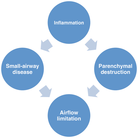
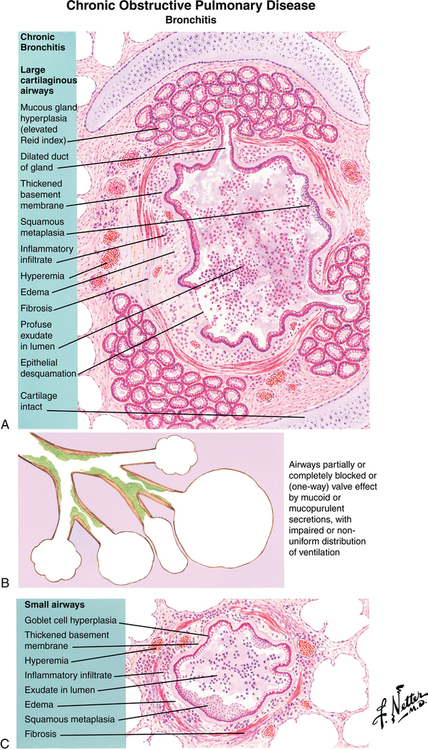
Patients with COPD frequently have a combination of obstructive bronchiolitis or small-airway disease and parenchymal destruction or emphysema (Figure 31-1). A ratio of forced expiratory volume in 1 sec (FEV1) to functional vital capacity (FVC) of less than 0.7 is essential to make the diagnosis of COPD. Total lung capacity, residual volume, and functional residual capacity are all increased in COPD, which is different from the pattern seen with restrictive lung disease.

Clinical features

The Global Initiative for Obstructive Lung Disease has calculated the prevalence of COPD to be 6.1% and 13.5%, respectively, after 9-year and 10-year cumulative studies. In young adults, the prevalence was found to be 2.2%, and in patients 40 to 44 years of age, it was 4.4%. The primary cause of COPD is cigarette smoking (with α1-antitrypsin deficiency as another cause in the young). Approximately half of patients older than 60 years of age who have a smoking history of at least 20 pack-years have a spirometry result consistent with COPD. Lung parenchymal destruction leads to loss of diffusing capacity and loss of the radial traction force on the airways. Airway inflammation produces increased mucus secretions and mucosal thickening, resulting in ventilation-perfusion mismatch and, finally, hypoxemia and CO2 retention. Airway collapse on expiration results in air trapping, leading to dynamic hyperinflation because of auto-positive end-expiratory pressure (Figure 31-2).

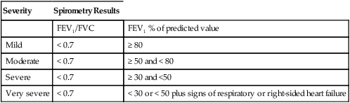
Signs and symptoms of COPD vary from asymptomatic to overt disease, depending on the severity of the disease (Table 31-1). Extrapulmonary symptoms include diaphragmatic dysfunction, right-sided heart failure, anxiety, depression, and weight loss with evidence of malnutrition.

Table 31-1

GOLD Classification of COPD Severity

Severity Spirometry Results
  FEV1/FVC FEV1 % of predicted value
Mild < 0.7 ≥ 80
Moderate < 0.7 ≥ 50 and < 80
Severe < 0.7 ≥ 30 and <50
Very severe < 0.7 < 30 or < 50 plus signs of respiratory or right-sided heart failure

image

COPD, Chronic obstructive pulmonary disease; FEV1, forced expiratory volume in 1 sec; FEV1/FVC, the ratio of FEV1 to FVC; FVC, forced vital capacity; GOLD, Global Initiative for Obstructive Lung Diseases.

Management

Smoking cessation can halt the decline of pulmonary function, and although it produces only a small improvement in FEV1, smoking cessation results in a subsequent rate of decline that eventually becomes equivalent to that of a nonsmoker. The use of short-acting β2-adrenergic receptor agonists and anticholinergic agents as combined bronchodilators is superior to the use of either agent alone. Long-acting bronchodilators provide sustained symptomatic relief. The use of inhaled corticosteroids is indicated for severe and repeated exacerbations but is not recommended as monotherapy. Theophylline may be tried in patients with severe COPD that is unresponsive to other regimens; blood levels of theophylline must be monitored to minimize potential side effects.

The Global Initiative for Obstructive Lung Disease defines an acute exacerbation of COPD as “an event in the natural course of the disease characterized by a change in the patient’s baseline dyspnea, cough, and/or sputum that is beyond normal day-to-day variations, is acute in onset, and may warrant a change in regular medications in a patient with underlying COPD.” Exacerbations are commonly caused by infection and air pollution and are treated with antibiotics. Bronchodilator therapy should be reviewed and changed if appropriate. Mechanical ventilation is sometimes required for acute exacerbations of COPD; noninvasive mechanical ventilation without tracheal intubation can be tried. Lung volume reduction surgery and lung transplantation have a limited role in the treatment of patients with COPD.

Perioperative considerations

Perioperative morbidity and mortality rates are not influenced by the use of general versus regional anesthetic techniques in patients with COPD. The main goal is to avoid excessive airway manipulation, thus lessening reflex-induced bronchospasm. Surgery should be postponed for symptomatic patients with severe dyspnea or acute exacerbations of symptoms who are scheduled to undergo elective operations.

Preoperative optimization of pulmonary function focuses on cessation of smoking, optimization of bronchodilator therapy, control of infections, and provision of chest physiotherapy, such as incentive spirometry, breathing exercises, and postural drainage techniques. Appropriate investigations such as arterial blood gas analysis, electrocardiography, echocardiography, and chest radiography provide information that is often helpful in determining gas exchange efficiency, right ventricular function, and the presence of asymptomatic bullae.

Intraoperative monitoring of airway pressure, O2 saturation, and end-tidal CO2 provides useful information about the degree of airflow obstruction. Anesthetics of choice include short-acting agents, such as propofol and remifentanyl, and drugs that do not stimulate histamine release. The respiratory rate of the ventilator should be lower than normal to allow for prolonged expiration and to minimize the chance of dynamic hyperinflation occurring. Postoperatively, the use of noninvasive positive-pressure ventilation is an attractive alternative to tracheal intubation and mechanical ventilation.

Restrictive lung disease

Clinical features

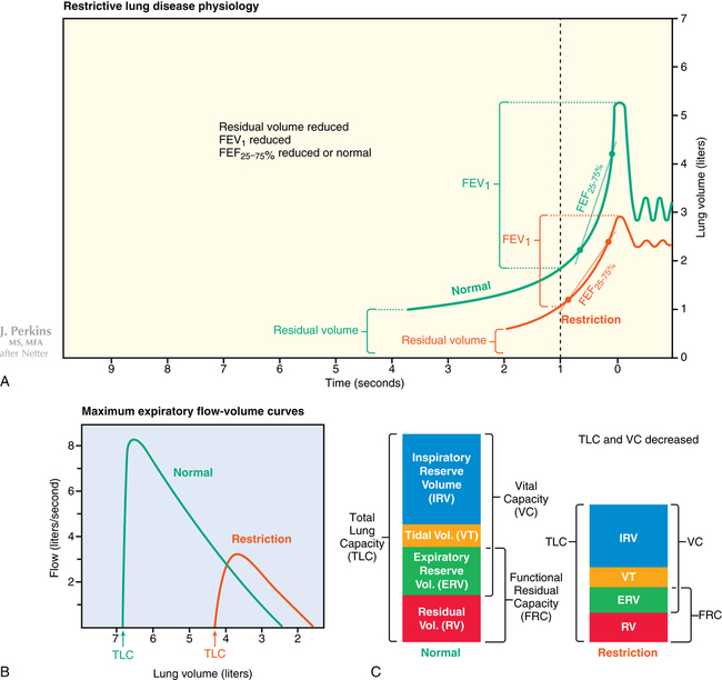
Limited lung expansion or restrictive lung disease may result from any of several pulmonary and extrapulmonary causes—pulmonary fibrosis, sarcoidosis, obesity, pleural effusion, scoliosis, or respiratory muscle weakness. Interstitial edema and acute lung injury/acute respiratory distress syndrome (ALI/ARDS) are classified as acute restrictive lung diseases. Spirometry can help differentiate between obstructive and restrictive patterns: in the latter, total lung capacity is reduced, airway resistance is normal, and airflow is preserved. Reduced FEV1 with a normal or increased FEV1/FVC suggests the presence of restrictive lung disease, but the diagnosis and severity grading are based on measurement of decreased total lung capacity (Figure 31-3). In patients who have restrictive lung disease with an intrinsic cause, reduced gas transfer is manifest as desaturation after exercise.

The prevalence rates of the mortality and morbidity associated with restrictive lung disease vary based on the underlying cause: idiopathic pulmonary fibrosis is found in 27 to 29 cases per 100,000 persons and has a median survival time of less than 3 years.

Perioperative considerations

For patients who have pulmonary lesions, preoperative spirometry, arterial blood gas analysis, and measurements of lung volume and gas transfer should be performed within 8 weeks before surgery to identify disease severity. Supplemental doses of corticosteroids and postoperative O2 therapy may be required; respiratory infections should be treated immediately. Patients who have extrapulmonary causes of their restrictive lung disease usually breathe rapidly and shallowly, which is not effective in clearing sputum, particularly after thoracic or upper abdominal operations. Providing vigorous chest physiotherapy and adequate analgesia are important. Patients who are mechanically ventilated during the operative and postoperative periods typically tolerate relatively low tidal volumes and high breathing rates.