Choroidal Hemangioma
Clinical Features:
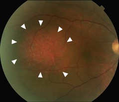
They may be identified as an incidental finding, but can affect visual acuity if there is associated subretinal fluid or cystic fluid involving the macula. Typical features include a reddish/orange coloration and location in the posterior pole (Fig. 21.3.1). RPE metaplasia on the surface is not rare. Occasionally, clinical findings are subtle and they are only identified on OCT.
OCT Features:
There is obscuration of the normal choriocapillaris by a hyporeflective signal and overlying round-shaped retinal elevation (Figs 21.3.2 and 21.3.3). Overlying subretinal and/or intraretinal fluid may also be present (Fig. 21.3.3), which can involve the macula (Fig. 21.3.4). Occasionally, fluid can occur subfoveally, even if the tumor is not located within the macula. Enhanced depth imaging techniques can be helpful for better visualization in larger tumors.
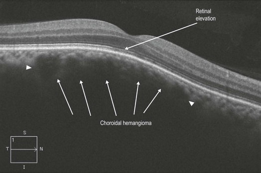
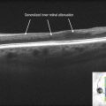
Figure 21.3.2 OCT of a subtle choroidal hemangioma that was not clearly visible clinically. The choriocapillaris is slightly obscured (abnormal hyporeflectivity between arrowheads) and there is elevation of the overlying retina.
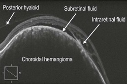
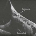
Figure 21.3.3 OCT of a more obvious choroidal hemangioma (corresponding to Figure 21.3.1). The choriocapillaris is completely obscured by the tumor and there is bullous overlying retinal elevation. Subretinal and intraretinal fluid are also present.
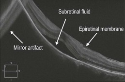
Figure 21.3.4 OCT of the macula (corresponding to Figure 21.3.3) shows the presence of subretinal fluid and a mild epiretinal membrane.
Ancillary Testing:
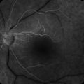
Indocyanine green angiography is useful in confirming the diagnosis, particularly in the early phase images 20–30 seconds following injection where prominent hyperfluorescence of the lesion is noted (Fig. 21.3.5). B-scan ultrasonography can also be helpful.
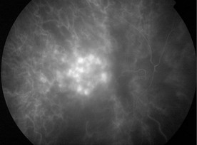
Figure 21.3.5 Indocyanine green angiography at 20 seconds (corresponding to Figure 21.3.1) shows intense early hyperfluorescence of the choroidal hemangioma, which is a characteristic feature distinguishing this lesion from other choroidal tumors.