Cholera

Published on 22/03/2015 by admin

Filed under Pediatrics

Last modified 22/04/2025

Print this page

rate 1 star rate 2 star rate 3 star rate 4 star rate 5 star
Your rating: none, Average: 0 (0 votes)

This article have been viewed 1633 times

Chapter 193 Cholera

Cholera is a rapidly dehydrating diarrheal disease that can lead to death, if appropriate treatment is not provided immediately. Although rare in industrialized countries, cholera has a propensity to cause outbreaks in areas with poor hygiene and inadequate sanitation and water facilities. These outbreaks may be explosive, especially when they occur in populations residing in crowded conditions, such as refugee camps. Reports from the World Health Organization (WHO) indicate that cholera is on the rise, with more cases being reported annually from 2006 and 2007 compared to the annual average of 2002-2005. In 2007, there were 177,963 cholera cases reported, with 4,031 deaths; actual figures may be higher.

Epidemiology

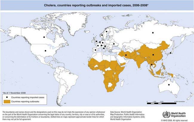
The first 6 cholera pandemics originated in the Indian subcontinent and were caused by classical O1 V. cholerae. The seventh pandemic is the most extensive of all and is caused by V. cholerae O1 El Tor. It began in 1961 in Sulawesi, Indonesia, and has spread to the Indian subcontinent, Southeast Asia, Africa, Oceania, Southern Europe, and the Americas. In 1991, V. cholerae O1 El Tor first appeared in Peru before rapidly spreading in the Americas. Cholera becomes endemic in areas following outbreaks when a large segment of the population develops immunity to the disease after recurrent exposure. The disease is now endemic in parts of East, Southern, and Northwest Africa, as well as in South and Southeast Asia (Fig. 193-1).

image

Figure 193-1 Countries reporting cholera outbreaks and imported cholera cases to WHO from 2006-2008.

(From World Health Organization: Cholera, areas reporting outbreaks, 2007-2009 (website). gamapserver.who.int/mapLibrary/Files/Maps/Global_ChoeraCases_ITHRiskMap.png. Accessed August 9, 2010.)

In 1992, the first non-O1 V. cholerae that resulted in epidemics was identified in India and Bangladesh and was designated V. cholerae O139. From 1992 to 1994, this organism replaced O1 as the predominant cause of cholera in South Asia but has since been an uncommon etiologic agent.

The hybrid El Tor strains were first identified sporadically in Bangladesh. In 2004, during routine surveillance in Mozambique, isolates of V. cholerae O1 El Tor carrying classical genes were identified. Since then, hybrid and variant El Tor strains have been reported in other parts of Asia and Africa and have caused outbreaks in India and Vietnam. Although the classical biotype has virtually disappeared, its genes remain within the El Tor biotype.

Humans are the only known hosts, but free-living and plankton-associated V. cholerae exist in the marine environment. The organism thrives best in moderately salty water but can survive in rivers and freshwater if nutrient levels are high, as occurs when there is organic pollution such as human feces. The formation of a biofilm on abiotic surfaces and the ability to enter a viable but nonculturable state has been hypothesized as factors that allow V. cholerae to persist in the environment. Surface sea temperature, pH, chlorophyll content, the presence of iron compounds and chitin, and climatic conditions such as amount of rainfall and sea level rise are all important environmental factors that influence the survival of V. cholerae in the environment and the expression of cholera toxin, an important virulence determinant.

Consumption of contaminated water and ingestion of undercooked shellfish are the main modes of transmission, with the latter more often seen in developed countries. Previous studies in Bangladesh revealed that children aged 2-4 yr have the highest incidence of the disease; data from two endemic areas in Jakarta, Indonesia, and Kolkata, India, revealed that the incidence of disease was highest among infants and children <2 yr of age. On the other hand, all age groups were commonly affected in areas where the disease has not gained a foothold. In epidemic and endemic settings, the disease usually first appears in men. Persons with blood group O, decreased gastric acidity, malnutrition, immunocompromised state, and absence of local intestinal immunity (prior exposure by infection or vaccination) are at increased risk for developing severe disease. Household contacts of cholera-infected patients are at high risk for the disease, because the stools of infected patients contain high concentrations of V. cholerae (up to 108/gof stool).

Pathogenesis

Following ingestion of V. cholerae from the environment, several changes occur in the vibrios while they traverse the human intestine: increased expression of genes required for nutrient acquisition, downregulation of chemotactic response, and expression of motility. Together these changes allow the vibrios to reach a hyperinfectious state, leading to lower infectious doses in secondarily infected persons.

Large inocula of bacteria (>108) are required for severe cholera to occur; however, for persons whose gastric barrier is disrupted, a much lower dose (105) is required. If the vibrios survive gastric acidity, they then colonize the small intestine through various factors such as toxin coregulated pili (TCP) and motility, leading to efficient delivery of cholera toxin. The cholera toxin consists of five binding B subunits and one active A subunit. The B subunits are responsible for binding to the GM1 ganglioside receptors located in the small intestinal epithelial cells. After binding, the A subunit is then released into the cell, where it stimulates adenylate cyclase and initiates a cascade of events. An increase in cyclic adenosine monophosphate (cAMP) leads to an increase in chloride secretion by the crypt cells, which in turn leads to inhibition of absorption of sodium and chloride by the microvilli. These events eventually lead to massive purging of electrolyte rich isotonic fluid in the small intestine that exceeds the absorptive capacity of the colon, resulting in rapid dehydration and depletion of electrolytes, including sodium, chloride, bicarbonate, and potassium. Metabolic acidosis and hypokalemia then ensues.

Clinical Manifestations

Most cases of cholera are mild or inapparent. Among symptomatic cases, around 20% develop severe dehydration that can rapidly lead to death. Following an incubation period of 1 to 3 days (range, several hours to 5 days), acute watery diarrhea and vomiting ensues. The onset may be sudden, with profuse watery diarrhea, but some patients have a prodrome of anorexia and abdominal discomfort and the stool may initially be brown. Diarrhea can progress to painless purging of profuse rice-water stools (suspended flecks of mucus) with a fishy smell, which is the hallmark of the disease. Vomiting with clear watery fluid is usually present at the onset of the disease.

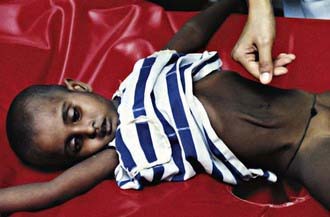
Cholera gravis, the most severe form of the disease, results when purging rates of 500-1000 mL/hr occur. This purging leads to dehydration manifested by decreased urine output, sunken fontanels (in infants), sunken eyes, absence of tears, dry oral mucosa, shriveled hands and feet (washerwoman’s hands), poor skin turgor, thready pulse, tachycardia, hypotension, and vascular collapse (Fig. 193-2). Patients with metabolic acidosis can present with typical Kussmaul breathing. Although patients may be initially thirsty and awake, they rapidly progress to obtundation and coma. If fluid losses are not rapidly corrected, death can occur within hours.

Diagnosis and Differential Diagnosis

In children who have acute watery diarrhea with severe dehydration and have recently traveled to an area known to have cholera, the disease may be suspected pending laboratory confirmation. Cholera differs from other diarrheal disease in that it often occurs in large outbreaks affecting both adults and children.

Treatment of dehydration should begin as soon as possible. Diarrhea due to other etiologic causes (e.g., enterotoxigenic Escherichia coli or rotavirus) may be difficult to distinguish from cholera clinically. Microbiologic isolation of V. cholerae remains the gold standard for diagnosis. Although definitive diagnosis is not required for treatment to be initiated, laboratory confirmation is necessary for epidemiologic surveillance. V. cholerae may be isolated from stools, vomitus, or rectal swabs. Specimens may be transported on Cary-Blair media, if they cannot be processed immediately. Selective media such as thiosulfate citrate bile salts sucrose (TCBS) agar that inhibit normal flora should be used. Because most laboratories in industrialized countries do not routinely culture for V. cholerae, clinicians should request appropriate cultures for clinically suspected cases.

Stool examination reveals few fecal leukocytes and erythrocytes because cholera does not cause inflammation. Dark-field microscopy may be used for rapid identification of typical “darting motility” in wet mounts of rice-water stools, which disappears once specific antibodies against V. cholerae O1 or O139 are added. Rapid diagnostic tests are currently being evaluated that will allow use at the bedside. Molecular identification with the use of polymerase chain reaction (PCR) and DNA probes are available but often not used in areas where cholera exists.

Treatment

Rehydration is the mainstay of therapy (Chapter 332). Effective and timely case management considerably decreases mortality. Children with mild or moderate dehydration may be treated with oral rehydration solution (ORS) unless the patient is in shock, is obtunded, or has intestinal ileus. Vomiting is not a contraindication to ORS. Severely dehydrated patients require intravenous fluid, ideally with lactated Ringer solution. When available, rice-based ORS should be used during rehydration, because this fluid has been shown to be superior to standard ORS in children and adults with cholera. Close monitoring is necessary, especially during the first 24 hr of illness, when large amounts of stool may be passed. After rehydration, patients have to be reassessed every 1-2 hr, or more frequently if profuse diarrhea is ongoing. Feeding should not be withheld during diarrhea. Frequent, small feedings are better tolerated than less frequent, large feedings.

As soon as vomiting stops (usually within 4-6 hr after initiation of rehydration therapy), an antibiotic to which local V. cholerae strains are sensitive must be administered. Antibiotics (Table 193-1) shorten the duration of illness, decrease fecal excretion of vibrios, decrease the volume of diarrhea, and reduce the fluid requirement during rehydration. Single-dose doxycycline increases compliance; there have been increasing reports of resistance to tetracyclines. Ciprofloxacin, azithromycin, and trimethoprim-sulfamethoxazole are also effective against cholera. Cephalosporins and aminoglycosides are not clinically effective against cholera and therefore should not be used, even if in vitro tests show strains to be sensitive.

Table 193-1 SUGGESTED ANTIMICROBIALS FOR SUSPECTED CHOLERA CASES WITH SEVERE DEHYDRATION

ANTIBIOTIC OF CHOICE* ALTERNATIVE
Doxycycline (adults and older children): 300 mg given as a single dose
or
Tetracycline 12.5 mg/kg/dose 4 times/day × 3 days (up to 500 mg per dose × 3 days)
Erythromycin 12.5 mg/kg/dose 4 times a day × 3 days (up to 250 mg 4 times a day × 3 days)

* Selection of an antimicrobial should be based on sensitivity patterns of strains of Vibrio cholerae O1 or O139 in the area.

Adapted from World Health Organization: The treatment of diarrhea: a manual for physicians and other senior health workers—4th revision, Geneva, 2005, World Health Organization.

Zinc should be given as soon as vomiting stops. Zinc deficiency is common among children in many developing countries. Zinc supplementation among children <5 yr of age has been shown to shorten the duration of diarrhea and reduce subsequent diarrhea episodes when given daily for 14 days at the time of the illness. For children <6 mo of age, 10 mg of oral zinc may be given daily for 2 wk, and for children aged 6 mo to 12 yr, 20 mg of oral zinc may be given daily for 2 wk.

Prevention

Improved personal hygiene, access to clean water, and sanitation are the mainstays of cholera control. Appropriate case management substantially decreases case fatalities to <1%. Travelers from developed countries often have no prior exposure to cholera and are therefore at risk of developing the disease. Children travelling to cholera-affected areas should avoid drinking potentially contaminated water and eating high-risk foods such as raw or undercooked fish and shellfish.

No country or territory requires vaccination against cholera as a condition for entry. There is no cholera vaccine licensed in the USA. An internationally licensed killed whole-cell oral cholera vaccine with recombinant B subunit (Dukoral, SBL/Crucell) has been available in more than 60 countries, including the European Union, and provides protection against cholera in endemic areas as well as cross-protection against certain strains of enterotoxigenic E. coli (ETEC). Older-generation parenteral cholera vaccines have not been recommended by WHO, due to the limited protection they confer and their high reactogenicity. Oral cholera vaccines (OCVs) have been available for >2 decades and are mostly used by travelers from industrialized countries going to cholera-affected areas. Although WHO has recommended the use of OCV in the control of cholera in certain endemic and epidemic situations since 2001, these vaccines have not been extensively adopted. Table 193-2 shows currently licensed vaccines and dosing schedules. Live-attenuated oral cholera vaccine (Orochol, Berna Biotech/Crucell) has not been shown to be protective against cholera in a clinical trial in an endemic area and is no longer manufactured.

Table 193-2 INTERNATIONALLY LICENSED ORAL CHOLERA VACCINES

VACCINE TRADE NAME CONTENTS DOSING SCHEDULE FOR CHOLERA
Dukoral (SBL/Crucell)

Orochol (Berna Biotech/Crucell) Live-attenuated classical V. cholerae O1 CVD 103- HgR One dose for children and adults aged ≥2 yr of age

Bibliography

Chaignat CL, Monti V. Use of oral cholera vaccine in complex emergencies: what next? Summary report of an expert meeting and recommendations of WHO. J Health Popul Nutr. 2007;25:244-261.

Constantin de Magny G, Murtugudde R, Sapiano MR, et al. Environmental signatures associated with cholera epidemics. Proc Natl Acad Sci U S A. 2008;105:17676-17681.

Deen JL, von Seidlein L, Sur D, et al. The high burden of cholera in children: comparison of incidence from endemic areas in Asia and Africa. PLoS Negl Trop Dis. 2008;2:e173.

Faruque AS, Alam K, Malek MA, et al. Emergence of multidrug-resistant strain of Vibrio cholerae O1 in Bangladesh and reversal of their susceptibility to tetracycline after two years. J Health Popul Nutr. 2007;25:241-243.

Griffith DC, Kelly-Hope LA, Miller MA. Review of reported cholera outbreaks worldwide, 1995–2005. Am J Trop Med Hyg. 2006;75:973-977.

Harris JB, LaRocque RC, Charles RC, et al. Cholera’s western front. Lancet. 2010;376:1961-1964.

Kumar P, Jain M, Goel AK, et al. A large cholera outbreak due to a new cholera toxin variant of the Vibrio cholerae O1 El Tor biotype in Orissa, Eastern India. J Med Microbiol. 2009;58(Pt 2):234-238.

Lopez AL, Clemens JD, Deen J, Jodar L. Cholera vaccines for the developing world. Hum Vaccin. 2008;4:165-169.

Nelson EJ, Nelson DS, Salam MA, et al. Antibiotics for both moderate and severe cholera. N Engl J Med. 2011;364(1):5-9.

World Health Organization. Cholera, 2007. Wkly Epidemiol Rec. 2008;83:269-283.

World Health Organization. The treatment of diarrhea: a manual for physicians and other senior health workers—4th revision. Geneva: World Health Organization; 2005.

Zuckerman JN, Rombo L, Fisch A. The true burden and risk of cholera: implications for prevention and control. Lancet Infect Dis. 2007;7:521-530.