Chemotherapy-Induced Cholangitis

Published on 19/07/2015 by admin

Filed under Radiology

Last modified 22/04/2025

Print this page

rate 1 star rate 2 star rate 3 star rate 4 star rate 5 star
Your rating: none, Average: 0 (0 votes)

This article have been viewed 2305 times

 ± dilation of upstream intrahepatic ducts, as associated periductal fibrosis may impede ductal dilatation

• Distribution of strictures in biliary tree reflects hepatic arterial supply to bile ducts

image Proximal extrahepatic duct and biliary confluence strictures are most common due to blood supply from hepatic artery
image Distal extrahepatic duct is supplied by gastroduodenal artery branches, and consequently usually not involved
image Gallbladder and cystic duct may be involved
image Rarely causes peripheral intrahepatic strictures
• CT or MR: Involved bile ducts may show periductal edema, mural thickening, and enhancement
• Biloma formation (± abscess formation) may reflect drug-induced necrosis of peripheral ducts

TOP DIFFERENTIAL DIAGNOSES

• Primary sclerosing cholangitis
• Autoimmune cholangitis
• Extrinsic compression by liver masses or lymphadenopathy
• Chemical or drug-induced liver injury

PATHOLOGY

• Results from either direct toxic effects of drug on biliary ducts or fibrosis/occlusion of peribiliary vascular plexus with resultant biliary ischemic cholangiopathy
• Risk factors: Preexisting biliary strictures, prior biliary surgery, portal vein occlusion, nonselective placement of catheter during chemoembolization, higher doses of chemotherapy
image
(Left) Axial CECT shows a liver metastasis image 
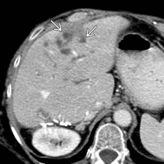
that is low in attenuation, likely as a result of necrosis. Note the dilated ducts image that resulted from a stricture of the biliary bifurcation and common hepatic duct, also due to chemotherapy.

image
(Right) Transhepatic cholangiogram in the same patient shows gross dilation of the intrahepatic ducts, with abrupt, high-grade stenosis image 
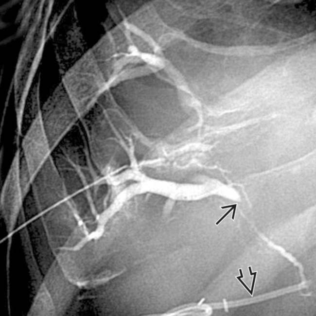
at the confluence of the right and left ducts. This patient had received floxuridine through an arterial catheter image.
image
(Left) CECT of a patient with carcinoid liver metastases after 8 courses of TACE shows irregular right periductal low attenuation image
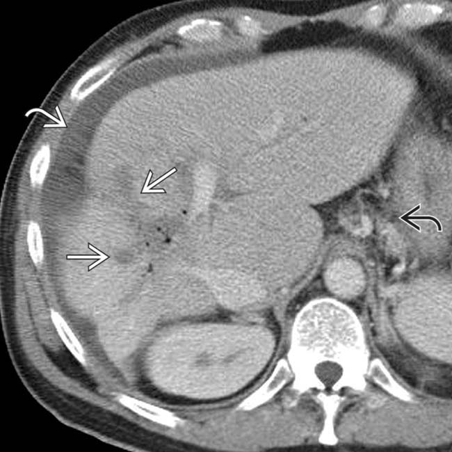
, pneumobilia, posterior segment atrophy, gastroesophageal varices image, and ascites image, compatible with chemotherapy-induced cholangitis and biliary cirrhosis.

image
(Right) ERCP of the same patient shows a proximal common duct stricture image 
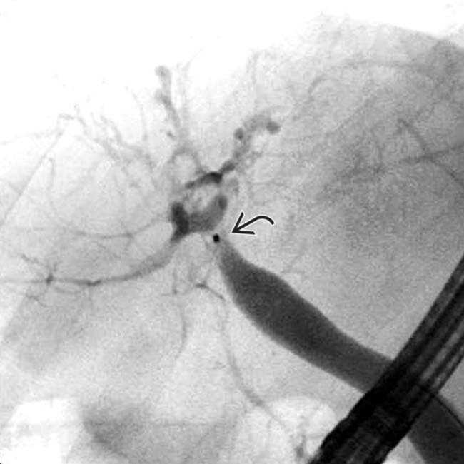
and irregular, strictured intrahepatic ducts. A liver transplant was performed with cholangitis, bilomas, and biliary cirrhosis identified within the explant.

TERMINOLOGY

Synonyms

• Chemotherapy-induced sclerosing cholangitis (CISC); biliary sclerosis

Definitions

• Iatrogenic cholangitis following intraarterial chemotherapy for hepatic malignancies or metastases

image Complication of hepatic artery infusion pump (HAIP) or transarterial chemoembolization (TACE)

IMAGING

General Features

• Location

image Distribution of strictures in biliary tree reflects hepatic arterial supply to bile ducts

– Proximal extrahepatic duct and central intrahepatic ducts/biliary confluence are most commonly involved (∼ 50%) due to blood supply from hepatic artery branches
– Distal extrahepatic duct supplied by gastroduodenal artery branches, and consequently not usually involved
– Rarely causes peripheral intrahepatic strictures
– Gallbladder and cystic duct may be involved
• Morphology

image Findings similar to primary sclerosing cholangitis, with stenosis or complete obstruction of involved ducts

– ± dilation of upstream intrahepatic ducts, as associated periductal fibrosis may impede ductal dilatation

Radiographic Findings

• ERCP

image Abnormalities range from minimal duct wall irregularity to marked duct wall thickening with near obliteration of lumen

CT Findings

• CT (or MR) essential to differentiate cholangitis from extrinsic duct compression by lymph nodes or tumor
• Mildly dilated intrahepatic bile ducts
• Affected bile ducts may show periductal edema, mural thickening, and enhancement with adjacent fat stranding in hepatoduodenal ligament
• Biloma formation (± abscess formation) may reflect drug-induced necrosis of peripheral ducts

MR Findings

• MRCP

image Segmental strictures of variable length (similar to those seen in primary sclerosing cholangitis)

Imaging Recommendations

• Best imaging tool

image CT, MRCP, and ERCP may be used in concert to differentiate this entity from other causes of jaundice

– e.g., compression by hepatic tumor, porta hepatis adenopathy, and chemotherapy hepatotoxicity

DIFFERENTIAL DIAGNOSIS

Primary Sclerosing Cholangitis (PSC)

• Multifocal “beaded” stenoses of intrahepatic and extrahepatic bile ducts with pruning, irregular wall thickening, and intervening sites of normal or dilated ducts
• Gallbladder and cystic duct are usually more severely involved in chemotherapy cholangitis than in PSC
• Associated with massive hypertrophy of caudate and marked peripheral liver atrophy in end-stage disease
• No history of intraarterial chemotherapy

Autoimmune Cholangitis

• Imaging findings are almost identical to those of PSC
• No history of prior chemotherapy

Extrinsic Compression by Liver Masses or Lymphadenopathy

• May compress extrahepatic or central intrahepatic ducts
• Extrinsic compression and narrowing of ducts without true strictures

Chemical or Drug-Induced Liver Injury

• Can result in hepatitis, cholestasis, or both
• Cholestatic pattern can result in injury to extrahepatic bile duct that is similar to PSC
• Chronic involvement can result in “vanishing bile duct syndrome”: Fibrosis and loss of intrahepatic ducts

PATHOLOGY

General Features

• Etiology

image Results from either direct toxic effects of drug on biliary ducts or fibrosis/occlusion of peribiliary vascular plexus with resultant biliary ischemic cholangiopathy

– Proximal extrahepatic duct and central intrahepatic ducts most commonly involved due to blood supply from hepatic artery branches
– Distal extrahepatic duct supplied by gastroduodenal artery branches, and consequently not usually involved
image Toxic effects of fluoropyrimidines (floxuridine and 5-FU)

– Rarely from other drugs (e.g., mitomycin-C)
image Risk factors

– Preexisting biliary strictures, prior liver/biliary surgery, or portal vein occlusion
– Nonselective placement of catheter during chemoembolization
– Small diameter embolization agents or higher doses of infused chemotherapy agents
• Associated abnormalities

image Aneurysm or pseudoaneurysm of hepatic artery at infusion site, pancreatitis, cholecystitis, gallbladder perforation

CLINICAL ISSUES

Presentation

• Most common signs/symptoms

image Intermittent or progressive jaundice and cholestasis
image Cholangitis (jaundice, pain, fever)
image ↑ bilirubin, alkaline phosphatase
image Symptoms as early as 2 months after chemoembolization

Demographics

• Epidemiology

image Cholangiographic abnormalities reported in 7-30% of patients undergoing intraarterial chemotherapy

Natural History & Prognosis

• Complications: Acute hepatic failure, ischemic cholecystitis, biliary cirrhosis (rare)

image Gangrenous cholecystitis in 1-2% after chemoembolization
• Mortality rate: 1%

Treatment

• Reduction or cessation of intraarterial chemotherapy

image Equivocal role of intraarterial steroids
• Biliary decompression with endoscopic balloon dilation of strictures ± stenting
• In rare cases, surgical resection of affected segment of bile duct or liver transplantation may be indicated

DIAGNOSTIC CHECKLIST

Consider

• When clinical signs of hepatic dysfunction occur in absence of tumor progression, biliary sclerosis from chemotherapy must be suspected
image
Axial CECT in a 50-year-old man with liver metastases from colon carcinoma, treated with intraarterial infusion of floxuridine, shows dilation of the intrahepatic ducts image in spite of a transhepatic internal-external biliary drainage catheter image.

image
Axial CECT in a patient who had a right hepatectomy for metastatic colon cancer shows mildly dilated intrahepatic bile ducts (IHBDs). Some focally dilated ducts image 
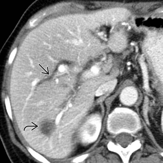
could be mistaken for metastases.
image
Axial CECT shows a centrally dilated IHBD image 
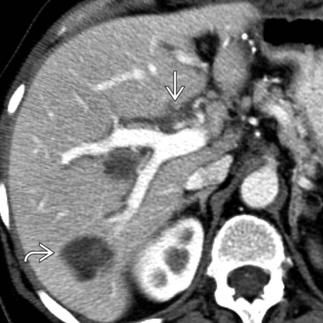
in a patient with metastatic colon carcinoma image who had undergone intraarterial chemotherapy.
image
Transhepatic cholangiogram in a patient who had intraarterial chemotherapy through an infusion pump catheter image 
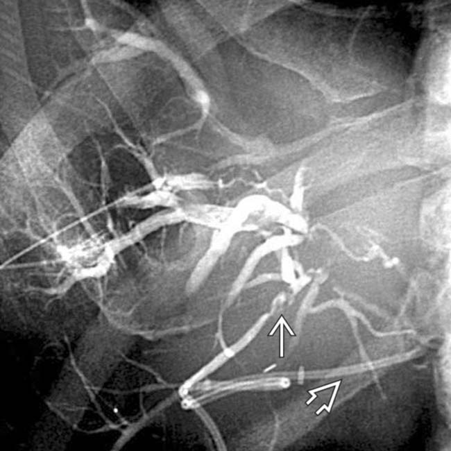
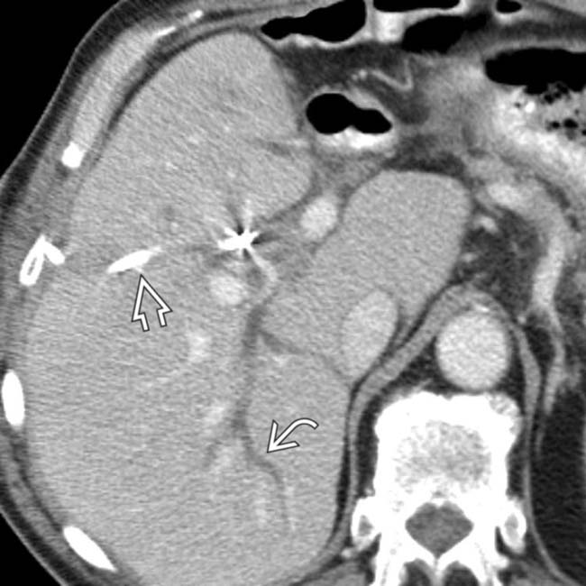
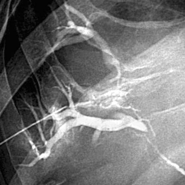
shows a dilated right IHBD with multiple central biliary strictures image.
image
Right transhepatic cholangiogram shows a dilated IHBD. High-grade central biliary strictures developed after intraarterial chemotherapy with FUDR for metastatic colon carcinoma to the liver.
image
Transhepatic cholangiogram in a patient after intraarterial chemotherapy demonstrates a high-grade stricture image 
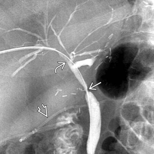
of the common hepatic duct with a milder degree of stricture of the intrahepatic ducts image as well. Note the indwelling hepatic arterial catheter image.

SELECTED REFERENCES

1. Nguyen, KD, et al. Atypical causes of cholestasis. World J Gastroenterol. 2014; 20(28):9418–9426.

Ito, K, et al. Biliary sclerosis after hepatic arterial infusion pump chemotherapy for patients with colorectal cancer liver metastasis: incidence, clinical features, and risk factors. Ann Surg Oncol. 2012; 19(5):1609–1617.

Pua, U. Transarterial chemoembolization-induced main duct stricture. Clin Gastroenterol Hepatol. 2011; 9(12):A22.

Torrisi, JM, et al. CT findings of chemotherapy-induced toxicity: what radiologists need to know about the clinical and radiologic manifestations of chemotherapy toxicity. Radiology. 2011; 258(1):41–56.

Kato, Y, et al. Chemotherapy-induced sclerosing cholangitis as a rare indication for resection: report of a case. Surg Today. 2009; 39(10):905–908.

Bang, BW, et al. Ischemic biliary stricture developed after repeated transcatheter arterial chemoembolization diagnosed by percutaneous transhepatic cholangioscopy in a patient with hepatocellular carcinoma. Gastrointest Endosc. 2008; 68(6):1224–1226.

Menias, CO, et al. Mimics of cholangiocarcinoma: spectrum of disease. Radiographics. 2008; 28(4):1115–1129.

Alazmi, WM, et al. Chemotherapy-induced sclerosing cholangitis: long-term response to endoscopic therapy. J Clin Gastroenterol. 2006; 40(4):353–357.

Phongkitkarun, S, et al. Bile duct complications of hepatic arterial infusion chemotherapy evaluated by helical CT. Clin Radiol. 2005; 60(6):700–709.

Aldrighetti, L, et al. Extrahepatic biliary stenoses after hepatic arterial infusion (HAI) of floxuridine (FUdR) for liver metastases from colorectal cancer. Hepatogastroenterology. 2001; 48(41):1302–1307.

Tarazov, PG, et al. Ischemic complications of transcatheter arterial chemoembolization in liver malignancies. Acta Radiol. 2000; 41(2):156–160.

Pozniak, MA, et al. Complications of hepatic arterial infusion chemotherapy. Radiographics. 1991; 11(1):67–79.

Shea, WJ, Jr., et al. Sclerosing cholangitis associated with hepatic arterial FUDR chemotherapy: radiographic-histologic correlation. AJR Am J Roentgenol. 1986; 146(4):717–721.

Botet, JF, et al. Cholangitis complicating intraarterial chemotherapy in liver metastasis. Radiology. 1985; 156(2):335–337.