[level-membership-for-neurosurgery-category]
Chapter 160 Cervical Total Disc Arthroplasty
Anterior cervical discectomy and fusion (ACDF) has been routinely used for the treatment of degenerative disease of the cervical spine since the 1950s.1 Interbody fusion has become the accepted method of treating cervical radiculopathy, myelopathy, segmental deformity, instability, and degenerative disc disease. With the evolution of technique and technology, fusion rates have exceeded 95%.2 Despite these results, concern for the development of adjacent-level degeneration and disease after fusion has prompted interest in and development of implants designed to preserve motion and maintain anatomic disc space height and normal segmental lordosis. In this chapter, we review the indications for cervical total disc arthroplasty and discuss the preliminary data, with an emphasis on complications and their avoidance. A full discussion of each of the developed artificial discs is beyond the scope of this chapter. Implants for which U.S. Food and Drug Administration (FDA) Investigational Device Exemptions (IDE) studies have been completed are discussed herein.
Rationale for Arthroplasty
The long-term clinical success of ACDF has been limited by the development of adjacent-level degeneration, leading to further symptoms and possibly additional surgery. Adjacent-level degeneration is defined as radiographic changes at a segment adjacent to a fusion and is not necessarily associated with symptoms. Adjacent-level disease, however, is associated with pain, radiculopathy, or myelopathy.3 Biomechanical studies have demonstrated that cervical fusion alters adjacent-level kinematics and that the loss of motion at the fused segment is compensated by an increase in motion and intradiscal pressure at adjacent levels.4–6 Some clinical studies have supported these findings, giving weight to this theory of fusion-induced degeneration. Hilibrand et al.7 reported that repeat surgical intervention was necessary for 2.9% of patients per year for symptomatic adjacent-segment disease after ACDF. They also found that 25% of patients reported symptoms from adjacent-segment disease 10 years after ACDF. In a comparative study, Goffin et al.8 showed that 92% of fused patients demonstrated radiographic evidence of adjacent-segment degeneration 5 years after surgery for both cervical spondylosis and trauma, suggesting that adjacent-level degeneration is accelerated by fusion and not necessarily the natural history of preexisting degenerative disc disease. It has been postulated that adjacent-level degeneration may be attributable to higher shear strains at levels adjacent to fusions.9
Debate continues as to whether adjacent-level degeneration is truly due to increased loads after fusion or to the natural history of degenerative disc disease. In a retrospective study of 303 patients undergoing single-level dorsal cervical foraminotomy for cervical radiculopathy, Clarke et al.10 showed that 15 (4.9%) of 303 developed symptomatic adjacent-level disease with a 0.7% annual rate for development of adjacent-level disease. The 10-year cumulative incidence of adjacent-level disease was 6.7%.
Biomechanical studies have suggested that cervical arthroplasty may allow for a more normal restoration of load transfer and kinematics at adjacent levels compared with fusion.5 After disc replacement, stress profiles in specimens at adjacent-level discs were similar to those of intact, nontreated levels, with reduced stresses in the adjacent-level anulus compared with spines with simulated fusions.11 Cervical disc arthroplasty may therefore be beneficial to patients because in theory it prevents the development of adjacent-segment disease by preserving normal neck motion.
Nomenclature and Design
With the development of numerous artificial discs in the last decade, the Cervical Spine Study Group developed a nomenclature system for cervical arthroplasty.12 Artificial discs are categorized by material, articulation, fixation, design, and kinematics.13 They can be classified as nonarticulating, uniarticulating, or biarticulating. Various designs include metal on metal (Prestige ST and LP; Medtronic Sofamor Danek, Memphis, TN), metal on polymer (Bryan, Medtronic Sofamor Danek; ProDisc-C, Synthes Spine, West Chester, PA), ceramic on polymer, or ceramic on ceramic. Discs are either modular with replaceable parts, or nonmodular. Some have points for vertebral body fixation and some have surfaces that promote bone ingrowth at the disc–end-plate interface. With regard to motion, they may be constrained, semiconstrained, or unconstrained. Constrained devices restrict motion to less than that seen physiologically, semiconstrained devices allow physiologic motion, and unconstrained devices rely on soft tissue and the inherent compression across the disc space to limit motion.14 Each of these devices is available in a range of heights and depths to accommodate individual variances in anatomy.
The Prestige ST artificial disc is constructed of stainless steel and consists of two articulating components attached to the cervical vertebrae with screws (Fig. 160-1). The ball-and-trough design provides relatively unconstrained motion compared with that of a normal cervical spinal segment. The surfaces of the device contacting the end plates are grit-blasted to promote bone osseointegration.
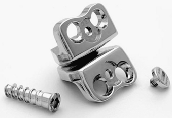
FIGURE 160-1 Prestige ST artificial cervical disc.
(Courtesy of Medtronic Sofamor Danek, Memphis, TN.)
The Prestige LP is the latest version of the Prestige artificial discs and differs from previous models in that fixation to the vertebral bodies is achieved by a set of rails that are imbedded in the vertebral end plates (Fig. 160-2). It is manufactured from a titanium-ceramic composite material that is highly durable and produces little artifact on CT and MRI. The ball-and-trough design is identical to that of the Prestige ST. A porous titanium plasma spray coating on the end-plate surface facilitates bone ingrowth and long-term fixation.
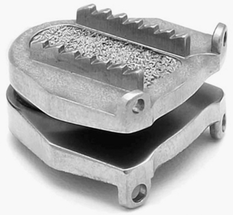
FIGURE 160-2 Prestige LP artificial cervical disc.
(Courtesy of Medtronic Sofamor Danek, Memphis, TN.)
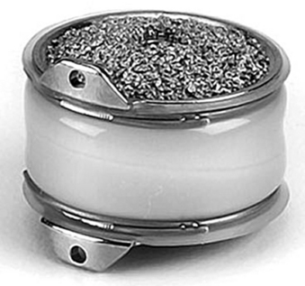
The Bryan cervical disc is designed to allow for motion similar to the normal cervical spine and resembles an actual disc (Fig. 160-3). It consists of two titanium alloy shells with a polyurethane nucleus. The bone-implant interface of each shell has an applied porous coating to facilitate ingrowth of bone and promote long-term stability. The nucleus is surrounded with a polyurethane sheath to establish a closed articulation. Sterile saline is injected into this sheath and functions as an initial lubricant. Titanium alloy seal plugs allow for retention of the saline lubricant. Small ventral flanges on the shells serve as handles to grasp the device for insertion.
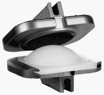
The ProDisc-C cervical disc is a metal-on-polyethylene articulating device (Fig. 160-4). This modular implant consists of two cobalt-chromium-molybdenum end plates and an ultra-high molecular weight polyethylene inlay that is preassembled into the inferior. The end plates of the prosthesis are initially secured to the vertebral bodies with central keels and have a plasma-sprayed titanium coating for long-term fixation stability.
Indications
Patients with cervical radiculopathy secondary to degenerative disc disease at one level with normal cervical spinal alignment and disc height who have failed conservative therapeutics are potential candidates for cervical arthroplasty.15 Factors to consider when selecting suitable candidates for arthroplasty include the disc height at the level of degeneration, inherent range of motion, and cervical alignment. Arthroplasty requires the dorsal elements to be intact and functional, so patients with suspected ligamentous or facet disease are not suitable candidates. Those with radiographic instability should be treated with an arthrodesis. Those with cervical kyphosis, cervical spondylolisthesis, severe multilevel spondylosis, previous laminectomy, osteoporosis, metabolic bone disease, or cervical trauma are not ideal either.16 Arthroplasty is contraindicated in the setting of significant segmental or global deformity. A recent history of infection or osteomyelitis precludes the use of a prosthetic disc device. Other relative contraindications include rheumatoid arthritis, renal failure, cancer, and chronic corticosteroid use. Multilevel cervical disc arthroplasty has not yet been evaluated prospectively.
Clinical Outcomes
There are currently seven artificial discs in the United States undergoing FDA IDE trials: Prestige ST, Prestige LP, Bryan, ProDisc-C, PCM (Cervitech, Inc., Rockaway, NJ), Kineflex (Spinal Motion, Inc., Mountain View, CA), and CerviCore (Stryker Spine, Allendale, NJ). To date, European and Australian clinical trials have demonstrated improved clinical outcomes with cervical arthroplasty compared with ACDF when using validated outcome measures such as the Neck Disability Index, an arm pain intensity visual analogue scale, and neurologic status.15,17–19 No reports of excessive wear debris, material fatigue, fracture, or failure have been reported in any of these trials.19–23 In the United States, published clinical data are available for the Prestige ST, Bryan, and ProDisc-C artificial discs.
The Prestige ST FDA IDE study was a prospective, multicenter, randomized trial comparing the Prestige ST artificial disc with an arthrodesis with allograft and ventral plate for symptomatic single-level cervical degenerative disc disease.24 Clinical, radiographic, and outcome measures were followed for a minimum of 2 years. Two hundred seventy-six patients with cervical disc replacement were compared with 265 patients with ACDF. At 2 years, the arthroplasty group had better clinical results and neurologic function than the ACDF group. At 12 and 24 months, Neck Disability Index scores improved in both treatment groups; however, scores for the arthroplasty group were two points higher than for the arthrodesis group. The arthroplasty group had a statistically higher rate of neurologic improvement as well as a lower rate of revision surgery and supplemental fixation. Implant removal was required in five patients with arthroplasty for persistent radiculopathy and neck pain. In each of these cases, the arthroplasty was revised to ACDF. The removal rate was lower in the arthroplasty group, but the difference was not statistically significant. The mean improvement in 36-Item Short Form Health Survey (SF-36) scores was greater in the arthroplasty group at 12 and 24 months. The rate of adjacent-segment reoperation was significantly lower in the arthroplasty group. Three patients in the arthroplasty group required surgery for adjacent-level disease. There were no cases of hardware fracture or migration during the follow-up period for the arthroplasty group. Arthroplasty was able to maintain sagittal motion on average more than 7 degrees.
Similarly, the Bryan cervical disc FDA IDE study was a prospective, multicenter, randomized trial comparing the Bryan artificial disc with an arthrodesis with allograft and ventral plate for symptomatic single-level cervical degenerative disc disease.25 In this study, 463 patients were randomized to arthroplasty (n = 242) or ACDF (n = 221). Clinical and radiographic evaluations were performed for 2 years after surgery. Pain and function were assessed using the Neck Disability Index, SF-36, and numeric rating scales for neck and arm pain. At 12 and 24 months, both groups showed improvement in all clinical outcome measures, although the arthroplasty group had a statistically greater improvement in the primary outcome measures, mainly the Neck Disability Index. Within the follow-up period, secondary surgical procedures at the treated level were performed in 2.5% of arthroplasty patients and 3.6% of ACDF patients. This difference was not statistically significant. In the arthroplasty group, there were three removals, two reoperations, and one revision. At 2 years after surgery, range of motion in the arthroplasty group was on average 8 degrees. No spontaneous fusions were noted in the arthroplasty group during the follow-up period. Fusion occurred in 94.3% of ACDF patients. Adjacent-level degeneration was not evaluated in this study.
Results from the prospective, randomized, multicenter ProDisc-C FDA IDE study were recently published.26 Similar to the previously discussed studies, the artificial disc was compared with ACDF for single-level cervical degenerative disease and patients were followed for 2 years. A total of 209 patients were enrolled, with 106 randomized to ACDF and 103 to ProDisc-C. Outcome measures used included neck and arm pain and intensity scale, Neck Disability Index, neurologic status, device success, adverse event occurrence, and SF-36 standardized questionnaires. Neck Disability Index and SF-36 scores were significantly lower compared with presurgery scores at all follow-up visits for both the treatment groups. Neck and arm pain intensity were statistically lower than preoperative levels but not different between treatments. Neurologic success was achieved at 24 months in 90.9% of ProDisc-C and 88.0% of ACDF patients. At 24 months after surgery, 84.4% of ProDisc-C patients achieved at least 4 degrees of motion or maintained motion relative to preoperative baseline at the operated level. There was a statistically significant difference in the number of revisions between the two groups: 8.5% of the ACDF group and 1.8% of the arthroplasty group underwent revisions. The ProDisc-C was successful in 73.5% of patients, versus 60.5% for ACDF at 2 years. Adjacent-level degeneration was not evaluated in this study.
As more FDA IDE studies continue to report their findings of efficacy, safety, and equivalence of various artificial discs to ACDF for single-level degenerative disc disease, further long-term follow-up studies are required to evaluate and scrutinize the ability of these implants to prevent adjacent-level degeneration and preserve motion. In the only published series with long-term follow-up, Robertson and Metcalf23 reported on the function of the Prestige I cervical disc after 4 years. The authors conducted clinical and radiographic examinations at 3 and 4 years after surgery to evaluate the long-term performance of the Prestige I device. Radiographic analysis in 14 patients showed that the Prestige I disc maintained motion at the treated segment at 3 and 4 years after surgery without the development of adjacent-level degeneration.
Avoidance of Complications
Complications related to surgical technique include those related to surgical approach and implantation technique. Approach-related complications are well known and are the same as those for ACDF, including infection, vascular damage, and dysphagia. Specific implantation technique-related complications have been reported and include implant migration, subsidence, sagittal split fractures, and dorsal vertebral avulsion fractures.27,28
Although published results of clinical trials have demonstrated and emphasized the clinical safety and efficacy of cervical arthroplasty, few reports have described complications associated with these implants. Few studies have shown heterotropic ossification as a potential complication of the artificial disc.29,30 Although this phenomenon has been well described in the orthopedic literature in the context of artificial hip and knee replacements, the exact cause has yet to be determined. In a European multicenter study of the Bryan cervical disc, 16 (17.8%) of the 90 studied patients developed prevertebral ossification at 12 months after implantation.29 Of these 16 patients, 6 developed heterotropic ossification extending across the vertebral end plate and eliminating motion at that segment. Less than 2 degrees of movement was shown in the other 10 patients. There was no association found between the development of these ossifications and clinical outcomes. It has been suggested that the amount of bone preparation involved with this implantation technique may predispose to heterotropic ossification. Although it has not been formally studied for cervical disc arthroplasty, the prophylactic use of nonsteroidal anti-inflammatory drugs has been suggested to reduce the incidence of heterotropic ossification.
Future Directions
The application of cervical arthroplasty continues to expand, with clinicians showing increasing interest in its use for the treatment of spondylotic myelopathy and multilevel degenerative disc disease. Novel applications for arthroplasty continue to be explored for an aging population with increasingly complex patterns of spinal degeneration. Reports have suggested a role for arthroplasty in treating adjacent-level disease for patients with previous multilevel anterior cervical fusions.31 Hybrid constructs consisting of arthroplasty and arthrodesis performed in the same setting for multilevel degenerative disease have also been reported, with early results suggesting safety and efficacy.32
With regard to spondylotic myelopathy, there is concern that motion preservation may maintain and perpetuate microtrauma to the spinal cord, thus defeating the advantage of arthroplasty. To date, the majority of the literature on cervical arthroplasty has been focused on the outcomes in patients with both radiculopathy and myelopathy as a mixed cohort.19,22,23 Although several studies have reported on the use of cervical arthroplasty for the treatment of myelopathy, these have been limited by a small number of patients and short follow-up.15,33,34 Sekhon reported a series of 11 patients who presented with cervical myelopathy and underwent Bryan disc replacement.33 Five patients were followed for 18 months or more and had improvements in Nurick grade by 1 and Neck Disability Index by 45%.
In a cross-sectional analysis of myelopathic patients who were enrolled in the FDA IDE prospective, randomized, multicenter trials of the Prestige ST and Bryan discs, myelopathic patients in both arthroplasty and arthrodesis groups had significant improvement after surgery, with no worsening myelopathy in the arthroplasty group.35 Patients included in the study were those with myelopathy and spondylosis or disc herniation at a single level from C3 to C7. General exclusion criteria included the presence of infection, metabolic bone disease, osteoporosis, ankylosing spondylitis, rheumatoid arthritis, and previous cervical surgery. Those with moderate to advanced cervical spondylosis, bridging syndesmophytes, marked reduction or absence of segmental spinal motion, disc space collapse greater than 50%, moderate or severe facet arthropathy, cervical kyphosis or reversal of lordosis, and 2 mm or more of spondylolisthesis and/or 11 degrees or more of angular instability relative to an adjacent segment or segments were also excluded. At 24 months, there were no significant differences in neurologic function or improvement in gait between the arthroplasty and arthrodesis groups. The Neck Disability Index, SF-36, arm and neck pain scores, and gait function improved at all time points. There were no revisions or implant failures in the arthroplasty groups.
Clarke M.J., Ecker R.D., Krauss W.E., et al. Same-segment and adjacent-segment disease following posterior cervical foraminotomy. J Neurosurg Spine. 2007;6:5-9.
Eck J.C., Humphreys S.C., Lim T.H., et al. Biomechanical study on the effect of cervical spine fusion on adjacent-level intradiscal pressure and segmental motion. Spine (Phila Pa 1976). 2002;27:2431-2434.
Goffin J., Geusens E., Vantomme N., et al. Long-term follow-up after interbody fusion of the cervical spine. J Spinal Disord Tech. 2004;17:79-85.
Hilibrand A.S., Carlson G.D., Palumbo M.A., et al. Radiculopathy and myelopathy at segments adjacent to the site of a previous anterior cervical arthrodesis. J Bone Joint Surg [Am]. 1999;81:519-528.
Maiman D.J., Kumaresan S., Yoganandan N., et al. Biomechanical effect of anterior cervical spine fusion on adjacent segments. Biomed Mater Eng. 1999;9:27-38.
Matsunaga S., Kabayama S., Yamamoto T., et al. Strain on intervertebral discs after anterior cervical decompression and fusion. Spine (Phila Pa 1976). 1999;24:670-675.
Robertson J.T., Metcalf N.H. Long-term outcome after implantation of the Prestige I disc in an end-stage indication: 4-year results from a pilot study. Neurosurg Focus. 2004;17:E10.
1. Smith G.W., Robinson R.A. The treatment of certain cervical-spine disorders by anterior removal of the intervertebral disc and interbody fusion. J Bone Joint Surg [Am]. 1958;40:607-624.
2. Kaiser M.G., Haid R.W.Jr., Subach B.R., et al. Anterior cervical plating enhances arthrodesis after discectomy and fusion with cortical allograft. Neurosurgery. 2002;50:229-236. discussion 236–238
3. Bartolomei J.C., Theodore N., Sonntag V.K. Adjacent level degeneration after anterior cervical fusion: a clinical review. Neurosurg Clin North Am. 2005;16:575-587. v
4. Maiman D.J., Kumaresan S., Yoganandan N., et al. Biomechanical effect of anterior cervical spine fusion on adjacent segments. Biomed Mater Eng. 1999;9:27-38.
5. DiAngelo D.J., Roberston J.T., Metcalf N.H., et al. Biomechanical testing of an artificial cervical joint and an anterior cervical plate. J Spinal Disord Tech. 2003;16:314-323.
6. Eck J.C., Humphreys S.C., Lim T.H., et al. Biomechanical study on the effect of cervical spine fusion on adjacent-level intradiscal pressure and segmental motion. Spine (Phila Pa 1976). 2002;27:2431-2434.
7. Hilibrand A.S., Carlson G.D., Palumbo M.A., et al. Radiculopathy and myelopathy at segments adjacent to the site of a previous anterior cervical arthrodesis. J Bone Joint Surg [Am]. 1999;81:519-528.
8. Goffin J., Geusens E., Vantomme N., et al. Long-term follow-up after interbody fusion of the cervical spine. J Spinal Disord Tech. 2004;17:79-85.
9. Matsunaga S., Kabayama S., Yamamoto T., et al. Strain on intervertebral discs after anterior cervical decompression and fusion. Spine (Phila Pa 1976). 1999;24:670-675.
10. Clarke M.J., Ecker R.D., Krauss W.E., et al. Same-segment and adjacent-segment disease following posterior cervical foraminotomy. J Neurosurg Spine. 2007;6:5-9.
11. Wigfield C.C., Skrzypiec D., Jackowski A., et al. Internal stress distribution in cervical intervertebral discs: the influence of an artificial cervical joint and simulated anterior interbody fusion. J Spinal Disord Tech. 2003;16:441-449.
12. Mummaneni P.V., Haid R.W. The future in the care of the cervical spine: interbody fusion and arthroplasty. Invited submission from the Joint Section Meeting on Disorders of the Spine and Peripheral Nerves, March 2004. J Neurosurg Spine. 2004;1:155-159.
13. Mummaneni P.V., Robinson J.C., Haid R.W.Jr. Cervical arthroplasty with the PRESTIGE LP cervical disc. Neurosurgery. 2007;60:310-314. discussion 314–315
14. Puttlitz C.M., DiAngelo D.J. Cervical spine arthroplasty biomechanics. Neurosurg Clin North Am. 2005;16:589-594. v
15. Sekhon L.H. Cervical arthroplasty in the management of spondylotic myelopathy. J Spinal Disord Tech. 2003;16:307-313.
16. Duggal N., Pickett G.E., Mitsis D.K., et al. Early clinical and biomechanical results following cervical arthroplasty. Neurosurg Focus. 2004;17:E9.
17. Cummins B.H., Robertson J.T., Gill S.S. Surgical experience with an implanted artificial cervical joint. J Neurosurg. 1998;88:943-948.
18. Goffin J., Casey A., Kehr P., et al. Preliminary clinical experience with the Bryan Cervical Disc Prosthesis. Neurosurgery. 2002;51:840-845. discussion 845–847
19. Goffin J., Van Calenbergh F., van Loon J., et al. Intermediate follow-up after treatment of degenerative disc disease with the Bryan Cervical Disc Prosthesis: single-level and bi-level. Spine (Phila Pa 1976). 2003;28:2673-2678.
20. Bertagnoli R., Yue J.J., Pfeiffer F., et al. Early results after ProDisc-C cervical disc replacement. J Neurosurg Spine. 2005;2:403-410.
21. Lafuente J., Casey A.T., Petzold A., et al. The Bryan cervical disc prosthesis as an alternative to arthrodesis in the treatment of cervical spondylosis: 46 consecutive cases. J Bone Joint Surg [Br]. 2005;87:508-512.
22. Porchet F., Metcalf N.H. Clinical outcomes with the Prestige II cervical disc: preliminary results from a prospective randomized clinical trial. Neurosurg Focus. 2004;17:E6.
23. Robertson J.T., Metcalf N.H. Long-term outcome after implantation of the Prestige I disc in an end-stage indication: 4-year results from a pilot study. Neurosurg Focus. 2004;17:E10.
24. Mummaneni P.V., Burkus J.K., Haid R.W., et al. Clinical and radiographic analysis of cervical disc arthroplasty compared with allograft fusion: a randomized controlled clinical trial. J Neurosurg Spine. 2007;6:198-209.
25. Heller J.G., Sasso R.C., Papadopoulos S.M., et al. Comparison of BRYAN cervical disc arthroplasty with anterior cervical decompression and fusion: clinical and radiographic results of a randomized, controlled, clinical trial. Spine (Phila Pa 1976). 2009;34:101-107.
26. Murrey D., Janssen M., Delamarter R., et al. Results of the prospective, randomized, controlled multicenter Food and Drug Administration investigational device exemption study of the ProDisc-C total disc replacement versus anterior discectomy and fusion for the treatment of 1-level symptomatic cervical disc disease. Spine J. 2009;9:275-286.
27. Datta J.C., Janssen M.E., Beckham R., et al. Sagittal split fractures in multilevel cervical arthroplasty using a keeled prosthesis. J Spinal Disord Tech. 2007;20:89-92.
28. Shim C.S., Shin H.D., Lee S.H. Posterior avulsion fracture at adjacent vertebral body during cervical disc replacement with ProDisc-C: a case report. J Spinal Disord Tech. 2007;20:468-472.
29. Leung C., Casey A.T., Goffin J., et al. Clinical significance of heterotopic ossification in cervical disc replacement: a prospective multicenter clinical trial. Neurosurgery. 2005;57:759-763. discussion 763
30. Mehren C., Suchomel P., Grochulla F., et al. Heterotopic ossification in total cervical artificial disc replacement. Spine (Phila Pa 1976). 2006;31:2802-2806.
31. Mobbs R.J., Mehan N., Khong P. Cervical arthroplasty for myelopathy adjacent to previous multisegmental fusion: early results and review of the literature of a novel hybrid surgical technique combining cervical arthrodesis and disc arthroplasty for treating multilevel degenerative disc disease: opposite or complementary techniques? Eur Spine J. 2009;18(Suppl 1):29-39.
33. Sekhon L.H. Cervical arthroplasty in the management of spondylotic myelopathy: 18-month results. Neurosurg Focus. 2004;17:E8.
34. Sekhon L.H. Two-level artificial disc placement for spondylotic cervical myelopathy. J Clin Neurosci. 2004;11:412-415.
35. Buchowski J.M., Anderson P.A., Sekhon L., et al. Cervical disc arthroplasty compared with arthrodesis for the treatment of myelopathy: surgical technique. J Bone Joint Surg [Am]. 2009;91(Suppl 2):223-232.
[/level-membership-for-neurosurgery-category][not-level-membership-for-neurosurgery-category]
Chapter 160 Cervical Total Disc Arthroplasty
Anterior cervical discectomy and fusion (ACDF) has been routinely used for the treatment of degenerative disease of the cervical spine since the 1950s.1 Interbody fusion has become the accepted method of treating cervical radiculopathy, myelopathy, segmental deformity, instability, and degenerative disc disease. With the evolution of technique and technology, fusion rates have exceeded 95%.2 Despite these results, concern for the development of adjacent-level degeneration and disease after fusion has prompted interest in and development of implants designed to preserve motion and maintain anatomic disc space height and normal segmental lordosis. In this chapter, we review the indications for cervical total disc arthroplasty and discuss the preliminary data, with an emphasis on complications and their avoidance. A full discussion of each of the developed artificial discs is beyond the scope of this chapter. Implants for which U.S. Food and Drug Administration (FDA) Investigational Device Exemptions (IDE) studies have been completed are discussed herein.
Rationale for Arthroplasty
The long-term clinical success of ACDF has been limited by the development of adjacent-level degeneration, leading to further symptoms and possibly additional surgery. Adjacent-level degeneration is defined as radiographic changes at a segment adjacent to a fusion and is not necessarily associated with symptoms. Adjacent-level disease, however, is associated with pain, radiculopathy, or myelopathy.3 Biomechanical studies have demonstrated that cervical fusion alters adjacent-level kinematics and that the loss of motion at the fused segment is compensated by an increase in motion and intradiscal pressure at adjacent levels.4–6 Some clinical studies have supported these findings, giving weight to this theory of fusion-induced degeneration. Hilibrand et al.7 reported that repeat surgical intervention was necessary for 2.9% of patients per year for symptomatic adjacent-segment disease after ACDF. They also found that 25% of patients reported symptoms from adjacent-segment disease 10 years after ACDF. In a comparative study, Goffin et al.8 showed that 92% of fused patients demonstrated radiographic evidence of adjacent-segment degeneration 5 years after surgery for both cervical spondylosis and trauma, suggesting that adjacent-level degeneration is accelerated by fusion and not necessarily the natural history of preexisting degenerative disc disease. It has been postulated that adjacent-level degeneration may be attributable to higher shear strains at levels adjacent to fusions.9
Debate continues as to whether adjacent-level degeneration is truly due to increased loads after fusion or to the natural history of degenerative disc disease. In a retrospective study of 303 patients undergoing single-level dorsal cervical foraminotomy for cervical radiculopathy, Clarke et al.10 showed that 15 (4.9%) of 303 developed symptomatic adjacent-level disease with a 0.7% annual rate for development of adjacent-level disease. The 10-year cumulative incidence of adjacent-level disease was 6.7%.
Biomechanical studies have suggested that cervical arthroplasty may allow for a more normal restoration of load transfer and kinematics at adjacent levels compared with fusion.5 After disc replacement, stress profiles in specimens at adjacent-level discs were similar to those of intact, nontreated levels, with reduced stresses in the adjacent-level anulus compared with spines with simulated fusions.11 Cervical disc arthroplasty may therefore be beneficial to patients because in theory it prevents the development of adjacent-segment disease by preserving normal neck motion.
Nomenclature and Design
With the development of numerous artificial discs in the last decade, the Cervical Spine Study Group developed a nomenclature system for cervical arthroplasty.12 Artificial discs are categorized by material, articulation, fixation, design, and kinematics.13 They can be classified as nonarticulating, uniarticulating, or biarticulating. Various designs include metal on metal (Prestige ST and LP; Medtronic Sofamor Danek, Memphis, TN), metal on polymer (Bryan, Medtronic Sofamor Danek; ProDisc-C, Synthes Spine, West Chester, PA), ceramic on polymer, or ceramic on ceramic. Discs are either modular with replaceable parts, or nonmodular. Some have points for vertebral body fixation and some have surfaces that promote bone ingrowth at the disc–end-plate interface. With regard to motion, they may be constrained, semiconstrained, or unconstrained. Constrained devices restrict motion to less than that seen physiologically, semiconstrained devices allow physiologic motion, and unconstrained devices rely on soft tissue and the inherent compression across the disc space to limit motion.14 Each of these devices is available in a range of heights and depths to accommodate individual variances in anatomy.
The Prestige ST artificial disc is constructed of stainless steel and consists of two articulating components attached to the cervical vertebrae with screws (Fig. 160-1). The ball-and-trough design provides relatively unconstrained motion compared with that of a normal cervical spinal segment. The surfaces of the device contacting the end plates are grit-blasted to promote bone osseointegration.
FIGURE 160-1 Prestige ST artificial cervical disc.
(Courtesy of Medtronic Sofamor Danek, Memphis, TN.)
The Prestige LP is the latest version of the Prestige artificial discs and differs from previous models in that fixation to the vertebral bodies is achieved by a set of rails that are imbedded in the vertebral end plates (Fig. 160-2). It is manufactured from a titanium-ceramic composite material that is highly durable and produces little artifact on CT and MRI. The ball-and-trough design is identical to that of the Prestige ST. A porous titanium plasma spray coating on the end-plate surface facilitates bone ingrowth and long-term fixation.
FIGURE 160-2 Prestige LP artificial cervical disc.
(Courtesy of Medtronic Sofamor Danek, Memphis, TN.)
The Bryan cervical disc is designed to allow for motion similar to the normal cervical spine and resembles an actual disc (Fig. 160-3). It consists of two titanium alloy shells with a polyurethane nucleus. The bone-implant interface of each shell has an applied porous coating to facilitate ingrowth of bone and promote long-term stability. The nucleus is surrounded with a polyurethane sheath to establish a closed articulation. Sterile saline is injected into this sheath and functions as an initial lubricant. Titanium alloy seal plugs allow for retention of the saline lubricant. Small ventral flanges on the shells serve as handles to grasp the device for insertion.
The ProDisc-C cervical disc is a metal-on-polyethylene articulating device (Fig. 160-4). This modular implant consists of two cobalt-chromium-molybdenum end plates and an ultra-high molecular weight polyethylene inlay that is preassembled into the inferior. The end plates of the prosthesis are initially secured to the vertebral bodies with central keels and have a plasma-sprayed titanium coating for long-term fixation stability.
Indications
Patients with cervical radiculopathy secondary to degenerative disc disease at one level with normal cervical spinal alignment and disc height who have failed conservative therapeutics are potential candidates for cervical arthroplasty.15 Factors to consider when selecting suitable candidates for arthroplasty include the disc height at the level of degeneration, inherent range of motion, and cervical alignment. Arthroplasty requires the dorsal elements to be intact and functional, so patients with suspected ligamentous or facet disease are not suitable candidates. Those with radiographic instability should be treated with an arthrodesis. Those with cervical kyphosis, cervical spondylolisthesis, severe multilevel spondylosis, previous laminectomy, osteoporosis, metabolic bone disease, or cervical trauma are not ideal either.16 Arthroplasty is contraindicated in the setting of significant segmental or global deformity. A recent history of infection or osteomyelitis precludes the use of a prosthetic disc device. Other relative contraindications include rheumatoid arthritis, renal failure, cancer, and chronic corticosteroid use. Multilevel cervical disc arthroplasty has not yet been evaluated prospectively.
Clinical Outcomes
There are currently seven artificial discs in the United States undergoing FDA IDE trials: Prestige ST, Prestige LP, Bryan, ProDisc-C, PCM (Cervitech, Inc., Rockaway, NJ), Kineflex (Spinal Motion, Inc., Mountain View, CA), and CerviCore (Stryker Spine, Allendale, NJ). To date, European and Australian clinical trials have demonstrated improved clinical outcomes with cervical arthroplasty compared with ACDF when using validated outcome measures such as the Neck Disability Index, an arm pain intensity visual analogue scale, and neurologic status.15,17–19 No reports of excessive wear debris, material fatigue, fracture, or failure have been reported in any of these trials.19–23 In the United States, published clinical data are available for the Prestige ST, Bryan, and ProDisc-C artificial discs.
[/not-level-membership-for-neurosurgery-category]
