Central Nervous System Infections

Published on 25/03/2015 by admin

Filed under Pediatrics

Last modified 22/04/2025

Print this page

rate 1 star rate 2 star rate 3 star rate 4 star rate 5 star
Your rating: none, Average: 0 (0 votes)

This article have been viewed 2081 times

Chapter 595 Central Nervous System Infections

Infection of the central nervous system (CNS) is the most common cause of fever associated with signs and symptoms of CNS disease in children. Many microorganisms can cause infection. Nonetheless, specific pathogens are identifiable and are influenced by the age and immune status of the host and the epidemiology of the pathogen. In general, viral infections of the CNS are much more common than bacterial infections, which, in turn, are more common than fungal and parasitic infections. Infections caused by rickettsiae (Rocky Mountain spotted fever, Ehrlichia) are relatively uncommon but assume important roles under certain epidemiologic circumstances. Mycoplasma spp. can also cause infections of the CNS, although their precise contribution is often difficult to determine.

Regardless of etiology, most patients with CNS infection have similar clinical manifestations. Common symptoms include headache, nausea, vomiting, anorexia, restlessness, altered state of consciousness, and irritability; most of these symptoms are nonspecific. Common signs of CNS infection, in addition to fever, include photophobia, neck pain and rigidity, obtundation, stupor, coma, seizures, and focal neurologic deficits. The severity and constellation of signs are determined by the specific pathogen, the host, and the area of the CNS affected.

Infection of the CNS may be diffuse or focal. Meningitis and encephalitis are examples of diffuse infection. Meningitis implies primary involvement of the meninges, whereas encephalitis indicates brain parenchymal involvement. Because these anatomic boundaries are often not distinct, many patients have evidence of both meningeal and parenchymal involvement and should be considered to have meningoencephalitis. Brain abscess is the best example of a focal infection of the CNS. The neurologic expression of this infection is determined by the site and extent of the abscess(es) (Chapter 596).

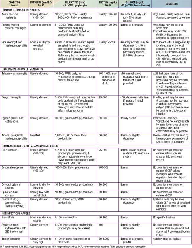
The diagnosis of diffuse CNS infections depends on examination of cerebrospinal fluid (CSF) obtained by lumbar puncture (LP). Table 595-1 provides an overview of the expected CSF abnormalities with various CNS disorders.

595.1 Acute Bacterial Meningitis Beyond the Neonatal Period

Bacterial meningitis is one of the most potentially serious infections occurring in infants and older children. This infection is associated with a high rate of acute complications and risk of long-term morbidity. The incidence of bacterial meningitis is sufficiently high in febrile infants that it should be included in the differential diagnosis of those with altered mental status and other evidence of neurologic dysfunction.

Epidemiology

A major risk factor for meningitis is the lack of immunity to specific pathogens associated with young age. Additional risks include recent colonization with pathogenic bacteria, close contact (household, daycare centers, college dormitories, military barracks) with individuals having invasive disease caused by N. meningitidis and H. influenzae type b, crowding, poverty, black or Native American race, and male gender. The mode of transmission is probably person-to-person contact through respiratory tract secretions or droplets. The risk of meningitis is increased among infants and young children with occult bacteremia; the odds ratio is greater for meningococcus (85 times) and H. influenzae type b (12 times) relative to that for pneumococcus.

Specific host defense defects due to altered immunoglobulin production in response to encapsulated pathogens may be responsible for the increased risk of bacterial meningitis in Native Americans and Eskimos. Defects of the complement system (C5-C8) have been associated with recurrent meningococcal infection, and defects of the properdin system have been associated with a significant risk of lethal meningococcal disease. Splenic dysfunction (sickle cell anemia) or asplenia (due to trauma, or congenital defect) is associated with an increased risk of pneumococcal, H. influenzae type b (to some extent), and, rarely, meningococcal sepsis and meningitis. T-lymphocyte defects (congenital or acquired by chemotherapy, AIDS, or malignancy) are associated with an increased risk of L. monocytogenes infections of the CNS.

Congenital or acquired CSF leak across a mucocutaneous barrier, such as cranial or midline facial defects (cribriform plate) and middle ear (stapedial foot plate) or inner ear fistulas (oval window, internal auditory canal, cochlear aqueduct), or CSF leakage through a rupture of the meninges due to a basal skull fracture into the cribriform plate or paranasal sinus, is associated with an increased risk of pneumococcal meningitis. The risk of bacterial meningitis, caused by S. pneumoniae, in children with cochlear implants, used for the treatment of hearing loss, is more than 30 times the risk in the general U.S. population. Lumbosacral dermal sinus and meningomyelocele are associated with staphylococcal and gram-negative enteric bacterial meningitis. CSF shunt infections increase the risk of meningitis due to staphylococci (especially coagulase-negative species) and other low virulence bacteria that typically colonize the skin.

Neisseria Meningitidis (Chapter 184)

Five serogroups of meningococcus, A, B, C, Y, and W-135, are responsible for disease. Meningococcal meningitis may be sporadic or may occur in epidemics. In the USA, serogroups B, C, and Y each account for ≈30% of cases, although serogroup distribution varies by location and time. Epidemic disease, especially in developing countries, is usually caused by serogroup A. Cases occur throughout the year but may be more common in the winter and spring and following influenza virus infections. Nasopharyngeal carriage of N. meningitidis occurs in 1-15% of adults. Colonization may last weeks to months; recent colonization places nonimmune younger children at greatest risk for meningitis. The incidence of disease occurring in association with an index case in the family is 1%, a rate that is 1,000-fold the risk in the general population. The risk of secondary cases occurring in contacts at daycare centers is about 1/1,000. Most infections of children are acquired from a contact in a daycare facility, a colonized adult family member, or an ill patient with meningococcal disease. Children younger than 5 yr have the highest rates of meningococcal infection. A 2nd peak in incidence occurs in persons between 15 and 24 yr of age. College freshmen living in dormitories have an increased incidence of infection compared to non–college-attending, age-matched controls.

The Centers for Disease Control and Prevention (CDC) recommends vaccination against meningococcus with 1 dose of a quadrivalent conjugate meningococcal vaccine between the ages of 11 and 18 yr and for persons 2-10 yr who are at increased risk for meningococcal disease. College freshmen living in dormitories who have not been previously vaccinated should also be vaccinated with MCV4.

Pathology and Pathophysiology

A meningeal purulent exudate of varying thickness may be distributed around the cerebral veins, venous sinuses, convexity of the brain, and cerebellum and in the sulci, sylvian fissures, basal cisterns, and spinal cord. Ventriculitis with bacteria and inflammatory cells in ventricular fluid may be present (more often in neonates), as may subdural effusions and, rarely, empyema. Perivascular inflammatory infiltrates also may be present, and the ependymal membrane may be disrupted. Vascular and parenchymal cerebral changes characterized by polymorphonuclear infiltrates extending to the subintimal region of the small arteries and veins, vasculitis, thrombosis of small cortical veins, occlusion of major venous sinuses, necrotizing arteritis producing subarachnoid hemorrhage, and, rarely, cerebral cortical necrosis in the absence of identifiable thrombosis have been described at autopsy. Cerebral infarction, resulting from vascular occlusion due to inflammation, vasospasm, and thrombosis, is a frequent sequela. Infarct size ranges from microscopic to involvement of an entire hemisphere.

Inflammation of spinal nerves and roots produces meningeal signs, and inflammation of the cranial nerves produces cranial neuropathies of optic, oculomotor, facial, and auditory nerves. Increased intracranial pressure (ICP) also produces oculomotor nerve palsy due to the presence of temporal lobe compression of the nerve during tentorial herniation. Abducens nerve palsy may be a nonlocalizing sign of elevated ICP.

Increased ICP is due to cell death (cytotoxic cerebral edema), cytokine-induced increased capillary vascular permeability (vasogenic cerebral edema), and, possibly, increased hydrostatic pressure (interstitial cerebral edema) after obstructed reabsorption of CSF in the arachnoid villus or obstruction of the flow of fluid from the ventricles. ICP may exceed 300 mm H2O; cerebral perfusion may be further compromised if the cerebral perfusion pressure (mean arterial pressure minus ICP) is <50 cm H2O due to systemic hypotension with reduced cerebral blood flow. The syndrome of inappropriate antidiuretic hormone secretion (SIADH) may produce excessive water retention and potentially increase the risk of elevated ICP (Chapter 553). Hypotonicity of brain extracellular spaces may cause cytotoxic edema after cell swelling and lysis. Tentorial, falx, or cerebellar herniation does not usually occur because the increased ICP is transmitted to the entire subarachnoid space and there is little structural displacement. Furthermore, if the fontanels are still patent, increased ICP is not always dissipated.

Hydrocephalus can occur as an acute complication of bacterial meningitis. It most often takes the form of a communicating hydrocephalus due to adhesive thickening of the arachnoid villi around the cisterns at the base of the brain. Thus, there is interference with the normal resorption of CSF. Less often, obstructive hydrocephalus develops after fibrosis and gliosis of the aqueduct of Sylvius or the foramina of Magendie and Luschka.

Raised CSF protein levels are due in part to increased vascular permeability of the blood-brain barrier and the loss of albumin-rich fluid from the capillaries and veins traversing the subdural space. Continued transudation may result in subdural effusions, usually found in the later phase of acute bacterial meningitis. Hypoglycorrhachia (reduced CSF glucose levels) is due to decreased glucose transport by the cerebral tissue.

Damage to the cerebral cortex may be due to the focal or diffuse effects of vascular occlusion (infarction, necrosis, lactic acidosis), hypoxia, bacterial invasion (cerebritis), toxic encephalopathy (bacterial toxins), elevated ICP, ventriculitis, and transudation (subdural effusions). These pathologic factors result in the clinical manifestations of impaired consciousness, seizures, cranial nerve deficits, motor and sensory deficits, and later psychomotor retardation.

Pathogenesis

Bacterial meningitis most commonly results from hematogenous dissemination of microorganisms from a distant site of infection; bacteremia usually precedes meningitis or occurs concomitantly. Bacterial colonization of the nasopharynx with a potentially pathogenic microorganism is the usual source of the bacteremia. There may be prolonged carriage of the colonizing organisms without disease or, more likely, rapid invasion after recent colonization. Prior or concurrent viral upper respiratory tract infection may enhance the pathogenicity of bacteria producing meningitis.

N. meningitidis and H. influenzae type b attach to mucosal epithelial cell receptors by pili. After attachment to epithelial cells, bacteria breach the mucosa and enter the circulation. N. meningitidis may be transported across the mucosal surface within a phagocytic vacuole after ingestion by the epithelial cell. Bacterial survival in the bloodstream is enhanced by large bacterial capsules that interfere with opsonic phagocytosis and are associated with increased virulence. Host-related developmental defects in bacterial opsonic phagocytosis also contribute to the bacteremia. In young, nonimmune hosts, the defect may be due to an absence of preformed IgM or IgG anticapsular antibodies, whereas in immunodeficient patients, various deficiencies of components of the complement or properdin system may interfere with effective opsonic phagocytosis. Splenic dysfunction may also reduce opsonic phagocytosis by the reticuloendothelial system.

Bacteria gain entry to the CSF through the choroid plexus of the lateral ventricles and the meninges and then circulate to the extracerebral CSF and subarachnoid space. Bacteria rapidly multiply because the CSF concentrations of complement and antibody are inadequate to contain bacterial proliferation. Chemotactic factors then incite a local inflammatory response characterized by polymorphonuclear cell infiltration. The presence of bacterial cell wall lipopolysaccharide (endotoxin) of gram-negative bacteria (H. influenzae type b, N. meningitidis) and of pneumococcal cell wall components (teichoic acid, peptidoglycan) stimulates a marked inflammatory response, with local production of tumor necrosis factor, interleukin 1, prostaglandin E, and other inflammatory mediators. The subsequent inflammatory response is characterized by neutrophilic infiltration, increased vascular permeability, alterations of the blood-brain barrier, and vascular thrombosis. Meningitis-associated brain injury is not simply caused by viable bacteria but occurs as a consequence of the host reaction to the inflammatory cascade initiated by bacterial components.

Rarely, meningitis may follow bacterial invasion from a contiguous focus of infection such as paranasal sinusitis, otitis media, mastoiditis, orbital cellulitis, or cranial or vertebral osteomyelitis or may occur after introduction of bacteria via penetrating cranial trauma, dermal sinus tracts, or meningomyeloceles.

Clinical Manifestations

The onset of acute meningitis has 2 predominant patterns. The more dramatic and, fortunately, less common presentation is sudden onset with rapidly progressive manifestations of shock, purpura, disseminated intravascular coagulation (DIC), and reduced levels of consciousness often resulting in progression to coma or death within 24 hr. More often, meningitis is preceded by several days of fever accompanied by upper respiratory tract or gastrointestinal symptoms, followed by nonspecific signs of CNS infection such as increasing lethargy and irritability.

The signs and symptoms of meningitis are related to the nonspecific findings associated with a systemic infection and to manifestations of meningeal irritation. Nonspecific findings include fever, anorexia and poor feeding, headache, symptoms of upper respiratory tract infection, myalgias, arthralgias, tachycardia, hypotension, and various cutaneous signs, such as petechiae, purpura, or an erythematous macular rash. Meningeal irritation is manifested as nuchal rigidity, back pain, Kernig sign (flexion of the hip 90 degrees with subsequent pain with extension of the leg), and Brudzinski sign (involuntary flexion of the knees and hips after passive flexion of the neck while supine). In some children, particularly in those younger than 12-18 mo, Kernig and Brudzinski signs are not consistently present. Indeed fever, headache, and nuchal rigidity are present in only 40% of adults with bacterial meningitis. Increased ICP is suggested by headache, emesis, bulging fontanel or diastasis (widening) of the sutures, oculomotor (anisocoria, ptosis) or abducens nerve paralysis, hypertension with bradycardia, apnea or hyperventilation, decorticate or decerebrate posturing, stupor, coma, or signs of herniation. Papilledema is uncommon in uncomplicated meningitis and should suggest a more chronic process, such as the presence of an intracranial abscess, subdural empyema, or occlusion of a dural venous sinus. Focal neurologic signs usually are due to vascular occlusion. Cranial neuropathies of the ocular, oculomotor, abducens, facial, and auditory nerves may also be due to focal inflammation. Overall, about 10-20% of children with bacterial meningitis have focal neurologic signs.

Seizures (focal or generalized) due to cerebritis, infarction, or electrolyte disturbances occur in 20-30% of patients with meningitis. Seizures that occur on presentation or within the 1st 4 days of onset are usually of no prognostic significance. Seizures that persist after the 4th day of illness and those that are difficult to treat may be associated with a poor prognosis.

Alterations of mental status are common among patients with meningitis and may be due to increased ICP, cerebritis, or hypotension; manifestations include irritability, lethargy, stupor, obtundation, and coma. Comatose patients have a poor prognosis. Additional manifestations of meningitis include photophobia and tache cérébrale, which is elicited by stroking the skin with a blunt object and observing a raised red streak within 30-60 sec.

Diagnosis

The diagnosis of acute pyogenic meningitis is confirmed by analysis of the CSF, which typically reveals microorganisms on Gram stain and culture, a neutrophilic pleocytosis, elevated protein, and reduced glucose concentrations (see Table 595-1). LP should be performed when bacterial meningitis is suspected. Contraindications for an immediate LP include (1) evidence of increased ICP (other than a bulging fontanel), such as 3rd or 6th cranial nerve palsy with a depressed level of consciousness, or hypertension and bradycardia with respiratory abnormalities (Chapter 584); (2) severe cardiopulmonary compromise requiring prompt resuscitative measures for shock or in patients in whom positioning for the LP would further compromise cardiopulmonary function; and (3) infection of the skin overlying the site of the LP. Thrombocytopenia is a relative contraindication for LP. If an LP is delayed, empirical antibiotic therapy should be initiated. CT scanning for evidence of a brain abscess or increased ICP should not delay therapy. LP may be performed after increased ICP has been treated or a brain abscess has been excluded.

Blood cultures should be performed in all patients with suspected meningitis. Blood cultures reveal the responsible bacteria in up to 80-90% of cases of meningitis.

Lumbar Puncture (Chapter 584)

The CSF leukocyte count in bacterial meningitis usually is elevated to >1,000/mm3 and, typically, there is a neutrophilic predominance (75-95%). Turbid CSF is present when the CSF leukocyte count exceeds 200-400/mm3. Normal healthy neonates may have as many as 30 leukocytes/mm3 (usually <10), but older children without viral or bacterial meningitis have <5 leukocytes/mm3 in the CSF; in both age groups there is a predominance of lymphocytes or monocytes.

A CSF leukocyte count <250/mm3 may be present in as many as 20% of patients with acute bacterial meningitis; pleocytosis may be absent in patients with severe overwhelming sepsis and meningitis and is a poor prognostic sign. Pleocytosis with a lymphocyte predominance may be present during the early stage of acute bacterial meningitis; conversely, neutrophilic pleocytosis may be present in patients in the early stages of acute viral meningitis. The shift to lymphocytic-monocytic predominance in viral meningitis invariably occurs within 8 to 24 hr of the initial LP. The Gram stain is positive in 70-90% of patients with untreated bacterial meningitis.

A diagnostic conundrum in the evaluation of children with suspected bacterial meningitis is the analysis of CSF obtained from children already receiving antibiotic (usually oral) therapy. This is an important issue, because 25-50% of children being evaluated for bacterial meningitis are receiving oral antibiotics when their CSF is obtained. CSF obtained from children with bacterial meningitis, after the initiation of antibiotics, may be negative on Gram stain and culture. Pleocytosis with a predominance of neutrophils, elevated protein level, and a reduced concentration of CSF glucose usually persist for several days after the administration of appropriate intravenous antibiotics. Therefore, despite negative cultures, the presumptive diagnosis of bacterial meningitis can be made. Some clinicians test CSF for the presence of bacterial antigens if the child has been pretreated with antibiotics and the diagnosis of bacterial meningitis is in doubt. These tests have technical limitations.

A traumatic LP may complicate the diagnosis of meningitis. Repeat LP at a higher interspace may produce less hemorrhagic fluid, but this fluid usually also contains red blood cells. Interpretation of CSF leukocytes and protein concentration are affected by LPs that are traumatic, although the Gram stain, culture, and glucose level may not be influenced. Although methods for correcting for the presence of red blood cells have been proposed, it is prudent to rely on the bacteriologic results rather than attempt to interpret the CSF leukocyte and protein results of a traumatic LP.

Differential Diagnosis

In addition to S. pneumoniae, N. meningitidis, and H. influenzae type b, many other microorganisms can cause generalized infection of the CNS with similar clinical manifestations. These organisms include less typical bacteria, such as Mycobacterium tuberculosis, Nocardia spp., Treponema pallidum (syphilis), and Borrelia burgdorferi (Lyme disease); fungi, such as those endemic to specific geographic areas (Coccidioides, Histoplasma, and Blastomyces) and those responsible for infections in compromised hosts (Candida, Cryptococcus, and Aspergillus); parasites, such as Toxoplasma gondii and those that cause cysticercosis and, most frequently, viruses (Chapter 595.2) (Table 595-2). Focal infections of the CNS including brain abscess and parameningeal abscess (subdural empyema, cranial and spinal epidural abscess) may also be confused with meningitis. In addition, noninfectious illnesses can cause generalized inflammation of the CNS. Relative to infections, these disorders are uncommon and include malignancy, collagen vascular syndromes, and exposure to toxins (see Table 595-2).

Table 595-2 CLINICAL CONDITIONS AND INFECTIOUS AGENTS ASSOCIATED WITH ASEPTIC MENINGITIS

VIRUSES

BACTERIA

BACTERIAL PARAMENINGEAL FOCUS

FUNGI

PARASITES (EOSINOPHILIC)

PARASITES (NONEOSINOPHILIC)

POSTINFECTIOUS

SYSTEMIC OR IMMUNOLOGICALLY MEDIATED

MALIGNANCY

DRUGS

MISCELLANEOUS

Compiled from Cherry JD: Aseptic meningitis and viral meningitis. In Feigin RD, Cherry JD, editors: Textbook of pediatric infectious diseases, ed 4, Philadelphia, 1998, WB Saunders, p 450; Davis LE: Aseptic and viral meningitis. In Long SS, Pickering LK, Prober CG, editors: Principles and practice of pediatric infectious disease, New York, 1997, Churchill Livingstone, p 329; Kliegman RM, Greenbaum LA, Lye PS: Practical strategies in pediatric diagnosis therapy, ed 2, Philadelphia, 2004, Elsevier, p 961.

Determining the specific cause of CNS infection is facilitated by careful examination of the CSF with specific stains (Kinyoun carbol fuchsin for mycobacteria, India ink for fungi), cytology, antigen detection (Cryptococcus), serology (syphilis, West Nile virus, arboviruses), viral culture (enterovirus), and polymerase chain reaction (herpes simplex, enterovirus, and others). Other potentially valuable diagnostic tests include blood cultures, CT or MRI of the brain, serologic tests, and, rarely, brain biopsy.

Acute viral meningoencephalitis is the most likely infection to be confused with bacterial meningitis (Tables 595-2 and 595-3). Although, in general, children with viral meningoencephalitis appear less ill than those with bacterial meningitis, both types of infection have a spectrum of severity. Some children with bacterial meningitis may have relatively mild signs and symptoms, whereas some with viral meningoencephalitis may be critically ill. Although classic CSF profiles associated with bacterial versus viral infection tend to be distinct (see Table 595-1), specific test results may have considerable overlap.

Table 595-3 CLASSIFICATION OF ENCEPHALITIS BY CAUSE AND SOURCE

CNS, central nervous system.

Modified from Behrman RE, editor: Nelson textbook of pediatrics, ed 14, Philadelphia, 1992, WB Saunders, p 667. From Kliegman RM, Greenbaum LA, Lye PS: Practical strategies in pediatric diagnosis and therapy, ed 2, Philadelphia, 2004, Elsevier, p 967.

Treatment

The therapeutic approach to patients with presumed bacterial meningitis depends on the nature of the initial manifestations of the illness. A child with rapidly progressing disease of less than 24 hr duration, in the absence of increased ICP, should receive antibiotics as soon as possible after an LP is performed. If there are signs of increased ICP or focal neurologic findings, antibiotics should be given without performing an LP and before obtaining a CT scan. Increased ICP should be treated simultaneously (Chapter 63). Immediate treatment of associated multiple organ system failure, shock (Chapter 64), and acute respiratory distress syndrome (Chapter 65) is also indicated.

Patients who have a more protracted subacute course and become ill over a 4-7 day period should also be evaluated for signs of increased ICP and focal neurologic deficits. Unilateral headache, papilledema, and other signs of increased ICP suggest a focal lesion such as a brain or epidural abscess, or subdural empyema. Under these circumstances, antibiotic therapy should be initiated before LP and CT scanning. If signs of increased ICP and/or focal neurologic signs are present, CT scanning should be performed first to determine the safety of performing an LP.

Initial Antibiotic Therapy

The initial (empirical) choice of therapy for meningitis in immunocompetent infants and children is primarily influenced by the antibiotic susceptibilities (Table 595-4) of S. pneumoniae. Selected antibiotics should achieve bactericidal levels in the CSF. Although there are substantial geographic differences in the frequency of resistance of S. pneumoniae to antibiotics, rates are increasing throughout the world. In the USA, 25-50% of strains of S. pneumoniae are currently resistant to penicillin; relative resistance (MIC = 0.1-1.0 µg/mL) is more common than high-level resistance (MIC = 2.0 µg/mL). Resistance to cefotaxime and ceftriaxone is also evident in up to 25% of isolates. In contrast, most strains of N. meningitidis are sensitive to penicillin and cephalosporins, although rare resistant isolates have been reported. Approximately 30-40% of isolates of H. influenzae type b produce β-lactamases and, therefore, are resistant to ampicillin. These β-lactamase–producing strains are sensitive to the extended-spectrum cephalosporins.

Based on the substantial rate of resistance of S. pneumoniae to β-lactam drugs, vancomycin (60 mg/kg/24 hr, given every 6 hr) is recommended as part of initial empirical therapy. Because of the efficacy of 3rd-generation cephalosporins in the therapy of meningitis caused by sensitive S. pneumoniae, N. meningitidis, and H. influenzae type b, cefotaxime (200 mg/kg/24 hr, given every 6 hr) or ceftriaxone (100 mg/kg/24 hr administered once per day or 50 mg/kg/dose, given every 12 hr) should also be used in initial empirical therapy. Patients allergic to β-lactam antibiotics and >1 mo of age can be treated with chloramphenicol, 100 mg/kg/24 hr, given every 6 hr. Alternatively, patients can be desensitized to the antibiotic (Chapter 146).

If L. monocytogenes infection is suspected, as in young infants or those with a T-lymphocyte deficiency, ampicillin (200 mg/kg/24 hr, given every 6 hr) also should also be given because cephalosporins are inactive against L. monocytogenes. Intravenous trimethoprim-sulfamethoxazole is an alternative treatment for L. monocytogenes.

If a patient is immunocompromised and gram-negative bacterial meningitis is suspected, initial therapy might include ceftazidime and an aminoglycoside.

Duration of Antibiotic Therapy

Therapy for uncomplicated penicillin-sensitive S. pneumoniae meningitis should be completed with 10 to 14 days with a 3rd-generation cephalosporin or intravenous penicillin (400,000 U/kg/24 hr, given every 4-6 hr). If the isolate is resistant to penicillin and the 3rd-generation cephalosporin, therapy should be completed with vancomycin. Intravenous penicillin (400,000 U/kg/24 hr) for 5-7 days is the treatment of choice for uncomplicated N. meningitidis meningitis. Uncomplicated H. influenzae type b meningitis should be treated for 7-10 days. Patients who receive intravenous or oral antibiotics before LP and who do not have an identifiable pathogen but do have evidence of an acute bacterial infection on the basis of their CSF profile should continue to receive therapy with ceftriaxone or cefotaxime for 7-10 days. If focal signs are present or the child does not respond to treatment, a parameningeal focus may be present and a CT or MRI scan should be performed.

A routine repeat LP is not indicated in patients with uncomplicated meningitis due to antibiotic-sensitive S. pneumoniae, N. meningitidis, or H. influenzae type b. Repeat examination of CSF is indicated in some neonates, in patients with gram-negative bacillary meningitis, or in infection caused by a β-lactam–resistant S. pneumoniae. The CSF should be sterile within 24-48 hr of initiation of appropriate antibiotic therapy.

Meningitis due to Escherichia coli or P. aeruginosa requires therapy with a 3rd-generation cephalosporin active against the isolate in vitro. Most isolates of E. coli are sensitive to cefotaxime or ceftriaxone, and most isolates of P. aeruginosa are sensitive to ceftazidime. Gram-negative bacillary meningitis should be treated for 3 wk or for at least 2 wk after CSF sterilization, which may occur after 2-10 days of treatment.

Side effects of antibiotic therapy of meningitis include phlebitis, drug fever, rash, emesis, oral candidiasis, and diarrhea. Ceftriaxone may cause reversible gallbladder pseudolithiasis, detectable by abdominal ultrasonography. This is usually asymptomatic but may be associated with emesis and upper right quadrant pain.

Corticosteroids

Rapid killing of bacteria in the CSF effectively sterilizes the meningeal infection but releases toxic cell products after cell lysis (cell wall endotoxin) that precipitate the cytokine-mediated inflammatory cascade. The resultant edema formation and neutrophilic infiltration may produce additional neurologic injury with worsening of CNS signs and symptoms. Therefore, agents that limit production of inflammatory mediators may be of benefit to patients with bacterial meningitis.

Data support the use of intravenous dexamethasone, 0.15 mg/kg/dose given every 6 hr for 2 days, in the treatment of children older than 6 wk with acute bacterial meningitis caused by H. influenzae type b. Among children with meningitis due to H. influenzae type b, corticosteroid recipients have a shorter duration of fever, lower CSF protein and lactate levels, and a reduction in sensorineural hearing loss. Data in children regarding benefits, if any, of corticosteroids in the treatment of meningitis caused by other bacteria are inconclusive. Early treatment of adults with bacterial meningitis, especially those with pneumococcal meningitis, results in improved outcome.

Corticosteroids appear to have maximum benefit if given 1-2 hr before antibiotics are initiated. They also may be effective if given concurrently with or soon after the 1st dose of antibiotics. Complications of corticosteroids include gastrointestinal bleeding, hypertension, hyperglycemia, leukocytosis, and rebound fever after the last dose.

Supportive Care

Repeated medical and neurologic assessments of patients with bacterial meningitis are essential to identify early signs of cardiovascular, CNS, and metabolic complications. Pulse rate, blood pressure, and respiratory rate should be monitored frequently. Neurologic assessment, including pupillary reflexes, level of consciousness, motor strength, cranial nerve signs, and evaluation for seizures, should be made frequently in the 1st 72 hr, when the risk of neurologic complications is greatest. Important laboratory studies include an assessment of blood urea nitrogen; serum sodium, chloride, potassium, and bicarbonate levels; urine output and specific gravity; complete blood and platelet counts; and, in the presence of petechiae, purpura, or abnormal bleeding, measures of coagulation function (fibrinogen, prothrombin, and partial thromboplastin times).

Patients should initially receive nothing by mouth. If a patient is judged to be normovolemic, with normal blood pressure, intravenous fluid administration should be restricted to one half to two thirds of maintenance, or 800-1,000 mL/m2/24 hr, until it can be established that increased ICP or SIADH is not present. Fluid administration may be returned to normal (1,500-1,700 mL/m2/24 hr) when serum sodium levels are normal. Fluid restriction is not appropriate in the presence of systemic hypotension because reduced blood pressure may result in reduced cerebral perfusion pressure and CNS ischemia. Therefore, shock must be treated aggressively to prevent brain and other organ dysfunction (acute tubular necrosis, acute respiratory distress syndrome). Patients with shock, a markedly elevated ICP, coma, and refractory seizures require intensive monitoring with central arterial and venous access and frequent vital signs, necessitating admission to a pediatric intensive care unit. Patients with septic shock may require fluid resuscitation and therapy with vasoactive agents such as dopamine and epinephrine. The goal of such therapy in patients with meningitis is to avoid excessive increases in ICP without compromising blood flow and oxygen delivery to vital organs.

Neurologic complications include increased ICP with subsequent herniation, seizures, and an enlarging head circumference due to a subdural effusion or hydrocephalus. Signs of increased ICP should be treated emergently with endotracheal intubation and hyperventilation (to maintain the pCO2 at approximately 25 mm Hg). In addition, intravenous furosemide (Lasix, 1 mg/kg) and mannitol (0.5-1.0 g/kg) osmotherapy may reduce ICP (Chapter 63). Furosemide reduces brain swelling by venodilation and diuresis without increasing intracranial blood volume, whereas mannitol produces an osmolar gradient between the brain and plasma, thus shifting fluid from the CNS to the plasma, with subsequent excretion during an osmotic diuresis.

Seizures are common during the course of bacterial meningitis. Immediate therapy for seizures includes intravenous diazepam (0.1-0.2 mg/kg/dose) or lorazepam (0.05-0.10 mg/kg/dose), and careful attention paid to the risk of respiratory suppression. Serum glucose, calcium, and sodium levels should be monitored. After immediate management of seizures, patients should receive phenytoin (15-20 mg/kg loading dose, 5 mg/kg/24 hr maintenance) to reduce the likelihood of recurrence. Phenytoin is preferred to phenobarbital because it produces less CNS depression and permits assessment of a patient’s level of consciousness. Serum phenytoin levels should be monitored to maintain them in the therapeutic range (10-20 µg/mL).

Complications

During the treatment of meningitis, acute CNS complications can include seizures, increased ICP, cranial nerve palsies, stroke, cerebral or cerebellar herniation, and thrombosis of the dural venous sinuses.

Collections of fluid in the subdural space develop in 10-30% of patients with meningitis and are asymptomatic in 85-90% of patients. Subdural effusions are especially common in infants. Symptomatic subdural effusions may result in a bulging fontanel, diastasis of sutures, enlarging head circumference, emesis, seizures, fever, and abnormal results of cranial transillumination. CT or MRI scanning confirms the presence of a subdural effusion. In the presence of increased ICP or a depressed level of consciousness, symptomatic subdural effusion should be treated by aspiration through the open fontanel (Chapters 63 and 584). Fever alone is not an indication for aspiration.

SIADH occurs in some patients with meningitis, resulting in hyponatremia and reduced serum osmolality. This may exacerbate cerebral edema or result in hyponatremic seizures (Chapter 52).

Fever associated with bacterial meningitis usually resolves within 5-7 days of the onset of therapy. Prolonged fever (>10 days) is noted in about 10% of patients. Prolonged fever is usually due to intercurrent viral infection, nosocomial or secondary bacterial infection, thrombophlebitis, or drug reaction. Secondary fever refers to the recrudescence of elevated temperature after an afebrile interval. Nosocomial infections are especially important to consider in the evaluation of these patients. Pericarditis or arthritis may occur in patients being treated for meningitis, especially that caused by N. meningitidis. Involvement of these sites may result either from bacterial dissemination or from immune complex deposition. In general, infectious pericarditis or arthritis occurs earlier in the course of treatment than does immune-mediated disease.

Thrombocytosis, eosinophilia, and anemia may develop during therapy for meningitis. Anemia may be due to hemolysis or bone marrow suppression. DIC is most often associated with the rapidly progressive pattern of presentation and is noted most commonly in patients with shock and purpura. The combination of endotoxemia and severe hypotension initiates the coagulation cascade; the coexistence of ongoing thrombosis may produce symmetric peripheral gangrene.

Prognosis

Appropriate antibiotic therapy and supportive care have reduced the mortality of bacterial meningitis after the neonatal period to <10%. The highest mortality rates are observed with pneumococcal meningitis. Severe neurodevelopmental sequelae may occur in 10-20% of patients recovering from bacterial meningitis, and as many as 50% have some, albeit subtle, neurobehavioral morbidity. The prognosis is poorest among infants younger than 6 mo and in those with high concentrations of bacteria/bacterial products in their CSF. Those with seizures occurring more than 4 days into therapy or with coma or focal neurologic signs on presentation have an increased risk of long-term sequelae. There does not appear to be a correlation between duration of symptoms before diagnosis of meningitis and outcome.

The most common neurologic sequelae include hearing loss, mental retardation, recurrent seizures, delay in acquisition of language, visual impairment, and behavioral problems. Sensorineural hearing loss is the most common sequela of bacterial meningitis and, usually, is already present at the time of initial presentation. It is due to cochlear infection and occurs in as many as 30% of patients with pneumococcal meningitis, 10% with meningococcal, and 5-20% of those with H. influenzae type b meningitis. Hearing loss may also be due to direct inflammation of the auditory nerve. All patients with bacterial meningitis should undergo careful audiologic assessment before or soon after discharge from the hospital. Frequent reassessment on an outpatient basis is indicated for patients who have a hearing deficit.

Prevention

Vaccination and antibiotic prophylaxis of susceptible at-risk contacts represent the 2 available means of reducing the likelihood of bacterial meningitis. The availability and application of each of these approaches depend on the specific infecting bacteria.

Neisseria meningitidis

Chemoprophylaxis is recommended for all close contacts of patients with meningococcal meningitis regardless of age or immunization status. Close contacts should be treated with rifampin 10 mg/kg/dose every 12 hr (maximum dose of 600 mg) for 2 days as soon as possible after identification of a case of suspected meningococcal meningitis or sepsis. Close contacts include household, daycare center, and nursery school contacts and health care workers who have direct exposure to oral secretions (mouth-to-mouth resuscitation, suctioning, intubation). Exposed contacts should be treated immediately on suspicion of infection in the index patient; bacteriologic confirmation of infection should not be awaited. In addition, all contacts should be educated about the early signs of meningococcal disease and the need to seek prompt medical attention if these signs develop.

Two quadrivalent (A, C, Y, W-135), conjugated vaccines (MCV-4; Menactra and Menueo) are licensed by the U.S. Food and Drug Administration. The Advisory Committee on Immunization Practices (ACIP) to the CDC recommends routine administration of this vaccine to 11-12 yr old adolescents. Meningococcal vaccine is also recommended for high-risk children older than 2 yr. High-risk patients include those with anatomic or functional asplenia or deficiencies of terminal complement proteins. Use of meningococcal vaccine should be considered for college freshmen, especially those who live in dormitories, because of an observed increased risk of invasive meningococcal infections compared to the risk in non–college-attending, age-matched controls. In the United Kingdom and Canada, a novel tetravalent meningococcal glycoconjugate vaccine (MenACWY) has been shown to be immunogenic in infancy. The vaccine also may be used as an adjunct with chemoprophylaxis for exposed contacts and during epidemics of meningococcal disease.

Haemophilus influenzae Type B

Rifampin prophylaxis should be given to all household contacts of patients with invasive disease caused by H. influenzae type b, if any close family member younger than 48 mo has not been fully immunized or if an immunocompromised person, of any age, resides in the household. A household contact is one who lives in the residence of the index case or who has spent a minimum of 4 hr with the index case for at least 5 of the 7 days preceding the patient’s hospitalization. Family members should receive rifampin prophylaxis immediately after the diagnosis is suspected in the index case because >50% of secondary family cases occur in the 1st wk after the index patient has been hospitalized.

The dose of rifampin is 20 mg/kg/24 hr (maximum dose of 600 mg) given once each day for 4 days. Rifampin colors the urine and perspiration red-orange, stains contact lenses, and reduces the serum concentrations of some drugs, including oral contraceptives. Rifampin is contraindicated during pregnancy.

The most striking advance in the prevention of childhood bacterial meningitis followed the development and licensure of conjugated vaccines against H. influenzae type b. Four conjugate vaccines are licensed in the USA. Although each vaccine elicits different profiles of antibody response in infants immunized at 2-6 mo of age, all result in protective levels of antibody with efficacy rates against invasive infections ranging from 70-100%. Efficacy is not as consistent in Native American populations, a group recognized as having an especially high incidence of disease. All children should be immunized with H. influenzae type b conjugate vaccine beginning at 2 mo of age (Chapter 165).

Bibliography

Aguilar J, Urday-Cornejo V, Donabedian S, et al. Staphylococcus aureus meningitis: case series and literature review. Medicine. 2010;89(2):117-125.

Assiri AM, Alasmari FA, Zimmerman VA, et al. Corticosteroid administration and outcome of adolescents and adults with acute bacterial meningitis: a meta-analysis. Mayo Clin Proc. 2009;84:403-409.

Blazer S, Berant M, Alon U. Bacterial meningitis: effect of antibiotic treatment on cerebrospinal fluid. J Clin Pathol. 1983;80:386-387.

Bonsu BK, Harper MB. Fever interval before diagnosis, prior antibiotic treatment, clinical outcome for young children with bacterial meningitis. Clin Infect Dis. 2001;32:566-572.

Brouwer MC, McIntyre P, de Gans J, et al: Corticosteroids for acute bacterial meningitis, Cochrane Database Syst Rev (9):CD004405, 2010.

Byingtom CL, Kendrick J, Sheng X. Normative cerebrospinal fluid profiles in febrile infants. J Pediatr. 2011;158:130-134.

Centers for Disease Control and Prevention. Invasive Haemophilus influenzae type b disease in five young children—Minnesota, 2008. MMWR Morb Mortal Wkly Rep. 2009;58:1-3.

Centers for Disease Control and Prevention. Pediatric bacterial meningitis surveillance—African region, 2002–2008. MMWR Morb Mortal Wkly Rep. 2009;58:493-497.

Chandran A, Herbert H, Misurski D, et al. Long-term sequelae of childhood bacterial meningitis. Pediatr Infect Dis J. 2011;30(1):3-6.

Curtis S, Stobart K, Vandermeer B, et al. Clinical features suggestive of meningitis in children: a systematic review of prospective data. Pediatrics. 2010;126:952-960.

De Gans J, Van De Beek D, for the Europena Dexamethasone in Adulthood Bacterial Meningitis Study Investigation. Dexamethasone in adults with bacterial meningitis. N Engl J Med. 2002;347:1549-1556.

Dubos F, Korczowski B, Aygun DA, et al. Distinguishing between bacterial and aseptic meningitis in children: European comparison of two clinical decision rules. Arch Dis Child. 2010;95:963-967.

Enders A, Pannicke U, Berner R, et al. Two siblings with lethal pneumococcal meningitis in a family with a mutation in interleukin-1 receptor-associated kinase 4. J Pediatr. 2004;145:698-700.

Gessner BD, Sutanto A, Linehan M, et al. Incidences of vaccine-preventable Haemophilus influenzae type b pneumonia and meningitis in Indonesian children: hamlet-randomised vaccine-probe trial. Lancet. 2005;365:43-52.

Grimwood K, Anderson P, Anderson V, et al. Twelve year outcome following bacterial meningitis: further evidence for persisting effects. Arch Dis Child. 2000;83:111-116.

Healy CM, Baker CJ. The future of meningococcal vaccines. Pediatr Infect Dis J. 2005;24:175-176.

Hsu HE, Shutt KA, Moore MR, et al. Effect of pneumococcal conjugate vaccine on pneumococcal meningitis. N Engl J Med. 2009;360:244-256.

Kestenbaum LA, Ebberson J, Zorc JJ, et al. Defining cerebrospinal fluid white blood cell count reference values in neonates and young infants. Pediatrics. 2010;125:257-264.

Kilpi T, Anttila M, Kallio MJ, et al. Length of prediagnostic history related to the course and sequelae of childhood bacterial meningitis. Pediatr Infect Dis J. 1993;12:184-188.

Koster-Rasmussen R, Korshin A, Meyer CN. Antibiotic treatment delay and outcome in acute bacterial meningitis. J Infect. 2008;57:449-454.

Kutz JW, Simon LM, Chennupati SK, et al. Clinical predictors for hearing loss in children with bacterial meningitis. Arch Otolaryngol Head Neck Surg. 2006;132:941-945.

Levy C, Taha MK, Olivier CW, et al. Association of meningococcal phenotypes and genotypes with clinical characteristics and mortality of meningitis in children. Pediatr Infect Dis J. 2010;29:618-623.

McIntyre PB, MacIntyre CR, Gilmour R, et al. A population based study of the impact of corticosteroid therapy and delayed diagnosis on the outcome of childhood pneumococcal meningitis. Arch Dis Child. 2005;90:391-396.

Mongelluzzo J, Mohamed Z, Ten Have TR, et al. Corticosteroids and mortality in children with bacterial meningitis. JAMA. 2008;299:2048-2055.

Nigrovic LE, Kuppermann N, Macias CG, et al. Clinical prediction rule for identifying children with cerebrospinal fluid pleocytosis at very low risk of bacterial meningitis. JAMA. 2007;297:52-60.

Ninis N, Phillips C, Bailey L, et al. The role of healthcare delivery in the outcome of meningococcal disease in children: case-control study of fatal and non-fatal cases. Br Med J. 2005;330:1475-1478.

Peltola H, Roine I, Fernandez J, et al. Adjuvant glycerol and/or dexamethasone to improve the outcomes of childhood bacterial meningitis: a prospective, randomized, double-blind, placebo-controlled trial. Clin Infect Dis. 2007;45:1287-1289.

Peltola H, Roine I, Leinonen M, et al. Diagnosis of Streptococcus pneumoniae and Haemophilus influenzae type b meningitis by identifying DNA from cerebrospinal fluid-impregnated filter paper strips. Pediatr Infect Dis J. 2010;29:111-114.

Radetsky M. Duration of symptoms and outcome in bacterial meningitis: An analysis of causation and the implications of a delay in diagnosis. Pediatr Infect Dis J. 1992;11:694-698.

Reefhuis J, Honein MA, Whitney CG, et al. Risk of bacterial meningitis in children with cochlear implants. N Engl J Med. 2003;349:435-445.

Ricard JD, Wolff M, Lacherade JC, et al. Levels of vancomycin in cerebrospinal fluid of adult patients receiving adjunctive corticosteroids to treat pneumococcal meningitis: a prospective multicenter observational study. Clin Infect Dis. 2007;44:250-255.

Roine I, Peltola H, Fernàndez J, et al. Influence of admission findings on death and neurological outcome from childhood bacterial meningitis. Clin Infect Dis. 2008;46:1248-1252.

Saha SK, Baqui AH, Darmstadt GL, et al. Invasive Haemophilus influenzae type B diseases in Bangladesh, with increased resistance to antibiotics. J Pediatr. 2005;146:227-233.

Schacham S, Kozer E, Bahat H, et al. Bulging fontanelle in febrile infants: is lumbar puncture mandatory? Arch Dis Child. 2009;94:690-692.

Scheld WM, Koedel U, Nathan B, et al. Pathophysiology of bacterial meningitis: mechanism(s) of neuronal injury. J Infect Dis. 2002;186(Suppl 2):S225-S233.

Snape MD, Perrett KP, Ford KJ, et al. Immunogenicity of a tetravalent meningococcal glycoconjugate vaccine in infants: a randomized control trial. JAMA. 2008;299:217-219.

Snape MD, Pollard AJ. Meningococcal polysaccharide-protein conjugate vaccines. Lancet Infect Dis. 2005;5:21-30.

Swartz MN. Bacterial meningitis—a view of the past 90 years. N Engl J Med. 2004;351:1826-1828.

Tunkel AR, Hartman BJ, Kaplan SL, et al. Practice guideline for the management of bacterial meningitis. Clin Infect Dis. 2004;39:1267-1284.

Van de Beek D, de Gans J, Spanjaard L, et al. Clinical features and prognostic factors in adults with bacterial meningitis. N Engl J Med. 2004;351:1849-1859.

Van de Beek D, de Gans J. Dexamethasone in adults with community-acquired bacterial meningitis. Drugs. 2006;66:415-427.

Van de Beek D, de Gans J, Tunkel AR, et al. Community-acquired bacterial meningitis in adults. N Engl J Med. 2006;354:44-52.

Van de Beek D, Farrar JJ, de Gans J, et al. Adjunctive dexamethasone in bacterial meningitis: a meta-analysis of individual patient data. Lancet Neurol. 2010;9:254-263.

Visintin C, Mugglestone MA, Fields EJ, et al. Management of bacterial meningitis and meningococcal septicemia in children and young people: summary of NICE guidance. BMJ. 2010;341:92-94.

Whitney CG, Pilishvili T, Farley MM, et al. Effectiveness of seven-valent pneumococcal conjugate vaccine against invasive pneumococcal disease: a matched case-control study. Lancet. 2006;368:1495-1502.

595.2 Viral Meningoencephalitis

Viral meningoencephalitis is an acute inflammatory process involving the meninges and, to a variable degree, brain tissue. These infections are relatively common and may be caused by a number of different agents. The CSF is characterized by pleocytosis and the absence of microorganisms on Gram stain and routine bacterial culture. In most instances, the infections are self-limited. In some cases, substantial morbidity and mortality occur.

Etiology

Enteroviruses are the most common cause of viral meningoencephalitis. To date, more than 80 serotypes of these small RNA viruses have been identified. The severity of infection caused by enteroviruses ranges from mild, self-limited illness with primarily meningeal involvement to severe encephalitis resulting in death or significant sequelae.

Arboviruses are arthropod-borne agents, responsible for some cases of meningoencephalitis during summer months. Mosquitoes and ticks are the most common vectors, spreading disease to humans and other vertebrates, such as horses, after biting infected birds or small animals. Encephalitis in horses (“blind staggers”) may be the 1st indication of an incipient epidemic. Although rural exposure is most common, urban and suburban outbreaks also are frequent. The most common arboviruses responsible for CNS infection in the USA are West Nile virus (WNV) and St. Louis and California encephalitis viruses (Chapter 259). West Nile virus made its appearance in the Western hemisphere in 1999. It has gradually made its way from the east to the west coast over successive summers. Cumulatively, from 1999 through 2005, a total of 46 states reported roughly 19,000 human infections caused by WNV. WNV may also be transmitted by blood transfusion, organ transplantation, or vertically across the placenta. Most children with WNV are either asymptomatic or have a nonspecific viral-like illness. Approximately 1% develop CNS disease; adults are more severely affected than children.

Several members of the herpes family of viruses can cause meningoencephalitis. Herpes simplex virus type 1 (HSV-1) is an important cause of severe, sporadic encephalitis in children and adults. Brain involvement usually is focal; progression to coma and death occurs in 70% of cases without antiviral therapy. Severe encephalitis with diffuse brain involvement is caused by herpes simplex virus type 2 (HSV-2) in neonates who usually contract the virus from their mothers at delivery. A mild transient form of meningoencephalitis may accompany genital herpes infection in sexually active adolescents; most of these infections are caused by HSV-2. Varicella-zoster virus (VZV) may cause CNS infection in close temporal relationship with chickenpox. The most common manifestation of CNS involvement is cerebellar ataxia, and the most severe is acute encephalitis. After primary infection, VZV becomes latent in spinal and cranial nerve roots and ganglia, expressing itself later as herpes zoster, sometimes with accompanying mild meningoencephalitis. Cytomegalovirus (CMV) infection of the CNS may be part of congenital infection or disseminated disease in immunocompromised hosts, but it does not cause meningoencephalitis in normal infants and children. Epstein-Barr virus (EBV) has been associated with myriad CNS syndromes (Chapter 246). Human herpes virus 6 (HHV-6) can cause encephalitis, especially among immunocompromised hosts.

Mumps is a common pathogen in regions where mumps vaccine is not widely used. Mumps meningoencephalitis is mild, but deafness due to damage of the 8th cranial nerve may be a sequela. Meningoencephalitis is caused occasionally by respiratory viruses (adenovirus, influenza virus, parainfluenza virus), rubeola, rubella, or rabies; it may follow live virus vaccinations against polio, measles, mumps, or rubella.

Clinical Manifestations

The progression and severity of disease are determined by the relative degree of meningeal and parenchymal involvement, which, in part, is determined by the specific etiology. The clinical course resulting from infection with the same pathogen varies widely. Some children may appear to be mildly affected initially, only to lapse into coma and die suddenly. In others, the illness may be ushered in by high fever, violent convulsions interspersed with bizarre movements, and hallucinations alternating with brief periods of clarity, followed by complete recovery.

The onset of illness is generally acute, although CNS signs and symptoms are often preceded by a nonspecific febrile illness of a few days’ duration. The presenting manifestations in older children are headache and hyperesthesia, and in infants, irritability and lethargy. Headache is most often frontal or generalized; adolescents frequently complain of retrobulbar pain. Fever, nausea and vomiting, photophobia, and pain in the neck, back, and legs are common. As body temperature increases, there may be mental dullness, progressing to stupor in combination with bizarre movements and convulsions. Focal neurologic signs may be stationary, progressive, or fluctuating. West Nile virus and nonpolio enteroviruses may cause anterior horn cell injury and a flaccid paralysis. Loss of bowel and bladder control and unprovoked emotional outbursts may occur.

Exanthems often precede or accompany the CNS signs, especially with echoviruses, coxsackieviruses, VZV, measles, rubella, and, occasionally, West Nile virus. Examination often reveals nuchal rigidity without significant localizing neurologic changes, at least at the onset.

Specific forms or complicating manifestations of CNS viral infection include Guillain-Barré syndrome, transverse myelitis, hemiplegia, and cerebellar ataxia.

Diagnosis

The diagnosis of viral encephalitis is usually made on the basis of the clinical presentation of nonspecific prodrome followed by progressive CNS symptoms. The diagnosis is supported by examination of the CSF, which usually shows a mild mononuclear predominance (see Table 595-1). Other tests of potential value in the evaluation of patients with suspected viral meningoencephalitis include an electroencephalogram (EEG) and neuroimaging studies. The EEG typically shows diffuse slow-wave activity, usually without focal changes. Neuroimaging studies (CT or MRI) may show swelling of the brain parenchyma. Focal seizures or focal findings on EEG, CT, or MRI, especially involving the temporal lobes, suggest HSV encephalitis.

Differential Diagnosis

A number of clinical conditions that cause CNS inflammation mimic viral meningoencephalitis (see Table 595-2). The most important group of alternative infectious agents to consider is bacteria. Most children with acute bacterial meningitis appear more critically ill than those with CNS viral infection. Parameningeal bacterial infections, such as brain abscess or subdural or epidural empyema, may have features similar to viral CNS infections. Infections caused by M. tuberculosis, T. pallidum (syphilis), B. burgdorferi (Lyme disease), and Bartonella henselae, the bacillus associated with cat scratch disease, tend to result in indolent courses. Analysis of CSF and appropriate serologic tests are necessary to differentiate these various pathogens.

Infections due to fungi, rickettsiae, mycoplasma, protozoa, and other parasites may also need to be included in the differential diagnosis. Consideration of these agents usually arises as a result of accompanying symptoms, geographic locality of infection, or host immune factors.

Various noninfectious disorders may be associated with CNS inflammation and have manifestations overlapping with those associated with viral meningoencephalitis. Some of these disorders include malignancy, collagen vascular diseases, intracranial hemorrhage, and exposure to certain drugs or toxins. Attention to history and other organ involvement usually allows elimination of these diagnostic possibilities.

Laboratory Findings

The CSF contains from a few to several thousand cells per cubic millimeter. Early in the disease, the cells are often polymorphonuclear; later, mononuclear cells predominate. This change in cellular type is often demonstrated in CSF samples obtained as little as 8-12 hr apart. The protein concentration in CSF tends to be normal or slightly elevated, but concentrations may be very high if brain destruction is extensive, such as that accompanying HSV encephalitis. The glucose level is usually normal, although with certain viruses, for example, mumps, a substantial depression of CSF glucose concentrations may be observed.

The CSF should be cultured for viruses, bacteria, fungi, and mycobacteria; in some instances, special examinations are indicated for protozoa, mycoplasma, and other pathogens. The success of isolating viruses from the CSF of children with viral meningoencephalitis is determined by the time in the clinical course that the specimen is obtained, the specific etiologic agent, whether the infection is a meningitic as opposed to a localized encephalitic process, and the skill of the diagnostic laboratory staff. Isolating a virus is most likely early in the illness, and the enteroviruses tend to be the easiest to isolate, although recovery of these agents from the CSF rarely exceeds 70%. To increase the likelihood of identifying the putative viral pathogen, specimens for culture should also be obtained from nasopharyngeal swabs, feces, and urine. Although isolating a virus from 1 or more of these sites does not prove causality, it is highly suggestive. Detection of viral DNA or RNA by polymerase chain reaction may be useful in the diagnosis of CNS infection caused by HSV and enteroviruses, respectively. CSF serology is the diagnostic test of choice for WNV.

A serum specimen should be obtained early in the course of illness and, if viral cultures are not diagnostic, again 2-3 wk later for serologic studies. Serologic methods are not practical for diagnosing CNS infections caused by the enteroviruses because there are too many serotypes. This approach may be useful in confirming that a case is caused by a known circulating serotype. Serologic tests may also be of value in determining the etiology of nonenteroviral CNS infection, such as arboviral infection.

Treatment

With the exception of the use of acyclovir for HSV encephalitis (Chapter 244), treatment of viral meningoencephalitis is supportive. Treatment of mild disease may require only symptomatic relief. Headache and hyperesthesia are treated with rest, non–aspirin-containing analgesics, and a reduction in room light, noise, and visitors. Acetaminophen is recommended for fever. Codeine, morphine, and medications to reduce nausea may be useful, but if possible, their use in children should be minimized because they may induce misleading signs and symptoms. Intravenous fluids are occasionally necessary because of poor oral intake. More severe disease may require hospitalization and intensive care.

It is important to monitor patients with severe encephalitis closely for convulsions, cerebral edema, inadequate respiratory exchange, disturbed fluid and electrolyte balance, aspiration and asphyxia, and cardiac or respiratory arrest of central origin. In patients with evidence of increased ICP, placement of a pressure transducer in the epidural space may be indicated. The risks of cardiac and respiratory failure or arrest are high with severe disease. All fluids, electrolytes, and medications are initially given parenterally. In prolonged states of coma, parenteral alimentation is indicated. SIADH is common in acute CNS disorders; monitoring of serum sodium concentrations is required for early detection (Chapter 553). Normal blood levels of glucose, magnesium, and calcium must be maintained to minimize the likelihood of convulsions. If cerebral edema or seizures become evident, vigorous treatment should be instituted.

Bibliography

Amin R, Ford-Jones E, Richardson SE, et al. Acute childhood encephalitis and encephalopathy associated with influenza. Pediatr Infect Dis J. 2008;27:390-394.

Arnold JC, Singh KK, Milder E, et al. Human metapneumovirus associated with central nervous system infection in children. Pediatr Infect Dis J. 2009;28:1057-1060.

Bernit E, de Lamballerie X, Zandotti C, et al. Prospective investigation of a large outbreak of meningitis due to echovirus 30 during summer 2000 in Marseilles, France. Medicine. 2004;83:245-253.

Centers for Disease Control and Prevention. West Nile Virus update—United States, January 1-August 19, 2008. MMWR Morb Mortal Wkly Rep. 2008;57:899-900.

Centers for Disease Control and Prevention. Japanese encephalitis among three U.S. travelers returning from Asia, 2003–2008. MMWR Morb Mortal Wkly Rep. 2009;58:737-739.

Fowlkes AL, Honarmand S, Glaser C, et al. Enterovirus-associated encephalitis in the California Encephalitis Project, 1998–2005. J Infect Dis. 2008;198:1685-1691.

Glaser CA, Gilliam S, Schnurr D, et al. California Encephalitis Project, 1998–2000. In search of encephalitis etiologies: diagnostic challenges in the California Encephalitis Project, 1998–2000. Clin Infect Dis. 2003;36:731-742.

Glaser CA, Honarmand S, Anderson LJ, et al. Beyond viruses: clinical profiles and etiologies associated with encephalitis. Clin Infect Dis. 2006;43:1565-1577.

Hayes EB, O’Leary DR. West Nile infection: a pediatric perspective. Pediatrics. 2004;113:1375-1381.

Kneen R, Jakka S, Mithyantha R, et al. The management of infants and children treated with acyclovir for suspected viral encephalitis. Arch Dis Child. 2010;95:100-106.

Lindsey NP, Hayes EB, Staples JE, et al. West Nile virus disease in children, United States, 1999–2007. Pediatrics. 2009;123:e1084-e1089.

Logan SAE, MacMahon E. Viral meningitis. BMJ. 2008;336:36-40.

Murray K, Walker C, Herrington E, et al. Persistent infection with West Nile virus years after initial infection. J Infect Dis. 2010;201:2-4.

Rorabaugh ML, Berlin LE, Heldrich F, et al. Aseptic meningitis in infants younger than 2 years of age: acute illness and neurologic complications. Pediatrics. 1993;92:206-211.

Solomon T. Flavivirus encephalitis. N Engl J Med. 2004;351:370-378.

Tyler KL. West Nile virus encephalitis in America. N Engl J Med. 2001;344:1858-1859.

Verboon-Maciolek MA, Groenendaal F, Hahn CD, et al. Human parechovirus causes encephalitis with white matter injury in neonates. Ann Neurol. 2008;64:266-273.

Yim R, Posfay-Barbe KM, Nolt D, et al. Spectrum of clinical manifestations of West Nile virus infection in children. Pediatrics. 2004;114:1673-1675.

595.3 Eosinophilic Meningitis

Eosinophilic meningitis is defined as 10 or more eosinophils/mm3 of CSF. The most common cause worldwide of eosinophilic pleocytosis is CNS infection with helminthic parasites. In countries such as the USA, where helminthic infestation is uncommon, however, the differential diagnosis of CSF eosinophilic pleocytosis is broad.

Etiology

Although any tissue-migrating helminth may cause eosinophilic meningitis, the most common cause is human infection with the rat lungworm, Angiostrongylus cantonensis (Chapter 289). Other parasites that can cause eosinophilic meningitis include Gnathostoma spinigerum (dog and cat roundworm) (Chapter 289), Baylisascaris procyonis (raccoon roundworm), Ascaris lumbricoides (human roundworm), Trichinella spiralis, Toxocara canis, T. gondii, Paragonimus westermani, Echinococcus granulosus, Schistosoma japonicum, Onchocerca volvulus, and Taenia solium. Eosinophilic meningitis may also occur as an unusual manifestation of more common viral, bacterial, or fungal infections of the CNS. Noninfectious causes of eosinophilic meningitis include multiple sclerosis, malignancy, hypereosinophilic syndrome, or a reaction to medications or a ventriculoperitoneal shunt.