Cellular Senescence

Published on 09/04/2015 by admin

Filed under Hematology, Oncology and Palliative Medicine

Last modified 09/04/2015

Print this page

rate 1 star rate 2 star rate 3 star rate 4 star rate 5 star
Your rating: none, Average: 0 (0 votes)

This article have been viewed 2993 times

Figure 15-1 Senescence stimuli and biological characteristics Senescence is induced by diverse endogenous and exogenous stresses that promote strong pro-proliferative signaling and DNA damage responses. Senescent cells undergo a permanent proliferative arrest and are identified by the combined presence of multiple biochemical and morphological characteristics. In vitro senescent cells display an enlarged and flattened morphology, have elevated senescence-associated β-galactosidase activity, and express markers consistent with RB and p53 tumor suppressor pathway activation, cell cycle arrest, and DNA damage response signaling. Senescent cells also undergo marked changes in chromatin organization (the formation of senescence-associated heterochromatic foci—SAHF), undergo epigenetic changes, and secrete a diverse collection of soluble and insoluble factors (the senescence-associated secretory phenotype—SASP).
The challenges associated with studying senescence in vivo have been a roadblock in the advancement of the senescence field. In contrast to apoptosis, another program that restricts proliferation and tumorigenesis, the morphological changes associated with senescence are difficult to visualize in whole tissues, and there are no simple assays (analogous to assessing DNA fragmentation or caspase activation during apoptosis) for definitively identifying senescent cells histologically. Thus senescent cells are predominantly identified in vivo by the presence of a collection of biochemical marks. Unfortunately, many “senescence markers” are not unique to the program; for example, upregulation of certain CDKi also occurs in quiescent cells. Moreover, the combination of biochemical marks expressed by given senescent cells can be cell-type and stimulus dependent. Despite these limitations, cells with combinations of senescent markers have been observed in aged, damaged and fibrotic tissues, in premalignant lesions, and in malignant tumors following chemotherapy, suggesting key processes in which the program might participate.

Replicative Senescence and the Hayflick Limit

Studies by Hayflick and colleagues 13 demonstrated that most normal human diploid cells could not be subcultured beyond about 50 passages in vitro—the so-called Hayflick limit. On reaching this limit, cells remained irreversibly arrested with a senescent morphology, even when presented with growth factors in an optimal proliferative environment. Although later reports demonstrated that the cellular lifespan of clones within a bulk population is more variable than originally proposed, 3 the original concept that normal cells invariably stop dividing in culture, even in optimal growth conditions, holds true.
Over the ensuing decades, the Hayflick limit was shown to occur as a consequence of accumulated telomere erosion and dysfunction. 4 Telomeres are complex structures consisting of repetitive DNA sequences ([TTAGGG]n in vertebrates) and specialized proteins that form protective caps on the ends of linear chromosomes to prevent their recognition as a DNA break. 5 The “directional” nature of DNA replication prevents the replication of the extreme ends of telomeres (the “end replication problem”); thus telomeric DNA shortens with every cell division. 5 With repeated divisions, telomeres can become critically short and fail to form the protective cap, resulting in activation of DNA damage signaling and the onset of replicative senescence 68 and thereby preventing cellular immortalization. Even when senescence is prevented through a variety of genetic perturbations in the program, ongoing telomere dysfunction creates a state of chromosomal instability called crisis that limits proliferation. 9
In order for cancer cells to bypass senescence and become immortal, they must acquire an ability to regulate telomere length and/or integrity. The addition of telomeric DNA repeats can be catalyzed by the enzyme telomerase. 10 On rare occasions cells can aberrantly upregulate the expression of telomerase (or elongate telomeres through alternative pathways), enabling bypass of replicative senescence and crisis. This effectively facilitates the unlimited propagation of cells with chromosomal fusions and genomic instability, a critical step preceding cellular transformation. 9 Indeed, expression of telomerase alone is sufficient to delay or completely abrogate the onset of replicative senescence in certain cell types, which provides definitive evidence linking telomere shortening to the onset of senescence. 11
Most normal cells do not express telomerase and are therefore susceptible to replicative senescence. However, telomerase is expressed by normal cells that are dependent on long-term proliferative potential for their biological function, such as germ, stem, and progenitor cells. 9 Although the expression of telomerase alone is insufficient to transform human cells, 12 telomerase activity is often associated with human cell immortalization and is upregulated in many cancer cells. 13 Moreover, expression of telomerase is a key factor in an oncogene “cocktail” capable of fully transforming normal human fibroblasts. 14 Hence, strategies to target telomerase for cancer control have received much attention.

Senescence and Viral Oncoproteins

The study of oncoproteins encoded by DNA tumor viruses has been instrumental in enabling the study of senescence and its contribution to cellular immortalization and transformation. Specifically, the expression of oncoproteins such as SV40 large T antigen, human papillomavirus (HPV) E6 and E7 proteins, and adenoviral E1A proteins can bypass cellular senescence under appropriate conditions. Moreover, such oncoproteins can collaborate with constitutively activated Ras proteins to transform normal cells, 1519 implying indirectly that senescence provides a barrier to malignant transformation, at least in vitro. Furthermore, sustained expression of these viral oncoproteins is required to maintain the immortalized/transformed state. For example, knockdown of HPV E6/E7 proteins is sufficient to induce senescence in HeLa and cervical cancer cell lines. 2023 It is now appreciated that these viral oncoproteins disable the retinoblastoma (RB) and p53 proteins, which are key tumor suppressors and central regulators of the senescence program (see later discussion). Collectively these studies helped reveal that the senescence program is genetically controlled and, indeed, might play a role in limiting cancer.

Premature Senescence

Senescence is more than merely a cell division clock that regulates the proliferative lifespan of normal cells. “Premature” senescence is an active cytostatic program that is triggered in response to proliferative or genotoxic stress, such as the expression of strong oncogenes, tumor suppressor loss, exposure to DNA damage, and reactivation of tumor suppressor pathways. Unlike replicative senescence, premature senescence can be induced irrespective of the replicative “age” of cells, is independent of telomere attrition, and cannot be overridden by restoration of telomerase activity. 24
The first example of oncogene-induced senescence was described in 1997, where forced expression of an oncogenic allele of Ras induced a senescence response in primary human and rodent cells that was accompanied by the induction of p53 and p16INK4A. 25 Inactivation of p53 or p16INK4A was sufficient to enable the proliferation of Ras-expressing rodent cells, and co-expression of adenoviral protein E1A and Ras was sufficient to enable senescence bypass in human cells. Of note, this study implied that oncogene-induced senescence acted as an important barrier to uncontrolled proliferation during multistep tumorigenesis and also provided a biological mechanism for the observed cooperation between Ras and immortalizing oncogenes (such as viral oncoproteins) alluded to earlier.
The Ras proteins are master regulators of pathways that cooperate to drive cell proliferation, growth, and survival. Dissection of Ras signaling through the use of engineered Ras mutants and activated forms of downstream signaling components identified the Raf/MEK/MAPK cascade as the major arm of Ras signaling that triggered senescence. 26,27 Hence, activation of the very same pathways that promote cellular transformation can also drive senescence when tumor suppressor genes are intact, thus implying that senescence can serve as an antiproliferative response to aberrant mitogenic cues.
Oncogene-induced senescence is not the only form of premature senescence that has been described. In fact, many forms of cellular damage, including exposure to ionizing radiation, cytotoxic drugs, and oxidative stress, can induce a cytostatic program that is phenotypically indistinguishable from senescence. Gene expression profiling studies support the notion that these senescence programs are related and demonstrate a strong similarity to the canonical replicative senescence program triggered by telomere erosion. Cellular senescence thus appears to represent a common response to cellular stress.
Interestingly, virtually all of the known stimuli that induce senescence—including telomere malfunction and hyperproliferation—can activate a DNA damage response (DDR), suggesting that some aspects of DDR signaling are crucial triggers of senescence. Accordingly, abrogation of DNA damage signaling through mutation/deletion of key regulators such as ATM, NBS1, CHK2, and ATR suppresses senescence, largely due to a failure to activate the p53 pathway. 7,28 Furthermore, DDR signaling can trigger generation of the SASP 29 and the global chromatin changes that are observed in senescent cells. 30
Generally, it appears that a substantial damage “threshold” must be reached before replicative or premature senescence responses can be triggered. For example, cells with minor DNA damage may arrest only transiently, providing an opportunity for the repair of genetic lesions. However, when DNA damage is extensive, cells may opt to undergo cell death or enter senescence. This notion of dose dependence holds true for oncogene-induced senescence, which is partly governed by the induction of negative feedback mechanisms that control mitogenic signaling. 31 High levels of Ras causing hyperreplication, extreme mitogenic stress, negative feedback signaling, and a strong DDR are required for Ras-induced senescence, whereas mutation of Ras without overexpression instead promotes proliferation and cellular transformation. 32,33

Senescence in Vivo

Traditionally, oncologists have relied on cytotoxic chemotherapy regimens for the treatment of patients with diverse tumor types. Because many of these agents cause widespread DNA damage, it is perhaps not surprising that senescence can be induced in tumor cells following treatment with chemotherapy. Early demonstrations of therapy-induced senescence in vivo employed a transgenic mouse model of lymphoma, where lymphomas engineered to be unable to apoptose underwent senescence in response to the chemotherapeutic agent cyclophosphamide, and the animals harboring these lymphomas showed prolonged survival. By contrast, mice harboring lymphomas in which senescence was also disabled responded much more poorly to chemotherapy and displayed a very poor prognosis. 34 Senescence markers are also detectable in patient tumor biopsies following neoadjuvant chemotherapy, 35,36 suggesting that therapy-induced-senescence is not just a phenomenon restricted to preclinical tumor models but an important determinant of therapeutic outcomes in patients.
Senescent cells have been identified in a variety of premalignant lesions, including lung adenomas, pancreatic intraductal neoplasias, chemically induced skin papillomas, 37 lymphocytes, 38 pituitary hyperplasia, 39 prostatic intraepithelial neoplasia, 40,41 and serrated colon hyperplasia. 42 A compelling example highlighting the potential importance of this program involves the occurrence of senescent melanocytes in benign melanocytic nevi (moles). 43 Most nevi harbor Ras pathway mutations, yet are capable of remaining in a growth-arrested state for decades. 44,45 In these nevi, melanocytes express hallmarks of senescent cells, including upregulation of p16INK4A and senescence-associated β-galactosidase activity. 43 Nevi are effectively poised for malignant progression following bypass of senescence (e.g., via deletion or silencing of p16INK4A); thus, in the context of melanoma, oncogene-induced senescence genuinely protects premalignant cells from tumor progression. The fact that Ras pathway mutations and INK4a loss commonly occur in human melanomas provides genetic support for the program as a barrier to cancer. 46
As occurs after activation of oncogenic Ras, senescence can also be triggered in vivo following the loss of tumor suppressor genes. For example, prostate-specific deletion of PTEN, a phosphatase responsible for suppressing mitogenic signals, evokes a senescence response that is dependent on p53 and opposes the development of late-stage invasive prostate tumors. 40 Irrespective of the proposed stimulus, the studies just mentioned strongly noted the conspicuous absence of senescent cells in malignant tumors, consistent with the notion that senescence acts as a barrier to tumorigenesis that must be disabled before tumors can progress. Ongoing studies of senescence continue to identify key regulators of the program that are also capable of influencing the tumorigenic process in animal models and human tumors.

Convergence of Senescence Stimuli on Two Major Pathways

The senescence-associated cell cycle arrest typically involves interplay between the RB and p53 tumor suppressor pathways (Figure 15-2 ), which are two of the most frequently disabled pathways in human cancer. Indeed, mutations compromising some aspect of each pathway may be a prerequisite for the formation of an advanced cancer. Crosstalk between RB and p53 occurs at multiple levels in their respective signaling hierarchies; thus the pathways actively modulate and reinforce each other to promote the senescence response. Accordingly, most human cells require defects in both pathways to efficiently bypass senescence. By contrast, inactivation of either p53 or RB is sufficient to bypass senescence in mouse embryo fibroblasts, though whether this reflects species differences in pathway wiring or merely variation between cell types is unclear.

p16INK4A/RB Pathway

RB is a member of a multigene family that also includes the structurally and functionally related proteins p107 and p130. 47 The tumor-suppressive capacity of RB predominantly arises in part from its ability to repress the E2F family of transcription factors and thereby regulate G1-S phase cell cycle transition. It is now appreciated that RB also controls many aspects of tumor biology, including apoptosis, differentiation, and the maintenance of chromosomal stability. 47
RB is frequently inactivated in diverse types of human cancer, and experiments with genetically modified mice have revealed a causal role for RB loss in tumor initiation and progression in a variety of different tissue types. 47 Hints that RB might be a senescence regulator came from studies that identified associations between RB and viral oncoproteins with immortalizing activity. Nevertheless, a clear-cut interpretation of these observations was confounded by the fact that these oncoproteins bind all three RB family members. 52 Moreover, RB, p107, and p130 have partly redundant functions and can often compensate for each other if one member is inactivated. 5355 Studies using conditional knockouts 56 and RNA interference 57,58 have since revealed a unique role for RB during senescence, such that specific depletion of RB (without affecting p130 or p107) can cooperate with p53 loss to bypass senescence. The fact that RB, and not p107 and p130, is a frequently mutated tumor suppressor gene suggests that control of senescence contributes to its tumor-suppressive role.
image
Figure 15-2 The RB and p53 tumor suppressor pathways cooperatively control the senescence cell cycle arrest In normal cells, p53 is maintained at low steady-state levels through activity of the E3 ligase MDM2 (HDM2 in humans). Hyperproliferative stresses, such as oncogene expression, can drive the induction of ARF, which sequesters MDM2, resulting in the stabilization of p53. Following DNA damage response signaling, p53 becomes hyperphosphorylated and stabilized, thus enabling its activity as a DNA-binding transcription factor. p53 transcriptionally upregulates the cyclin-dependent kinase (CDK) inhibitor p21CIP1/WAF1, which promotes cell cycle arrest by inhibiting cyclin/cdk2 complexes, and thereby activating RB. RB is expressed in proliferating and noncycling cells, and its activity is primarily regulated at the posttranslational level. 47 As dividing cells approach the G1/S boundary of the cell cycle, RB is phosphorylated by cyclin D/CDK4 and cyclin E/cdk2 complexes, causing the release of activator E2F factors (E2FA) from RB and transcriptional activation of E2F target genes required for DNA synthesis. During senescence, elevation of CDK inhibitors including p16INK4A and p21CIP1/WAF1 ultimately promotes hypophosphorylation of RB, inhibition of E2F-responsive gene expression, and arrest of cells at the G1/S checkpoint. Multiple opportunities exist for crosstalk between the pathways, including the p53-mediated induction of p21CIP1/WAF1 and E2F7 (a repressor E2F family member), which operate at different levels in the signaling hierarchy to reinforce RB pathway activity. In addition, deregulation of E2FA activity leads to upregulation of ARF, thus activating p53 signaling to initiate a secondary proliferative block. 48 Viral oncoproteins disable both p53 and RB to bypass senescence and facilitate cell immortalization. Some components of the pathways are tumor suppressors in their own right. CDKN2A (encoding p16INK4A) is frequently mutated and/or lost in a variety of human tumors, and there is extensive evidence for gene silencing through methylation of the p16INK4A promoter in tumors. 49 p16INK4A-null mouse embryonic fibroblasts (MEFs) possess normal growth characteristics and can senesce in response to oncogenic Ras but have increased immortalization rates in comparison with wild-type MEFs, and p16INK4A-null mice are predisposed to tumorigenesis. 50 Consistent with ARF acting as a major regulator of p53 during oncogene-induced senescence, fibroblasts isolated from ARF-deficient embryos do not senesce and are transformed by oncogenic Ras alone. 51 ARF-deficient mice are also tumor prone. 51
In human cells, RB inhibits senescence by binding to E2F factors and preventing the expression of a series of E2F-responsive genes linked to DNA replication. 58 RB achieves this by inhibiting E2F proteins capable of otherwise activating growth-promoting genes and/or by recruiting chromatin-modifying factors to E2F-responsive promoters that repress gene transcription. 59 During senescence, RB also inhibits E2F-induced gene expression by incorporating E2F target genes into dense heterochromatic regions known as SAHF. 57 Each SAHF is thought to consist of a single condensed chromosome, enriched in histone modifications and proteins that are typically associated with transcriptionally silent heterochromatin, and largely devoid of sites of active transcription. 57,60 Consistent with these observations, proteomic analyses of the histone content of senescent cells indicate that they generally harbor modifications linked with gene repression, and certain enzymes (e.g., Jarid histone demethylases) associated with gene repression are required for the repression of some E2F target genes during senescence. 61
The RB-directed changes just described appear to buffer E2F target genes from activation by normal mitogenic cues, thereby contributing to the stable cell cycle arrest that is a hallmark of senescence. Accordingly, disruption of RB prevents SAHF formation and gene silencing in cells given a senescence stimulus. 57 However, inactivation of RB alone is insufficient to bypass senescence in many cell types. This is largely due to compensatory activation of the p53 pathway, which modulates an independent set of antiproliferative genes and also reinforces RB signaling to provide a secondary proliferative block.

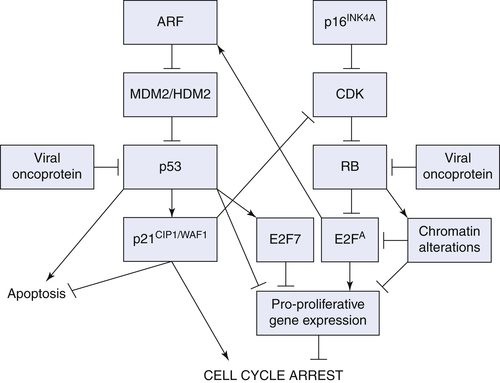
ARF/p53/p21CIP1/WAF1 Pathway

The ARF/p53/p21CIP1/WAF1 tumor suppressor pathway is a major regulator of cellular responses to oncogenic stress and DNA damage, resulting in the induction of cell cycle arrest, apoptosis, and senescence, depending on the stimulus. Cells isolated from p53-deficient mice are largely resistant to stimuli that would promote growth arrest or apoptosis under normal culture conditions, 62 and p53-deficient mouse embryo fibroblasts bypass the senescence response that usually occurs following extensive DNA damage or forced expression of oncogenic Ras. 25 Not only is loss of p53 required for bypass of senescence; restoration of p53 in Ras-transformed cells causes the induction of senescence and the subsequent regression of established tumors, demonstrating that continual suppression of p53 is required for tumor maintenance. 6365 Beyond their biological significance, these data suggest that p53-reactivation “therapies” may hold promise for the treatment of established tumors by promoting a senescence response.
p53 primarily exerts its antitumor effects by acting as a DNA-binding transcription factor that directly regulates the expression of many genes involved in apoptosis and cell cycle arrest. The effect of p53 during senescence is due, in part, to the direct transcriptional upregulation of the cyclin/cdk2 inhibitor p21CIP1/WAF1. By inhibiting cyclin/cdk2, p21CIP1/WAF1 promotes activation of the p16INK4A/RB pathway and subsequent cell cycle arrest, thus serving as an important point of crosstalk between the two central pathways of the senescence program. In support of this hypothesis, cosuppression of p21CIP1/WAF1 and RB pathway components (RB or p16INK4A) by RNA interference inhibits senescence in human fibroblasts, highlighting p21CIP1/WAF1 as a critical component of the p53 pathway. 58
Deletion of p21CIP1/WAF1 alone is not sufficient to bypass senescence in mouse embryo fibroblasts, 66 suggesting that additional p53 targets contribute to the senescence response. Such targets include the p53-responsive gene PML, which is induced by oncogenic Ras and promotes senescence by localizing RB/E2F proteins into PML bodies, thereby repressing E2F transcriptional activity. 6769
Another recently described point of crosstalk between the p53 and RB pathways involves the p53 transcriptional target E2F7 (an atypical E2F family member), which represses a subset of E2F target genes required for mitotic progression. 70,71 As seen for p21CIP1/WAF1, co-inhibition of E2F7 and RB is sufficient to circumvent Ras-mediated senescence and enable transformation of mouse embryo fibroblasts. 70 Collectively, these studies highlight the interplay between the p53 and RB pathways that govern institution of the senescence proliferative block and thereby the suppression of tumorigenesis. The extent to which the control of senescence contributes to the tumor-suppressive function in human cancer remains to be determined, but the co-occurrence of Ras, p53, and RB mutations in human tumors suggests that disruption of senescence may be required for cells to tolerate high levels of oncogenic signaling and become fully malignant.

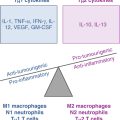
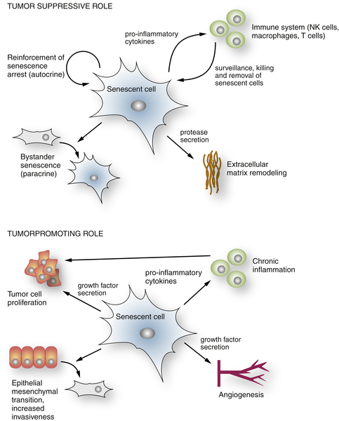
The Senescence-Associated Secretory Phenotype

Previously, we have focused largely on the cell-intrinsic mechanisms that govern senescence responses and tumorigenesis—these mechanistically explain much of the biology underlying the cell cycle arrest associated with the Hayflick limit. Senescent cells also secrete a multitude of factors, predominantly proinflammatory cytokines, chemokines, growth factors, and extracellular matrix remodeling factors, that have collectively been referred to as the senescence-associated secretory phenotype (SASP—Figure 15-3 ). Initially, SASP factors were used as biomarkers to confirm the presence of senescent cells. Now, emerging evidence indicates that the SASP acts in an autocrine and paracrine manner to influence the senescence program and profoundly alter the behavior of neighboring cells. This secretory program may contribute to the natural role of senescence in aged or damaged tissues.
The contribution of the SASP toward the biology of senescence and tissues is complex and can promote and suppress tumorigenesis depending on the context. Perhaps the greatest consequence of the SASP in vivo, in terms of tumor suppression, chemotherapy-induced senescence, and disease resolution, is enhanced immune surveillance and subsequent clearance of senescent cells. In such scenarios, the secretion of proinflammatory cytokines and chemokines by senescent cells attracts innate and adaptive immune cells (including natural killer cells, macrophages, and T cells) to the site of senescence and cell/tissue damage. The immune cells subsequently kill and clear the senescent cells, restoring tissue homeostasis. 63,72
Senescent cells also upregulate the expression of cell surface receptors, ligands, and intracellular signaling components that facilitate recognition of senescent cells and aid their elimination by immune cells. 72,73 Indeed, immune surveillance of senescent cells has been positively linked to regression of established tumors, 63 the prevention of tumor development, 74 and the improved survival of animals following treatment with chemotherapy. 75 Of course, some senescent cells—for example, those occurring in benign melanocytic nevi—remain in tissues for reasons that are yet to be elucidated.
image
Figure 15-3 Tumor suppressive and tumor-promoting activities of the SASP Senescent cells secrete a range of factors that profoundly affect the tissue microenvironment. In the context of tumor suppression, wound healing, and disease resolution, factors secreted by senescent cells attract components of the innate and adaptive immune system, which recognize, kill, and clear the senescent cells. This results in removal of damaged/stressed cells and restoration of tissue integrity. Senescent cells secrete molecules that actively reinforce the senescence arrest and can induce secondary “bystander” senescence in neighboring cells. The secretion of proteases enables remodeling of the extracellular matrix, which facilitates resolution of tissue disease (such as fibrosis) and thereby promotes wound healing. The consequence of the SASP is context dependent. Senescent cells secrete proinflammatory cytokines and growth factors known to enhance cell proliferation and transformation. Pro-angiogenic factors from senescent cells can promote tissue vascularization, while additional factors drive epithelial-to-mesenchymal transitions and increased invasiveness of premalignant cells.
Perhaps counterintuitively, SASP factors previously appreciated for their protumorigenic activity contribute to tumor-suppressive aspects of the senescence response. For example, the proinflammatory cytokines interleukin-6 (IL6) and interleukin-8 (IL8) reinforce the senescence growth arrest, presumably by continuing to drive pro-proliferative and DDR signaling within the senescent cells. 76,77 The nuclear factor-κB (NFκB) family of transcription factors (master regulators of immune signaling traditionally regarded for their protumorigenic activity) is responsible for transcription of a substantial part of the SASP signature, including the expression of IL6 and IL8. 75,76,78,79
Gene expression profiling indicates that NFκB controls the expression of more SASP genes during senescence than either p53 or RB, cementing its role as a key regulator of the senescence program. 75 During therapy-induced senescence, canonical NFκB activity was required both for the induction of senescence in tumor cells and for tumor regression following administration of chemotherapy, implying that the NFκB-driven SASP contributes to the senescent cell-intrinsic and -extrinsic aspects of the program and that NFκB can have remarkable antitumor activity. 75,78 These observations also suggest an important role for the SASP in facilitating chemotherapy responses, a possibility that is consistent with clinical data. 78
Secreted factors are capable of influencing their microenvironment for as long as they persist. It is therefore possible that promitogenic SASP factors can promote the hyperproliferation and subsequent “bystander” or “secondary” senescence of normal and/or premalignant neighboring cells. In agreement, secondary senescence can be conferred on incubating normal growing cells in “conditioned” media from senescent cells. 80
Alternatively, SASP factors might simultaneously drive the proliferation/invasiveness of neighboring malignant cells in which the senescence response is defective. For example, factors such as growth-related oncogene (GRO)-α and VEGF can facilitate the proliferation and transformation of premalignant cells 81 or promote tumor vascularization, 82 respectively. SASP factors can also promote invasiveness of premalignant cells. 79,8385 Hence, in some circumstances, the continued presence of senescent cells (and thus production of SASP factors) in aged, damaged, and premalignant tissues may exacerbate the decline of tissue integrity and/or promote tumor development or relapse. 79,86,87 The precise contexts in which SASP factors are pro- or antitumorigenic remain to be determined.

Senescence and Noncancer Disease States

Because senescence limited the replicative capacity of cells in vitro, Hayflick and colleagues proposed that replicative senescence might generally reflect the process of aging at the cellular level. 2,3 Although there is perhaps little evidence to demonstrate that organismal aging occurs as a direct consequence of cellular aging, senescent cells accumulate in aging tissues at sites of age-related diseases, such as in atherosclerotic plaques, skin ulcers, and arthritic joints, 88,89 and expression of senescence markers such as p16INK4A increases in aging tissues. 9092
Consistent with an active contribution of senescent cells to aging, elimination of p16INK4A-expressing cells from mouse tissues delays aging-related phenotypes such as sarcopenia, cataracts, and loss of adipose tissue. 93 Moreover, the late-life removal of p16INK4A-expressing cells was sufficient to attenuate the progression of already established age-related disorders, suggesting that senescent cells contribute to the initiation and maintenance of the aging phenotype. 93 Although the etiology of the senescent cells in aging tissues is often unclear, at least some cases have been attributed to telomere dysfunction, 91,92 and experimental manipulations to produce telomere dysfunction can reproduce aging phenotypes.
Senescent cells may contribute to organismal aging in a number of ways. For example, because senescent cells are irreversibly arrested, they may be incapable of effectively repopulating aged/damaged tissue and thus promote tissue decline. At the same time, senescent cells remain metabolically active and have a SASP that may also influence the behavior of neighboring cells and promote tissue dysfunction. 94 They may also promote chronic inflammation, which is associated with diverse age-related diseases including cancer. 95 Conversely, senescence can positively facilitate wound-healing responses and promote the resolution of disease states, which may produce positive effects on tissue function. For example, in a murine model of chemical-induced liver fibrosis, the senescence program limited the extent of fibrosis by halting the proliferation of fibrogenic (activated stellate) cells, reducing extracellular matrix production, and promoting immune clearance of the senescent cells, suggesting a positive role in this wound-healing response. Other studies imply that senescence restricts fibrosis in cutaneous wound healing. 96 In both models, the SASP played a prominent role in the elimination of senescent cells and tissue restoration.
Emerging evidence thus suggests that the impact of senescence on tissue function may be influenced by acute versus chronic induction. Hence, senescence can facilitate tissue homeostasis following acute damage through a two-pronged mechanism, resulting in the proliferative arrest and changes to the microenvironment that lead to the elimination of damaged cells and the remodeling of the surrounding tissue to restore tissue structure and function. However, persistent cellular damage would continue to produce excessive presenescent or senescent cells that might contribute to tissue degeneration, the onset of age-related disease, or tumor formation.

Conclusions and Perspectives

The true physiological role for senescence appears to be the prevention of the propagation of damaged cells, where autonomous aspects control the proliferative arrest and the SASP reinforces arrest and further directs the immune system to promote their elimination. In a speculative model, the original evolutionary purpose of senescence might be to help coordinate wound-healing responses, and these actions may be co-opted, directly or indirectly, for tumor suppression. Ironically, when persistent, some of these activities may be protumorigenic and contribute to age-related pathologies.
Mechanistically, the program involves active interplay among at least three major transcription factors (Rb, p53, and NFκB) whose activities are known to play an important roles in cancer. Because senescence can be influenced by extracellular factors and molecules, for which drugs and inhibitors are available, the possibilities for therapeutic modulation of the program in various settings exist. Such therapies could, in principle, harness the cell-autonomous aspects of the program to promote arrest or, alternatively, aid the immune recognition and clearance of senescent cells. Imperative for the success of such treatments is a thorough understanding of the molecules that govern senescence. Gaining new insight into senescence markers and biological aspects of the program in normal and pathological states is therefore of key importance for current and future biomedical research.
References

1. Hayflick L. , Moorhead P.S. The serial cultivation of human diploid cell strains . Exp Cell Res . 1961 ; 25 : 585 621 .

2. Hayflick L. The limited in vitro lifetime of human diploid cell strains . Exp Cell Res . 1965 ; 37 : 614 636 .

3. Smith J.R. , Hayflick L. Variation in the life-span of clones derived from human diploid cell strains . J Cell Biol . 1974 ; 62 : 48 53 .

4. Harley C.B. , Futcher A.B. , Greider C.W.

Buy Membership for Hematology, Oncology and Palliative Medicine Category to continue reading. Learn more here