Mucosal hyperenhancement accompanies active ulceration
Reversal of jejunoileal fold patterns (atrophied jejunal, thickened ileal)
Submucosal edema, fat, or gas
Small bowel intussusception
Eccentric soft tissue density mass in bowel wall (tumor)
Mesenteric adenopathy (may be cavitated)
• Excess fluid within SB lumen
Conformation of flaccid SB segments
Distends lumen and dilutes contrast medium
• Colonic luminal dilation
Excess gas, fluid, fat within lumen
• Eccentric soft tissue density mass in bowel wall
Strongly suggests lymphoma or carcinoma
TOP DIFFERENTIAL DIAGNOSES
• Whipple disease
• Crohn disease
• Intestinal opportunistic infections
CLINICAL ISSUES
• Common: Affects 1 in 200 in USA, but < 10% are currently diagnosed
Most common cause of SB disease and malabsorption
• Steatorrhea, abdominal distension, flatulence
Diarrhea, weight loss, glossitis, anemia
• Refractory disease
Enteritis that does not respond to at least 6 months of gluten-free diet
GI malignancies are main cause of death in celiac disease
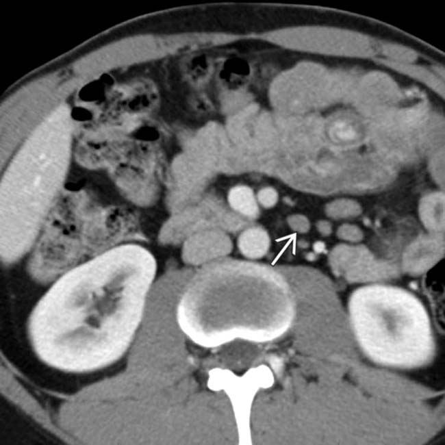
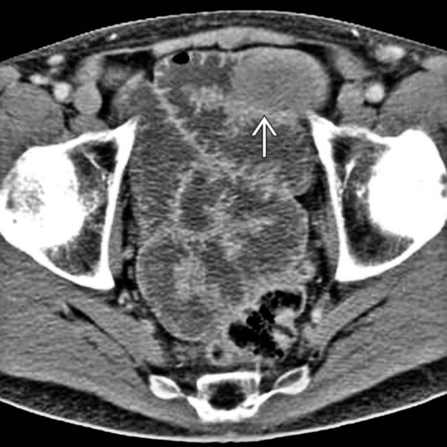
(Left) Axial CECT in a 37-year-old man with painful abdominal cramps shows 1 of several sites of intussusception , typically short segment and nonobstructing.
(Right) Axial CECT in the same patient demonstrates that the jejunal fold pattern seems blunted. Also noted is mesenteric lymphadenopathy .
(Left) Axial CECT in the same patient shows more mesenteric lymphadenopathy along with the abnormally blunted jejunal fold pattern.
(Right) Axial CECT in the same patient shows another intussusception . There is a suggestion of abnormal fold prominence in the ileum . The flaccid, dilated pelvic SB loops press on each other without intervening space, known as the conformation sign.
TERMINOLOGY
Synonyms
• Nontropical sprue or celiac-sprue disease, gluten-sensitive enteropathy
Definitions
• Celiac disease: Chronic intolerance of gluten that induces intestinal injury in genetically predisposed individuals
• Tropical sprue: Malabsorption seen in inhabitants of tropical countries
IMAGING
General Features
• Best diagnostic clue
CT enterography: Evidence of reversed fold pattern, multifocal intussusception
• Location
Celiac disease: More proximal small bowel
Tropical sprue: Entire small bowel
• Other general features
Most common small bowel disease producing malabsorption syndrome
Due to sensitivity of small bowel to α-gliadin
– Component of gluten
Has familial susceptibility with genetic basis
Radiographic Findings
• Barium small bowel follow-through (SBFT)
Dilatation of small bowel (jejunum): > 3 cm
Valvulae conniventes: May exhibit 5 patterns
– Valvulae look normal in most patients
– Ends at margin that are squared off rather than rounded
– Reversed jejunoileal fold pattern: ↓ number of jejunal folds and ↑ ileal folds
– Blunted or absent valvulae: “Moulage” sign (cast): Characteristic of sprue
– Thickening: In severe disease and hypoproteinemia
“Colonization of jejunum”: Loss of jejunal folds → colon-like haustrations
Hypersecretion-related artifacts: Due to excess fluid
– Flocculation: Coarse granular appearance of small clumps of disintegrated barium due to excess fluid; mainly in patients with steatorrhea
– Segmentation: Break up of normal continuous column of barium, creating large clumps of barium separated by string-like strands
Transit time: May be long, short, or normal
Nonpropulsive peristalsis (flaccid and poorly contracting bowel loops)
Painless, transient intussusceptions often seen on fluoroscopic studies
• Fluoroscopic-guided enteroclysis
More accurate than SBFT in diagnosing celiac disease
Jejunal folds
– Decreased number of proximal jejunal folds (< 3/inch; normal: ≥ 5/inch)
– Increased separation and absence of folds; “ileal” appearance
Ileal folds
– Increased number of folds in distal ileum (4-6/inch; normal: 2-4/inch)
– Increased fold thickness ≥ 1 mm: “Jejunization” of ileum in 78% of cases
Mosaic pattern: Due to total villous atrophy
– 1-2 mm islands of mucosa surrounded by barium-filled grooves
Duodenal changes
– Decreased number and irregular folds, especially in distal duodenum
– “Bubbly” duodenum: Nodular pattern in mucosa
CT Findings
• Excess fluid within SB lumen
Distends lumen and dilutes positive enteric contrast medium
• SB wall may be thick or thinned
Mucosal hyperenhancement accompanies active ulceration
Submucosal edema; halo sign
Submucosal fat in wall of duodenum and jejunum
Pneumatosis has been reported (not due to ischemia)
• Conformation of SB segments
Dilated, flaccid loops press against each other (especially in pelvis)
Common: Affects 1 in 200 in USA, but < 10% are currently diagnosed
Most common SB disease and cause of malabsorption
Celiac disease: Increased incidence in Ireland and Northern Europe; unknown in Africa, China, Japan
Tropical sprue: Increased incidence in tropics, especially in Vietnam and Puerto Rico
Natural History & Prognosis
• Natural history
Adult disease: Extension of childhood form or new onset
• Complications
Refractory disease
– Symptomatic severe enteritis that does not respond to at least 6 months of gluten-free diet
– These are patients at most risk for complications
Ulcerative jejunoileitis
↑ risk of T-cell lymphoma and carcinoma of jejunum
– GI malignancies are main cause of death in celiac disease
Cause 50% of deaths in patients with refractory disease
Deep venous thrombosis
• Prognosis
Celiac disease: Improvement within 48 hours; full remission (weeks to months)
– Response to gluten-free diet
Tropical sprue: Improvement in 4-7 days; complete recovery (6-8 weeks)
Treatment
• Nontropical sprue or celiac disease
Lifelong gluten-free diet
• Tropical sprue
Broad spectrum antibiotics (tetracycline) and folates
DIAGNOSTIC CHECKLIST
Image Interpretation Pearls
• Imaging findings will vary according to duration and severity of disease
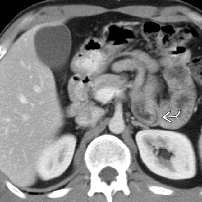
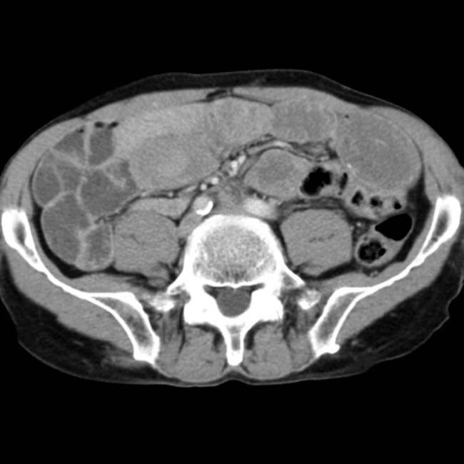
(Left) Axial CT in a 69-year-old woman with chronic diarrhea & pain shows fluid distention of the SB with conformation of the flaccid segments. The fold pattern of the jejunum is blunted.
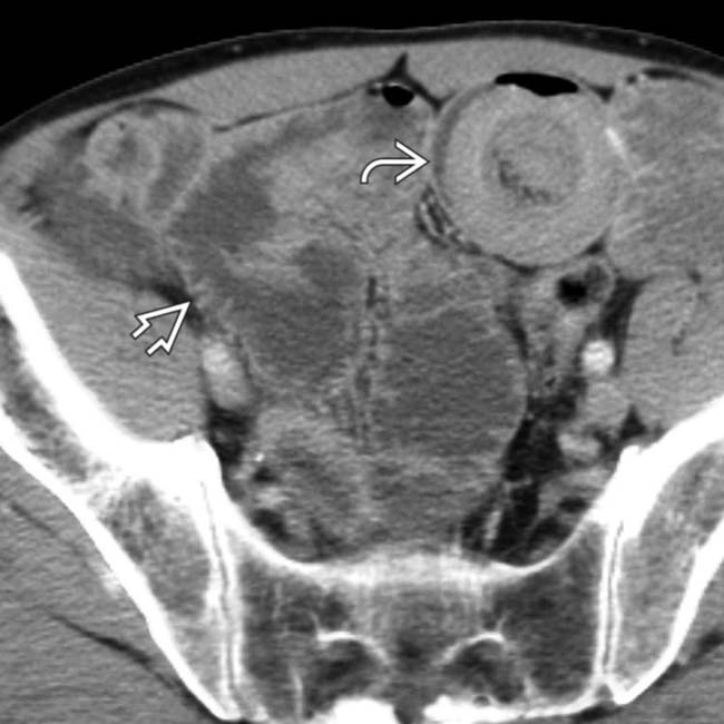
(Right) Axial CT in the same patient shows a short segment, nonobstructing intussusception with a “target” appearance and intraluminal mesenteric fat. The fold pattern of the ileum is more prominent than that of the jejunum, a reversal of the normal situation. Biopsy & response to a gluten-free diet confirmed the diagnosis of celiac disease.
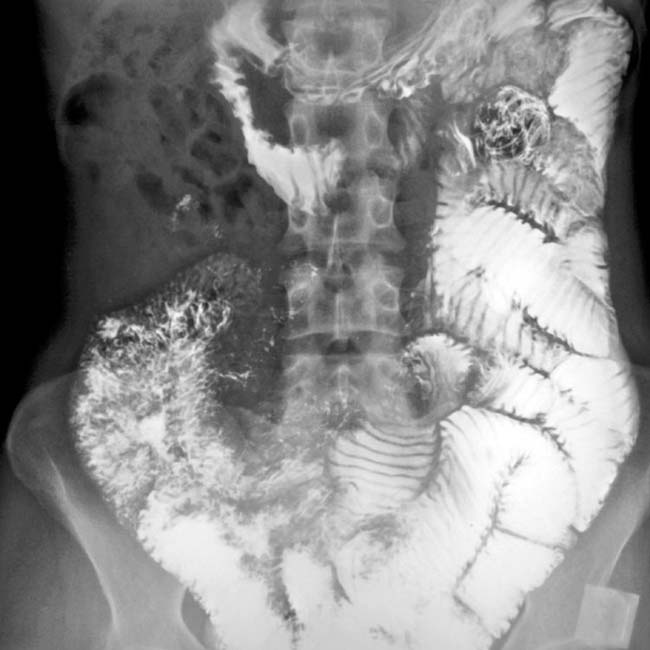
(Left) Films from a barium SBFT in a 30-year-old woman with steatorrhea show a typical malabsorption pattern, consisting of dilution of the barium and dilation of the lumen. The folds within the jejunum appear blunted. There is poor coating of the mucosa by the barium.
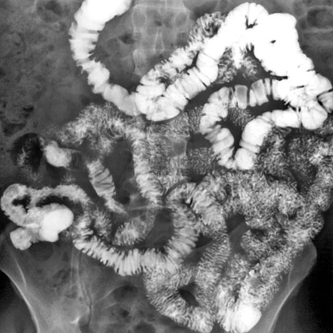
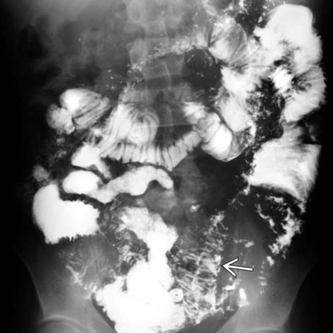
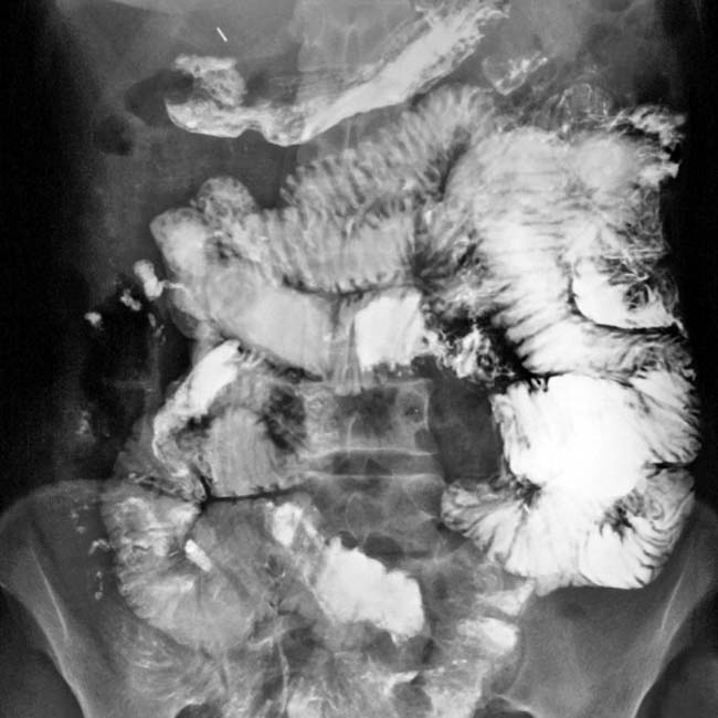
(Right) SBFT in the same patient illustrates intermittent intussusception, with a coiled “spring” appearance in the mid jejunum . Celiac disease is the most common specific cause of malabsorption.
(Left) Axial CECT in a patient with sprue shows fluid-distended bowel. One segment of jejunum has focal thickening of the wall, which was found to be due to lymphoma.
(Right) Axial CECT in the same patient shows multifocal gastric wall thickening , which was also due to lymphoma. Patients with refractory sprue are at increased risk for both lymphoma and carcinoma of the bowel.
SBFT shows severe loss of folds in the duodenum and jejunum, representing the “moulage” pattern.
SBFT shows dilated bowel, dilution of barium, and the reduced number and size of jejunal folds.
Axial CECT shows dilated fluid-distended bowel and the bottom of an intussusception . Note the prominent folds in the ileum.
Axial CECT shows fluid-distended small bowel, prominent ileum folds, and short segment intussusception .
SBFT shows dilated lumen plus dilution of barium within the small intestine and a reversal of the normal jejunal and ileal fold patterns.
Small bowel follow-through (SBFT) shows a decreased size and number of jejunal folds and an increased number and size of ileal folds (reversal pattern).


 , typically short segment and nonobstructing.
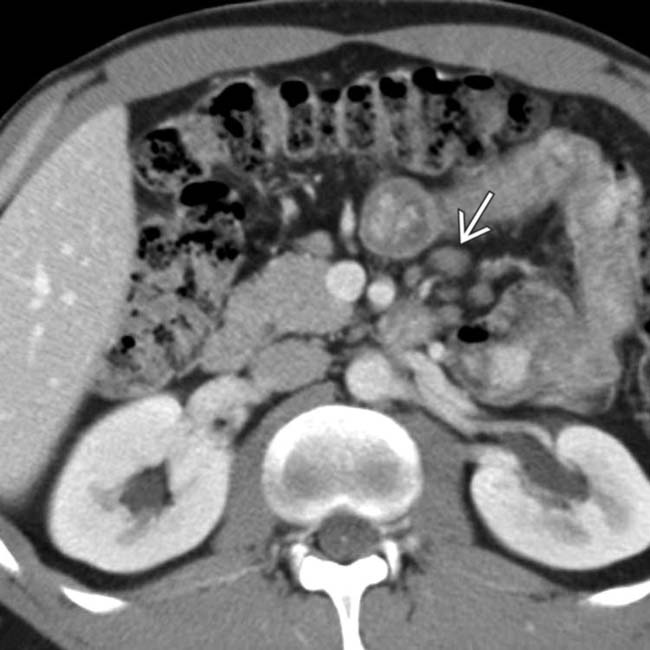
 .
.
 along with the abnormally blunted jejunal fold pattern.
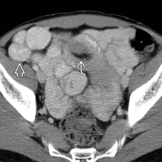
 . There is a suggestion of abnormal fold prominence in the ileum
  . The flaccid, dilated pelvic SB loops press on each other without intervening space, known as the conformation sign.


 with a “target” appearance and intraluminal mesenteric fat. The fold pattern of the ileum
  is more prominent than that of the jejunum, a reversal of the normal situation. Biopsy & response to a gluten-free diet confirmed the diagnosis of celiac disease.
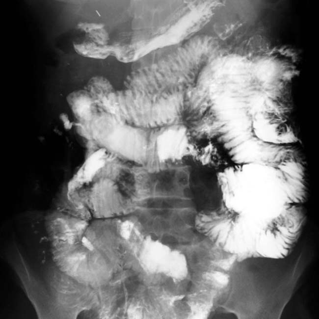
 . Celiac disease is the most common specific cause of malabsorption.
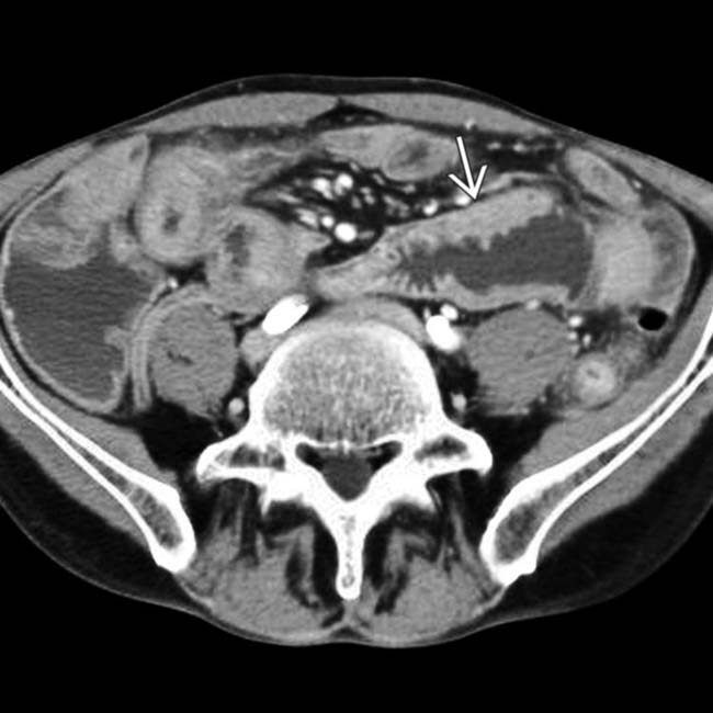
 of the wall, which was found to be due to lymphoma.
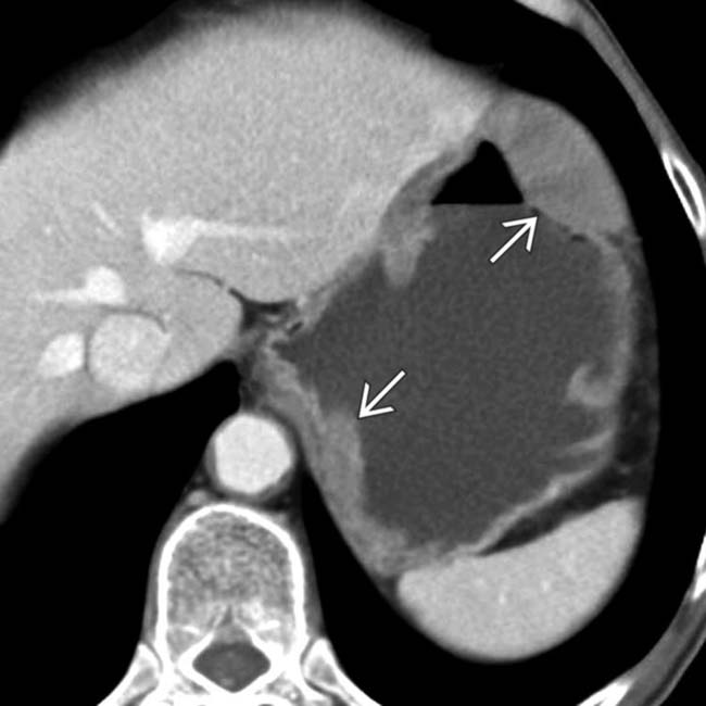
 , which was also due to lymphoma. Patients with refractory sprue are at increased risk for both lymphoma and carcinoma of the bowel.
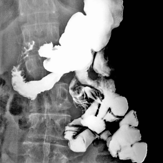
 . Note the prominent folds in the ileum.
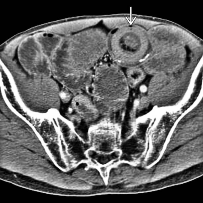
 .
.