INTRODUCTION
CASE IN DETAIL
SF’s current problems started with the gradual onset of lower abdominal pain 1 week ago. This was accompanied by abdominal distension. His pain gradually progressed in intensity and became generalised. The pain was dull and constant in nature and he could not identify any precipitating or relieving factors. Associated with these symptoms he developed anorexia and nausea, but did not vomit. This was followed by black, tarry bowel motions, which he first noticed 3 days ago. He presented to the emergency department of the hospital 1 day ago, where he was investigated with multiple blood tests, abdominal ultrasonography and upper gastrointestinal endoscopy. He was significantly anaemic and was transfused with 4 units of packed red cells. He was also commenced on an octreotide infusion and kept nil by mouth. On endoscopic examination he was diagnosed with bleeding varices as well as erosive gastritis. He was treated with banding of the varices and high-dose intravenous omeprazole therapy at 20 mg twice daily. He was also commenced on lactulose 10 mL three times daily for the prevention of hepatic encephalopathy. With this therapy he experienced diarrhoea with liquid motions on average five times a day. He underwent a diagnostic paracentesis and was subsequently commenced on intravenous ceftriaxone 1 g daily. However, he denied any fevers, rigors or drenching sweats.
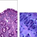
SF was diagnosed with hepatic cirrhosis 1 year ago when he presented with abdominal distension and haematemesis. He was also noted to be hepatitis C antibody-positive on this presentation. He admitted having had body piercing and tattoos as a possible risk factor but denied any previous blood transfusions or intravenous drug use. Subsequent upper gastrointestinal endoscopy showed oesophageal varices, which were managed with banding. During the same admission he was commenced on a low-salt diet and advised against further alcohol consumption, but he did not comply with either instruction. He was followed up on discharge with four more 3-weekly endoscopic bandings.
Eight months ago he was readmitted with severe abdominal distension, dyspnoea and ascites, which was treated with abdominal paracentesis. He was then commenced on spironolactone 100 mg daily and propranolol 40 mg daily. Subsequently he has presented on three more occasions with tense abdominal distension for paracentesis, and the last episode was 6 weeks ago. He is currently on spironolactone 200 mg daily and frusemide 40 mg daily.
He was diagnosed with a duodenal ulcer 2 years ago when he presented with severe epigastric pain, nausea and vomiting. Diagnosis was made on gastroscopy. He was commenced on ranitidine 150 mg twice daily. Five months ago his symptoms recurred and his general practitioner changed his treatment to omeprazole 20 mg daily. He denies any knowledge of his Helicobacter pylori status and denies ever having been treated for H. pylori.
In summary, his current medications are frusemide, spironolactone, omeprazole, propranolol, lactulose and ceftriaxone.
He denies any allergies.
His family history is significant, with his father dying of bowel carcinoma at the age of 52. His mother is alive at the age of 65 and is well. He has three brothers: the oldest, aged 44, suffers from alcoholic cirrhosis and the second brother, aged 42, was operated on this year for bowel carcinoma; his youngest brother, aged 36, is well.
He lives by himself in a local hotel and has a flight of 10 steps to negotiate to get to his room. He is independent with the activities of daily living. He was married once but he divorced his wife 5 years ago. He has two daughters, aged 10 and 8, both of whom are well and living with his former wife. He rarely sees his daughters, and the last time was 3 years ago. His main social support is a group of friends that he meets in the pub. He does not visit his GP regularly. He spends most of the day in the pub, with his friends.
He worked as a seaman and a waterfront labourer before retiring 5 years ago after a back injury. But he denies any symptoms associated with it currently. He survives on a disability pension and claims it is adequate for his survival.
He smokes approximately one packet of cigarettes a day and has a history of 30 pack-years. He consumes in excess of 100 g alcohol a day in the form of cheap wine or rum. My directed assessment (CAGE questionnaire) confirmed that he is alcohol dependent. He has tried giving up drinking on previous occasions without any success. He has not undergone alcohol detoxification previously. He had his last drink prior to hospitalisation, 1 week ago. He denies any other previous or current recreational drug use.
My nutritional history suggests inadequate nutrition. He usually has his meals at the local fast-food outlet and often neglects his main meals.
He demonstrates very poor insight into his medical and social conditions. He has suffered from depression in the past and has been treated medically. He denies any ongoing vegetative symptoms of depression and is currently not on any antidepressant medication.
He complains of daytime somnolence and nocturnal insomnia.
ON EXAMINATION
SF was drowsy but rousable and cooperative. He appeared cachectic and had mild scleral icterus. He had tattoos on the ventral and dorsal aspects of his thorax and an intravenous cannula in the dorsal aspect of his right hand; the cannula site was not inflamed. He had multiple ecchymoses in the forearms bilaterally and in the forehead.
His pulse rate was 90 beats per minute and respiratory rate 20 per minute. He was afebrile, and his blood pressure was 130/95 mmHg with no postural drop.
His estimated body mass index was 20. His cognitive function was preserved, with a Mini-Mental State score of 28/30.
In the gastrointestinal examination there was no clubbing of the fingers, but there was palmar erythema bilaterally and a Dupuytren’s contracture in the left palm. There was a mild hepatic flap. He had parotidomegaly but no fetor hepaticus. His oral hygiene was poor. There were multiple blistering lesions in the perioral region, some filled with clear fluid and some crusted. There were multiple spider naevi in the upper thorax, both anteriorly and posteriorly. There was gynaecomastia. He had generalised abdominal distension without superficial venous dilatation. There were multiple scars in the left lateral aspect of the abdomen—possible evidence of previous paracentesis. On palpation the abdomen was firm and non-tender. There was firm-to-hard, non-tender hepatomegaly with an irregular edge. The hepatic span was 19 cm along the right anterior clavicular line. His spleen was just tippable in the left upper quadrant. There was a positive fluid thrill and a shifting dullness of 3 cm in the flanks. The kidneys were not ballotable and his bowel sounds were positive and normal.
There was evidence of testicular atrophy, and I would like to confirm this with formal orchidometry.
His cardiovascular examination demonstrated a parasternal heave and two heart sounds. The pulmonary component of the second sound was louder. Jugular venous pressure was not elevated but was visible 3 cm above the angle of Louis. There was bilateral pitting ankle oedema up to a level midway from the ankle. All peripheral pulses were well palpable.
Respiratory system examination showed normal lung expansion bilaterally, and there was no dullness to percussion. There were medium coarse crepitations in both bases and the breath sounds were vesicular.
Neurological examination showed normal cranial nerve examination, with preserved visual acuity of 6/6 bilaterally without correction. Fundoscopy was normal. In the upper limbs, motor function was preserved and the coordination was normal. But there was impaired proprioception, soft touch and pinprick sensation in the hands in a glove distribution to the level of the wrists bilaterally. Otherwise, the sensory examination was unremarkable.
In the lower limb examination, his gait was wide-based with bilateral foot drop. He demonstrated a positive Romberg’s test. There was significant wasting of the musculature distal to the knee bilaterally but without pes cavus. The tone was normal. There was significant weakness at the level of ankle bilaterally in all directions of movement. There was sensory impairment to vibration, joint position, soft touch and pinprick sensation bilaterally in a stocking distribution to a level just above the ankles. The deep tendon reflexes and coordination were preserved. He demonstrated flexor plantar response bilaterally.
His rheumatological examination was unremarkable and there was no lymphadenopathy.
I would like to see the result of his per rectum examination.
In summary, this is a 38-year-old man presenting with decompensated cirrhosis of the liver with portal hypertension, associated with chronic alcoholism and hepatitis C infection on a background of peptic ulcer disease, depression and peripheral neuropathy. He also has the significant problems of alcohol dependency, poor compliance, poor social support and a family history of carcinoma of the bowel.
His main problem currently is the management of multiple acute complications of cirrhosis, namely gastrointestinal bleeding, portal hypertension, sepsis and hepatic encephalopathy.
In addition to the above, I have identified four medical problems and a social problem:
Approaching the management of this man’s main acute problem of decompensated cirrhosis, I would like to see the results of the following investigations:
1. Full blood count, looking for leucocytosis, anaemia with macrocytosis, and thrombocytopenia
2. His temperature chart, looking for recent fevers
3. Coagulation profile, looking for a high international normalised ratio
4. Electrolyte profile, looking for hyponatraemia, hyperkalaemia and renal failure, and the liver function indices, looking for abnormalities
5. Microscopy and culture of ascitic fluid, and the serum-to-fluid albumin ratio
6. Serum ammonia level.
Questions and answers
Q: How would you manage this patient when he initially presents?
A: The immediate objective in the management of this patient is to control the gastrointestinal bleeding and achieve haemodynamic stability. I would quickly assess the patient’s haemodynamic status by looking at the oral mucosa and skin turgor to assess the level of hydration, and check the arterial pulse and the blood pressure and look for any postural drop in blood pressure. Then I would insert large-bore cannulae in two large peripheral veins (e.g. the cubital fossa) and perform urgent blood tests for the haemoglobin level, haematocrit, and coagulation and electrolyte profiles. I would cross-match six units of blood and platelets and commence an intravenous infusion of colloids or crystalloids to maintain vascular volume. Given his history of bleeding varices and duodenal ulcer disease, I would start intravenous infusions of octreotide as well as omeprazole. I would keep the patient nil by mouth, organise urgent gastroscopy and also notify the general surgeon.
Q: He has been afebrile. Do you still think that he has spontaneous bacterial peritonitis?
Q: How would you manage spontaneous bacterial peritonitis?
A: I would perform a diagnostic paracentesis for microscopy and culture and assess the pH of the ascitic fluid. Even if microbiological tests were unyielding, an acidic pH would suggest infection. The most likely organism is E. coli, so I would commence this man on 1 g daily intravenous ceftriaxone. On recovery I would maintain him on long-term prophylactic antibiotic therapy with norfloxacin.
Q: Would you give him gentamicin?
A: No, I would not. Gentamicin has the potential to cause renal damage and in this setting it can lead to hepatorenal syndrome.
Q: How would you manage his encephalopathy?
A: He is drowsy and has a hepatic flap, so he has grade 1 hepatic encephalopathy. First I would address the possible precipitating causes—control and treat gastrointestinal bleeding and treat spontaneous bacterial peritonitis. I would give him oral lactulose, commencing with a dose of 30 mL twice daily and gradually increasing the dose so as to cause diarrhoea at least four times a day. I would also put the patient on a low-protein diet. If these measures did not achieve adequate control of the encephalopathy, I would commence the patient on oral neomycin.
Q: Why did you say that you wanted to further investigate his hepatomegaly?
A: This man has known cirrhosis of the liver, and usually cirrhosis causes shrinkage of the liver, so it is not usual to present with hepatomegaly in the clinical setting of hepatic cirrhosis. This man runs the risk of developing a hepatocellular carcinoma in the cirrhotic liver, which could very well lead to decompensation of his cirrhosis. Also, with his significant family history of large bowel carcinoma, there is an indication to exclude metastatic bowel cancer. To approach this issue I would first like to check his serum alpha-fetoprotein levels and the results of an ultrasound scan of his liver. If there was a mass lesion I would proceed with a liver biopsy once the patient was haemodynamically stable and his coagulopathy treated.
Q: How would you manage this man if he has a hepatocellular carcinoma?
A: If there was a biopsy-proven hepatoma, my approach to its management would be first to stage the disease. Staging would be done with following investigations:
If the patient has a tumour confined to one lobe of the liver, he should be considered for hepatectomy and liver transplantation in consultation with a liver transplant unit. But given his decompensated condition, his prospects of being a candidate for liver transplantation are grim. Depending on the stage, the other therapeutic options include alcohol injection, intraarterial chemoembolisation or intraarterial chemotherapy. If he has disseminated disease, the management option would be palliative.
Q: If endoscopy shows a duodenal ulcer, how would you manage it?
A: If there is a duodenal ulcer I would like to know whether there is evidence of recent bleeding in the ulcer and also features that would suggest a high likelihood of a rebleed, such as a visible vessel in the ulcer base or a blood clot. If there is evident bleeding, it should be treated with adrenaline injection or electrocautery.
On securing haemostasis, I would perform a rapid urease test and a biopsy to ascertain the H. pylori status of the patient. If the patient was positive for H. pylori I would treat him with a 10-day course of triple therapy, consisting of omeprazole 20 mg twice daily, amoxycillin 500 mg twice daily and clarithromycin 100 mg twice daily. I would follow the patient up 4 weeks later with a urease breath test to confirm eradication of the organism. If the patient tested negative for H. pylori I would treat him with oral omeprazole 20 mg daily.
Q: What do you think about this patient’s peripheral neuropathy?
A: His neuropathy is most likely to be due to the chronic alcoholism. The other differential diagnoses that should be entertained are vitamin B12 deficiency and peripheral neuropathy, possibly associated with as yet undiagnosed diabetes or mononeuritis multiplex due to hepatitis-C-induced cryoglobulinaemia. To approach the management of this neuropathy, I would like to perform nerve conduction studies to ascertain the exact nature of the neuropathy and perform blood tests looking for vitamin B12 deficiency, hyperglycaemia (fasting blood sugar levels) and cryoglobulins. I would also refer the patient to a podiatrist for foot care and for foot orthotics to facilitate safe mobilisation. I would educate the patient in the implications of the peripheral neuropathy, the dangers involved, and stress the need for good foot care for the prevention of complications such as burns, pressure ulcers and Charcot’s joints.
It is likely that this neuropathy is irreversible, but progression can be controlled by addressing the causative factor, if possible, once definitively diagnosed.
Q: What are you going to do about this man’s alcoholism?
A: My objective is to achieve full abstinence. To arrive at this goal I would formulate a plan of action in collaboration with the drug and alcohol facility of the hospital, the social worker and the patient’s GP. First I would educate the patient in the absolute need for abstinence and inform the patient of the various supportive facilities available to people with problems associated with alcohol. I would refer the patient to the local drug and alcohol outpatients clinic and, if the patient was interested, to organisations that help people overcome alcohol abuse, such as the Salvation Army and Alcoholics Anonymous. If the patient cannot maintain the detoxification achieved during this admission to hospital, consideration may be given to anti-craving medications such as acamprosate in collaboration with the alcohol or liver clinic.
Q: What would you do to optimise his social support network?
A: I would assess his home situation and the support available in collaboration with the social worker. I would also call a family conference to explain the patient’s situation and to ascertain what level of care the family can provide. He may need community resources, such as home care and the community nursing sister. I would also formulate a shared-care program for the ongoing medical management of the patient with his GP.
Q: Would you continue to treat his hepatitis C infection?
A: No, not at this stage. He would not tolerate interferon alpha treatment because of decompensated cirrhosis. A further deterrent to antihepatitis C therapy is his continued alcohol abuse.
Q: Is this man a candidate for liver transplantation?
A: No. He has to be abstinent prior to being considered for transplantation. Therefore, once he was stable I would first refer him for psychological assessment and counselling.
Q: What else would you want to do for this man?
A: I would strongly advise him on lifestyle measures, to curb the spread of his hepatitis C infection to others. Advice would be given not to share razors etc. I would educate him in the need for proper compliance with medical therapy and in proper nutrition with the help of the dietitian. I would also commence him on regular oral thiamine supplements.