INTRODUCTION
CASE IN DETAIL
LS is a 45-year-old woman who presented 3 weeks ago with fevers, rigors and drenching sweats of 4 days’ duration. She also had a productive cough with green, rusty sputum and pleuritic chest pain in the left subscapular region. She was subsequently diagnosed with pneumonia and treated with intravenous antibiotics until 2 weeks ago. Her symptoms settled on this treatment. This was her third admission for pneumonia within the past 5 months.
During this admission she was also diagnosed with significant anaemia. However, she could not remember her haemoglobin level at the time of diagnosis. She denied any symptoms suggestive of anaemia prior to presentation. She was investigated thoroughly with multiple blood tests, upper and lower gastrointestinal endoscopy and a bone marrow biopsy. She was not aware of the outcomes of any of the above investigations. She was treated with the transfusion of two units of packed cells and discharged from hospital 10 days ago. She denies any known source of bleeding. She is currently stable but feels very lethargic.
LS was diagnosed with end-stage renal failure 5 years ago when she presented with frank haematuria, bipedal pitting oedema, dyspnoea at rest, persistent nausea and vomiting. She had multiple investigations including a renal biopsy. A diagnosis of end-stage renal failure secondary to IgA nephropathy was established. Almost immediately she was commenced on haemodialysis. She was dialysed for 2 years, initially at the hospital and then at home until 3 years ago, when she received a cadaveric renal allograft. Three months later she developed severe rejection non-responsive to pulse methylprednisolone therapy and she was recommenced on dialysis. Two years ago she received her second cadaveric allograft, which has been functioning well to date.
She has been treated with multiple immunosuppressant medications, including prednisone at various doses, azathioprine and cyclosporin, for 3 years.
She has received oral prednisolone as well as parenteral pulsed methylprednisolone at different times. The maximum dose has been 500 mg methylprednisolone intravenously for 3 days during the rejection episode of her first allograft. The minimum is 10 mg oral prednisolone, which is her current maintenance dose. She denies any adverse effects associated with her corticosteroid therapy, but she has never had her bone densitometry measured.
She also takes 150 mg cyclosporin twice daily. With her cyclosporin therapy she has experienced side effects of gum hypertrophy, hypertension and recurrent gouty arthritis. The first gout attack was 1 year ago. On controlling the acute gout attack with colchicine and local corticosteroid injection, she was commenced on allopurinol maintenance therapy, and the azathioprine she was on was replaced with mycophenolate mofetil. Since the commencement of maintenance therapy with allopurinol, she has not suffered from any acute exacerbations of gouty arthritis. Her cyclosporin level is monitored at the local hospital’s renal clinic at 2-weekly intervals and the most recent trough level has been 175 ng/L. Her cyclosporin-induced hypertension is managed with long-acting nifedipine 30 mg daily and she denies any side effects associated with this therapy. Her blood pressure is monitored at the local renal clinic every second week and has been well controlled at around 130/85 mmHg.
She is currently managed on 1 g mycophenolate mofetil twice a day. She denies any side effects associated with this therapy.
She has had multiple skin malignancies associated with her immunosuppression, excised over the past 3 years by her general practitioner and a plastic surgeon. The most recent was a squamous cell carcinoma removed from her forehead 2 months ago.
In summary, her medications are: cyclosporin-A, mycophenolate mofetil, prednisolone, nifedipine and allopurinol. She denies any allergy.
LS’s mother is 68 years old and suffers from emphysema and diabetes mellitus. Her father is 70 years old and has ischaemic heart disease. She is an only child.
She lives with her partner, aged 47, who is well and supportive. She has one daughter, aged 12, who is well. She lives in a farmhouse with only one step at the entrance. There are many animals living on the farm, including swine, cattle and various birds. She is independent with activities of daily living.
She gave up smoking 4 years ago, but prior to that she had a significant smoking history of 30 pack-years. She smokes marijuana once a month and consumes alcohol only very rarely.
She works as a gardener at a local nursery and has prolonged periods of sun exposure.
My dietary history of LS suggests satisfactory nutrition. She denies any significant weight loss in the recent past. She has no problems with sleep.
She had an episode of depression about 4 months ago, due to a dispute with her partner. She believes that he is getting tired of all her medical problems. She is also anxious about the current developments with her medical condition. She denies any vegetative symptoms of depression or anxiety.
ON EXAMINATION
LS was alert and cooperative. She had a clinically functioning arteriovenous fistula in her right forearm. There were multiple healed scars of previously excised cutaneous malignancies in her left forehead, left cheek and over the right shoulder. There was an ulcerative lesion in her left thigh of 1.5 cm diameter with a hard, everted edge and a dry, erythematous base. There was no evidence of any granulation tissue. This lesion was not tender and the features were very suggestive of a squamous cell skin malignancy.
Her pulse rate was 50 beats per minute, regular in rhythm and normal in character. Her blood pressure was 140/95 mmHg and there was no postural drop. Her respiratory rate was 10 per minute. The estimated body mass index was 20. She was afebrile and her cognition was preserved.
Respiratory tract examination showed normal chest expansion, in both the apical and basal regions. There was no area of dullness to percussion, and breath sounds were vesicular throughout. There were fine crepitations in the right lung base. There was no sputum mug available for inspection.
Cardiovascular system examination showed no palmar crease or conjunctival pallor. Her heart sounds were dual and normal in character and there was no murmur. There was no peripheral oedema.
In the gastrointestinal system examination, her abdomen was not distended or tender. There was a longitudinal scar of 4 cm in the left lower quadrant, underneath which a non-tender, firm mass suggestive of the renal transplant was palpable. There was no organomegaly and the rest of the abdominal examination was unremarkable.
Neurological examination showed an unremarkable cranial nerve examination. Her visual acuity was preserved at 6/6 without correction. Fundoscopy showed silver wiring of the arteries and arteriovenous nipping with no haemorrhage or exudates. Fundoscopic findings suggest grade 2 hypertensive changes according to the Keith-Wagener-Barker classification.
Upper and lower limb examinations showed normal tone, power, reflexes and coordination with an unremarkable sensory examination. Importantly, there was no proximal muscle wasting or weakness noticed.
Her rheumatological examination was unremarkable and there was no lymphadenopathy.
In summary, LS is a 45-year-old renal transplant patient with a recent diagnosis of anaemia. She is on multiple immunosuppressive medications with associated complications of hypertension, gout, recurrent infections and cutaneous malignancy. She suffers from occasional depression and anxiety. She also has the important problem of hazardous occupational sun exposure.
The main problem with this woman is the management of immunosuppression appropriately so as to minimise complications and maximise graft survival and function.
In approaching the management of this patient, I have identified the following five problems:
1. Diagnosis of the aetiology of this woman’s anaemia and the treatment of same
2. Ascertaining the cause of her recurrent pneumonia and taking appropriate preventive measures
3. Reviewing her immunosuppressive medication regimen and optimising efficacy while minimising the complications
4. Tackling the issue of her skin malignancies and protection from further sun exposure
5. Addressing her current state of anxiety and helping the family with effective coping strategies.
First I would address the problem of anaemia. The first differential diagnosis I would consider is bone marrow suppression associated with immunosuppressive therapy. Other differential diagnoses that I would consider in this situation are:
To ascertain the severity of the anaemia and to exclude the above differential diagnoses, I would like to see the results of the following investigations:
1. Full blood count, looking at the haemoglobin levels. I would like to see both the current haemoglobin level and the haemoglobin level on presentation.
2. Blood film, looking at red cell morphology and reticulocyte count
3. Iron studies in the form of serum iron, serum ferritin and serum transferrin levels, together with the serum vitamin B12 level and red cell and serum folate levels
4. Results of the recent endoscopic procedures, gastroscopy results looking at peptic ulcer disease or gastritis, and colonoscopy result looking for colonic mass lesions
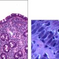
5. Results of the recent bone marrow biopsy, looking for marrow suppression showing hypocellularity, marrow infiltration or ring sideroblasts
6. A haemolytic screen: the blood film, looking for spherocytes, red cell fragments and reticulocytes, together with the serum haptoglobin, serum bilirubin and serum lactate dehydrogenase levels
7. Urine analysis, looking for haematuria to exclude glomerulonephritis, recurrent IgA nephropathy, uroepithelial malignancy and paroxysmal nocturnal haemoglobinuria.
Management of this patient’s anaemia will be guided by the results of the above tests. Depending on the haemoglobin level and the clinical symptoms, I would decide on the need for blood transfusion.
Questions and answers
Q: Please interpret this full blood count and the blood film report of this patient on presentation:
A: There is significant anaemia together with thrombocytopenia. The severity of the anaemia warrants blood transfusion, and I would transfuse this woman with three units of packed cells. I would use leucocyte-filtered blood for the purpose of transfusion in order to prevent immune sensitisation, which might preclude future transplant prospects if needed. The suboptimal reticulocyte count suggests inadequate bone marrow response to the anaemia, and I would be keen to follow this up with the bone marrow biopsy, looking at both the aspirate and the trephine pictures.
Cytomegalovirus disease is another possibility with anaemia and thrombocytopenia. Although this tends to be more common in the early post-transplant period, I would like to rule it out with CMV PCR.
Q: The bone marrow biopsy shows hypocellularity. How does this finding influence your management plan for this woman?
A: I believe that the anaemia is likely to be due to bone marrow suppression associated with the immunosuppressive therapy. However, to exclude aplastic anaemia and myelofibrosis, I would like to see whether there was any evidence of cellular atypia. Cyclosporin or corticosteroids are not known to cause bone marrow suppression, as would azathioprine and mycophenolate mofetil. In this situation the likely drug is mycophenolate. I would do further research into the adverse effects of this drug. I would cut her current dose of mycophenolate mofetil to 500 mg twice daily and increase her prednisolone to 15 mg daily to compensate.
Q: What options do you have if the proposed dose reduction does not work?
A: In such a situation I would consider withdrawing mycophenolate and replacing it with an alternative agent, such as sirolimus or tacrolimus. Unfortunately, the full side-effect profiles of these drugs have not yet been described. Therefore it would be a decision that I would take after extensive consultation with colleagues who have practical experience with the use of these agents.
Q: Please interpret the electrolyte profile of this patient:
A: This picture is consistent with moderate renal failure. I would like to see some previous figures to see whether this is acute or chronic renal failure, and, if it is chronic, to see whether it is progressive.
Q: Six months ago her urea level was 7.1 mmol/L and creatinine level was 180 mmol/L.
A: There is chronic renal failure and it is progressive. The possible differential diagnoses are chronic allograft nephropathy, recurrent glomerulonephritis or viral infections of graft due to CMV or BK virus. I would also do an ultrasound of graft and, if it is normal, I would proceed with a renal biopsy for a definitive diagnosis.
Q: The renal biopsy done 2 weeks ago shows striped fibrosis and evidence of chronic rejection. How would you approach this?
A: This pathology report suggests cyclosporin toxicity as evidenced by striped fibrosis in combination with chronic rejection. Cyclosporin toxicity should be addressed with dose reduction, which is possible with either the addition of an agent like sirolimus or substitution of cyclosporin with sirolimus. Sirolimus is known to have a synergic effect with cyclosporin.
To address the issue of rejection, there is a need to increase immunosuppression. It is difficult to increase mycophenolate mofetil in this case due to the anaemia it has possibly caused. Therefore I would increase the prednisolone dose while introducing sirolimus to the regimen, as I described before.
Q: What is the haemolytic screen?
A: Haemolytic screen includes blood film, serum bilirubin (particularly the unconjugated fraction), reticulocyte count, serum haptoglobulin level, Coombs’ test and cryoglobulins. Urine should be tested for bilirubin, haemosiderin and urobilinogen levels.
Q: Why do you suspect bone marrow infiltration?
A: With her current immunosuppressed status, she is susceptible to various malignancies. She has already been diagnosed with cutaneous malignancies, and therefore bone marrow metastases with an occult or inadequately excised squamous cell carcinoma or a melanoma should be considered high on the list of differential diagnoses. Immunosuppressed individuals also tend to suffer from a high incidence of lymphomas and epithelial malignancies, so I believe malignant infiltration of the bone marrow needs to be excluded.
Q: How would you approach the management of her recurrent chest infections?
A: I would consider the following differential diagnoses:
1. Recurrent community-acquired pneumonia associated with immunosuppression
2. Recurrent exacerbation of chronic airway limitation, considering her significant past smoking history
3. Atypical pneumonitis due to avian exposure in the form of psittacosis or bird fancier’s lung
4. Neumocystis carinii (PCP) pneumonia associated with immunosuppression.
I would like to see the results of the following investigations to formulate my approach to the management of her recurrent chest infections:
Whatever the diagnosis, I would regularly immunise this woman against Pneumococcus pneumoniae.
Q: What do you know about transplant rejection?
A: There are three types of transplant rejection: hyperacute, acute and chronic. Hyperacute rejection happens immediately after transplantation, is not treatable, and the prognosis is grave. This form of rejection is mediated by preformed HLA antibodies against the HLA antigens of the donor. Management is immediate transplant nephrectomy. Another early form of rejection mediated by preformed antibodies is accelerated acute rejection. This takes place over the first few weeks post transplantation.
Acute rejection can be classified into acute cellular rejection or acute humoral (antibody-mediated) rejection. Acute cellular rejection is characterised by tubulitis and interstitial infiltration with mononuclear cells, whereas humoral rejection shows vascular changes. The former has a better prognosis if treated promptly with immunosuppressive agents, such as high-dose corticosteroids or anti-thymocyte globulin (ATG). Vascular rejection in the form of intimal arteritis or transmural arteritis has a poor prognosis. The most common form of chronic graft failure is chronic allograft nephropathy, which is due to a combination of previous episodes of rejection, calcineurin inhibitor toxicity, hypertension and dyslipidaemia.
Q: Would ongoing sun exposure associated with her occupation put her at risk of further cutaneous malignancy?
A: She suffers from recurrent skin malignancies. This may be significantly contributed to by previous prolonged sun exposure. It is the sun exposure during the first two decades of life that places later immunosuppressed transplant recipients at risk of skin malignancy. Therefore I would not advise her to change her job, but I would definitely advise her to wear sunscreen and adequate protection against the sun’s radiation. I would also prescribe retinoids, which have some proven efficacy in treating skin cancers in transplant patients.
Q: How would you propose to manage her depression and anxiety?
A: This woman suffers from anxiety and occasional depression. She denies vegetative symptoms of anxiety or depression, but I feel that she is distressed and needs therapy to preserve the quality of her life. Also, given the significant complications associated with the medical condition she suffers from at present, and the potential for deterioration of the disease in the future, she needs treatment before she develops significant depression. I would commence her on a selective serotonin reuptake inhibitor agent such as fluoxetine 30 mg daily. I would advise her that it takes about a week for the agent to start acting and warn her of the possible side effects, such as agitation and insomnia.
In addition I would consult the social worker to assess her home situation and the family’s ability to cope with her problems. I would like to investigate the level of support that is available to her. I would also refer the patient to a counsellor or a psychologist with expertise in the field of renal disease, to help her with advice and reassurance. Her partner’s coping strategies need to be improved, and a group therapy approach with the attendance of the patient, her partner and other close family or friends would be helpful in this regard.