[level-membership-for-internal-medicine-category]Chapter 48
Cardiac Tumors
1. Which are more common, primary cardiac tumors or metastatic tumors to the heart?
2. What are the most common tumors that metastasize to the heart?
3. What are the most common primary cardiac tumors?
Benign tumors are more common than malignant tumors, occurring approximately three times as often as malignant tumors. In children, 90% of primary cardiac tumors are benign. The most common benign cardiac tumors in adults are myxomas, accounting for approximately half of all primary cardiac neoplasms; other common benign cardiac tumors are lipomas and papillary fibroelastomas. Rhabdomyomas are the most common benign tumor occurring in infants and children. Interestingly, rhabdomyomas usually regress over time and do not require specific treatment in asymptomatic individuals. Primary and secondary (metastatic) tumors involving the heart are listed in Table 48-1.
TABLE 48-1
PRIMARY AND SECONDARY (METASTATIC) TUMORS INVOLVING THE HEART
Primary Tumors
Myxoma
Lipoma
Papillary fibroelastoma
Rhabdomyoma
Fibroma
Angiosarcoma
Rhabdomyosarcoma
Lymphoma
Lipomatous hypertrophy
Secondary (Metastatic) Tumors
Lung (bronchogenic) cancer
Breast cancer
Esophageal cancer
Thyroid cancer
Melanoma
Lymphoma
Leukemia
Renal cell carcinoma
4. In what chamber do most myxomas occur?
Approximately 75% to 80% of myxomas occur in the left atrium (Fig. 48-1), with 15% to 20% occurring in the right atrium. Only 3% to 4% of myxomas arise in the left ventricle and 3% to 4% arise in the right ventricle. Myxomas are usually pedunculated and typically arise from the interatrial septum via a stalk. They are described on gross pathological examination as gelatinous in consistency. They most commonly occur between the third and sixth decades of life, and more frequently occur in women. They can cause effective obstruction of filling of the left or right ventricles, leading to left or right heart failure symptoms and findings, mimicking the symptoms and findings of mitral or tricuspid valve stenosis. Systemic embolism occurs in 30% to 40% of patients. Constitutional symptoms and findings (see Question 6) are also common. Most myxomas occur sporadically, but approximately 7% to 10% may be familial (see Question 10).
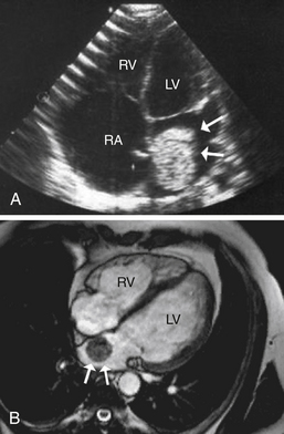
Figure 48-1 Left atrial myxoma (arrows), as visualized on (A) transthoracic echocardiogram and (B) on cardiac MRI. (A modified from Erdol C, Ozturk C, Ocal A, et al: Contralateral recurrence of atrial myxoma—case report and review of the literature. Images Paediatr Cardiol 8:3-9, 2001.) LV, Left ventricle; RA, right atrium; RV, right ventricle.
5. What are the most common primary malignant tumors?
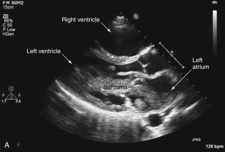
The most common primary malignant tumors are sarcomas (Figs. 48-2 and 48-3). Such sarcomas include angiosarcomas (the most common), rhabdomyosarcomas, fibrosarcomas, and leiomyosarcomas. Upon imaging, sarcomas often appear as large heterogeneous, infiltrative masses that frequently occupy most of the affected chamber (see Fig. 48-3). The results of surgery or chemotherapy in the treatment of cardiac sarcomas have been generally poor, with mean survival of only 6 to 12 months.
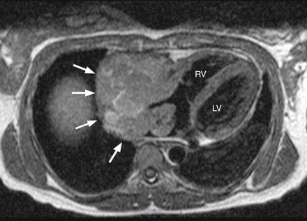
Figure 48-2 Massive angiosarcoma (arrows) arising from the right atrium, as visualized by MRI. (Modified from Sparrow PJ, Kurian JB, Jones TR, et al: MR imaging of cardiac tumors, Radiographics 25(5):1255-1276, 2005.) LV, Left ventricle; RV, right ventricle.
6. What symptoms do cardiac tumors cause?
7. What is the workup for suspected cardiac tumors?
A tumor plop is a sound heard in early diastole during auscultation. It is produced when a left atrial myxoma prolapses into the left ventricle during diastole (Fig. 48-4). The sound may be due to the tumor striking the left ventricular wall or to tension created on the tumor stalk.
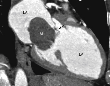
Figure 48-4 A left atrial myxoma (M) transiting across the mitral valve (arrow) in to the left ventricle. This may cause an audible “tumor plop.” LA, Left atrium; LV, left ventricle. (From Haaga JR: CT and MRI of the Whole body, ed 5, Philadelphia, PA, 2008, Mosby, fig. 28-24.)
9. What is lipomatous hypertrophy of the interatrial septum?
10. What is the most common valvular tumor?
11. What is the Carney complex?
Known by various names and acronyms, the Carney complex is an autosomal-dominant syndrome consisting of cardiac myxomas, cutaneous myxomas, spotty pigmentation of the skin, endocrinopathy, and other tumors. Myxomas occurring as part of the Carney complex are reported to account for 7% of all cardiac myxomas. Myxomas occurring as part of the Carney complex often present earlier in life, tend to be located in atypical locations, are often multiple, and often have a higher rate of recurrence after surgical resection compared with sporadic myxomas.
Bibliography, Suggested Readings, and Websites
1. Basson, C.T. Carney complex. Available at http://www.emedicine.com. Accessed March 23, 2013
2. Bruce, C.J. Cardiac tumours: diagnosis and management. Heart. 2011;97:151–160.
3. Meuller, D.K. Benign cardiac tumors. Available at http://www.emedicine.com. Accessed March 25, 2013
4. Goodkind, M.J. Cardiac tumors. Available at http://www.merck.com/mmpe. Accessed March 28, 2013
5. Kapoor, A. Cancer of the heart. New York: Springer Verlag; 1986.
6. Reardon, M.J., Walkes, J.C., Benjamin, R. Therapy insight: malignant primary cardiac tumors. Nat Clin Pract Cardiovasc Med. 2006;3:548–553.
7. Reynen, K. Cardiac myxomas. N Engl J Med. 1995;333:1610–1617.
8. Reynen, K., Kockeritz, U., Strasser, R.H. Metastases to the heart. Ann Oncol. 2004;15:375–381.
9. Sharma, G.K. Atrial myxoma. Available at http://www.emedicine.com. Accessed March 28, 2013
10. Sparrow, P.J., Kurian, J.B., Jones, T.R., et al. MR imaging of cardiac tumors. Radiographics. 2005;25:1255–1276.
[/level-membership-for-internal-medicine-category][not-level-membership-for-internal-medicine-category]Chapter 48
Cardiac Tumors
1. Which are more common, primary cardiac tumors or metastatic tumors to the heart?
2. What are the most common tumors that metastasize to the heart?
3. What are the most common primary cardiac tumors?
Benign tumors are more common than malignant tumors, occurring approximately three times as often as malignant tumors. In children, 90% of primary cardiac tumors are benign. The most common benign cardiac tumors in adults are myxomas, accounting for approximately half of all primary cardiac neoplasms; other common benign cardiac tumors are lipomas and papillary fibroelastomas. Rhabdomyomas are the most common benign tumor occurring in infants and children. Interestingly, rhabdomyomas usually regress over time and do not require specific treatment in asymptomatic individuals. Primary and secondary (metastatic) tumors involving the heart are listed in Table 48-1.
TABLE 48-1
PRIMARY AND SECONDARY (METASTATIC) TUMORS INVOLVING THE HEART
Primary Tumors
Myxoma
Lipoma
Papillary fibroelastoma
Rhabdomyoma
Fibroma
Angiosarcoma
Rhabdomyosarcoma
Lymphoma
Lipomatous hypertrophy
Secondary (Metastatic) Tumors
Lung (bronchogenic) cancer
Breast cancer
Esophageal cancer
Thyroid cancer
Melanoma
Lymphoma
Leukemia
Renal cell carcinoma
4. In what chamber do most myxomas occur?
Approximately 75% to 80% of myxomas occur in the left atrium (Fig. 48-1), with 15% to 20% occurring in the right atrium. Only 3% to 4% of myxomas arise in the left ventricle and 3% to 4% arise in the right ventricle. Myxomas are usually pedunculated and typically arise from the interatrial septum via a stalk. They are described on gross pathological examination as gelatinous in consistency. They most commonly occur between the third and sixth decades of life, and more frequently occur in women. They can cause effective obstruction of filling of the left or right ventricles, leading to left or right heart failure symptoms and findings, mimicking the symptoms and findings of mitral or tricuspid valve stenosis. Systemic embolism occurs in 30% to 40% of patients. Constitutional symptoms and findings (see Question 6) are also common. Most myxomas occur sporadically, but approximately 7% to 10% may be familial (see Question 10).
Figure 48-1 Left atrial myxoma (arrows), as visualized on (A) transthoracic echocardiogram and (B)
[/not-level-membership-for-internal-medicine-category]
