Cardiac Arrhythmias

Published on 23/06/2015 by admin

Filed under Emergency Medicine

Last modified 22/04/2025

Print this page

rate 1 star rate 2 star rate 3 star rate 4 star rate 5 star
Your rating: none, Average: 0 (0 votes)

This article have been viewed 1889 times

5.9 Cardiac Arrhythmias

Normal conduction system

The normal heartbeat is initiated by an impulse which originates from the sinoatrial (SA) node located in the wall of the right atrium near the superior vena cava junction. The impulse is then propagated via conducting cells that form a specialised system throughout the heart. Initially the impulse travels across the atria and via transatrial internodal pathways that converge on the atrioventricular (AV) node. The impulse proceeds down the bundle of His to the right and left bundle branches; the impulse then finally spreads throughout the Purkinje fibres to depolarise the ventricular muscle. The conducting cells of this specialised system have a rapid conduction velocity to rapidly propagate the electrical impulse throughout the heart. The various parts of this conduction system are also capable of spontaneous depolarisation and impulse generation under abnormal conditions.

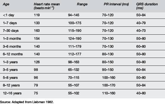
The SA node is normally the dominant (fastest) cardiac pacemaker but this can change if sinus node dysfunction occurs or if other parts of the conduction system develop increased automaticity. As stated above, the SA node is located near the junction of the superior vena cava (SVC) and the right atrium. The SA node is innervated by both sympathetic and parasympathetic nerve endings. Parasympathetic tone predominates during rest. Children are known to have relatively greater parasympathetic tone than adults and are also known to have developmental and age-dependent differences in action potential amplitude and conduction speed. Accordingly, there are age-dependent normal values for resting heart rate as well as PR interval and QRS duration, in addition to many other electrophysiology parameters (see Table 5.9.1).

image

Depolarisation initiated in the SA node spreads rapidly through the internodal pathways to converge on the AV node. Usually, the atria and ventricles are electrically separated from one another by a ring of fibrous tissue at the atrioventricular junction (the annulus fibrosis). Accordingly, in usual circumstances, the impulse must pass to the His bundle via the AV node. The AV node is located in the inferomedial wall of the right atrium near the insertion of the septal leaflet of the tricuspid valve. Conduction through the AV node is slow to allow completion of atrial systole (and ventricular diastole). This delay (and therefore potentially the completeness of ventricular filling) can be reduced during sympathetic stimulation.

After leaving the AV node the impulse travels along the bundle of His for 1–2 cm along the posteroinferior margin of the membranous portion of the interventricular septum, before dividing into the right and left bundle branches. These extensions of the conducting system spread subendocardially across the ventricular chambers to the base of the papillary muscles. The left bundle separates into two distinct fascicles (anterior and posterior). From these specialised fibres the impulse spreads from endocardium to epicardium throughout the right and left ventricles.

Abnormally situated embryonic remnants of conducting tissue can persist as accessory tracts. These are most commonly found around the AV node and are capable of conducting electrical activity between the atria and ventricles thus bypassing the AV node. These remnants are the anatomical substrate for re-entrant tachyarrhythmias.

The cardiac action potential

The cardiac action potential is divided into five phases: 0, 1, 2, 3, 4.

Vaughan Williams antiarrhythmia drug classification

Antiarrhythmic drugs are classified into five classes in the Vaughan Williams classification by the mechanism or channel that they most reliably affect during in vitro studies. There is, however, considerable overlap.

Class 1 drugs block the fast inward sodium channels and thereby increase the refractory period. This class is further subdivided into:

It is fair to say that these are infrequently used in paediatrics, though lignocaine is still an option in the ventricular fibrillation protocol and flecainide may sometimes be used in supraventricular tachyarrhythmias.

Class 2 agents are β-blockers, which act by a combination of β-antagonism and a quinidine-like membrane stabilising effect. There is a slowing of conduction velocity, prolonging of action potential duration and a reduction of automaticity Their main clinical use is in treating supraventricular tachyarrhythmias (SVT, atrial fibrillation, atrial flutter) by increasing the refractory period of the AV node. Propranolol or atenolol may be used but in acute situations many clinicians favour esmolol, a very short-acting parenteral β-1-antagonist (half life 9 minutes), which can be useful in supraventricular tachycardia (SVT) and possibly ventricular tachycardia (VT).

Class 3 drugs act primarily as potassium channel blockers and basically prolong the phase 3 repolarisation phase, thereby causing a prolongation of the action potential and increase in the effective refractory period. This class of drug is particularly useful in ventricular tachyarrhythmias and includes amiodarone and sotalol.

Amiodarone is one of these antiarrhythmics with a plethora of effects, in that it is primarily class 3 (i.e. potassium channel blocker) but also has sodium channel blocking, β-antagonism and calcium channel blocking effects. Oral amiodarone has unusual pharmacokinetics, with clinical effect only apparent after several days of treatment and a half life of 3–15 weeks. Side effects are frequent and serious (corneal photosensitivity, hyper- or hypothyroidism, pulmonary fibrosis and pro-arrhythmia). Intravenous amiodarone is used in the acute management of post-operative tachyarrhythmias (usually junctional ectopic tachycardia, JET). In the adult ventricular fibrillation protocol, a randomised comparison between amiodarone and lignocaine found a greater chance of successful resuscitation (but not survival) with amiodarone. Therefore some authorities have recommended amiodarone as the agent of choice to help effect defibrillation after adrenaline (epinephrine). In haemodynamically stable VT the agent of choice is probably sotalol unless there is impaired ventricular function (EF <40%, signs of congestive heart failure) when amiodarone is recommended. In unstable or polymorphic VT the recommended agent depends a little on the initial trace. If it looks like torsade de pointes with the trace oscillating around the baseline (or in polymorphic VT with a normal QT interval), the clear recommendation is intravenous (IV) magnesium. If it is not torsade-like and the QT interval can be determined and is not prolonged, amiodarone is a second-line agent to β-blockers. If the QT interval is prolonged, then IV magnesium, followed if necessary by lignocaine, would be the recommended approach.

Sotalol is classified as a class 3 drug, although it is a non-selective β-blocker with additional class 3 properties. It therefore combines the β-blocking effect on the SA and AV nodes with prolongation of AP duration and lengthening of refractory period elsewhere in the heart. It is valuable acutely and long term in preventing SVT of all sorts and is the agent of choice in monomorphic stable VT with apparent normal ventricular function. Like amiodarone, it does increase the QT interval and is therefore pro-arrhythmic as well as having the other risks of hypotension and bradycardia, presumably resulting from its β-blocking effects.

Class 4 agents are calcium channel blockers and the prototype remains verapamil, which has wide-ranging effects on shortening the plateau phase and reducing contractility. Verapamil is primarily used in the adult setting because of its reduction of conduction velocity in the AV node, thereby controlling the ventricular response rate to SVT, atrial flutter or atrial fibrillation. Even in that situation it is still a second-line agent to adenosine for SVT termination in adults, provided there is good left ventricular (LV) function. However, because of serious bradycardia, hypotension and cardiac arrest in infants, it is not used in children. Instead, β-blockers are used in Wolff–Parkinson–White (WPW) syndrome and digoxin in non-WPW patients.

Finally, Class 5 agents are a miscellaneous group and include adenosine and digoxin. Adenosine is an endogenous purine nucleoside with a very rapid half life (less than 5 seconds). It acts by depressing slow calcium channels and enhancing potassium conduction. Its main effects are to depress sinus node and AV node activity with a shortening of atrial refractoriness. Because of its effect in blocking AV nodal activity, adenosine is superbly suited to block re-entry phenomena, the most frequent cause of SVT in children. Series show that adenosine terminates 90–100% of SVTs but the arrhythmia re-initiates in approximately 25%. Other atrial tachycardias like multifocal atrial tachycardia, atrial flutter or atrial fibrillation are almost always resistant to adenosine. Adenosine is given in increasing doses from 50–200 mcg kg–1 every 2 minutes, using a two-syringe technique so that it can be flushed rapidly into the circulation. Side effects can be facial flushing, bronchospasm or sinus arrest. Adenosine is essentially very safe, although there is a single case report of VF occurring after a dose of adenosine in a neonate with a concealed WPW syndrome previously treated with digoxin.

Although adenosine is unlikely to be controlling in supraventricular tachycardias other than re-entrant SVT, the acute injection of adenosine may transiently slow AV nodal activity and allow the flutter wave or P wave morphology to be assessed. This may be very valuable in diagnosing the specific rhythm. In a haemodynamically stable wide complex tachycardia of uncertain origin, the differential diagnosis is primarily between VT and SVT with aberrant conduction. Adenosine administration might be discussed as a way of making this distinction (i.e. effective in the latter but not in the former). In the adult literature, unless SVT origin is strongly suspected, this practice is discouraged because of the potential for brief hypotension, and accelerated accessory pathway conduction following adenosine injection.

Digoxin is still very commonly used parenterally, acutely, and as a maintenance medication, primarily to slow AV conduction and decrease the ventricular response to atrial dysrhythmia like flutter or fibrillation. It provides rate control and sometimes converts supraventricular tachyarrhythmias. There is an important caveat with digoxin, that it may shorten accessory pathway refractoriness and increase the resulting ventricular response. Therefore, it should not be used in accessory pathway dysrhythmias like WPW syndrome. Virtually any arrhythmia can arise from intoxication with digoxin. This risk is greater with hypokalaemia and the acute treatment is intravenous Digitalis antibody.

Pathogenesis of arrhythmias

Tachyarrhythmias

There are three fundamental mechanisms proposed for the generation of tachyarrhythmias:

Re-entry

Re-entry exists when a closed loop of specialised conducting tissue allows an electrical impulse to travel in a circular fashion and permits atrial or ventricular electrical activation with each pass around the circuit. Re-entry may occur on a large (macro) or small (micro) scale. Atrial flutter and ventricular fibrillation are examples of micro re-entry; paroxysmal supraventricular tachycardia is an example of macro re-entry.

Macro re-entry usually involves the participation of an accessory conduction pathway. Accessory pathway conduction characteristics vary widely among patients. The accessory pathway in some patients conducts antegrade during sinus rhythm, whereas in others it conducts only retrograde during tachycardia. When anterograde conduction is possible down the accessory pathway the standard ECG will show pre-excitation with a short PR interval and wide QRS, including an initial delta wave resulting from the pre-excitation of the ventricle from the sinus impulse conducting through the accessory pathway before the impulse has passed through the normal conducting system. Patients with WPW usually manifest orthodromic tachycardia (forward excitation through AV node and rapid retrograde conduction via accessory pathway to create a circuit) but also (rarely) display antidromic tachycardia involving retrograde conduction up the usual atrioventricular route or via an alternative accessory connection.

Supraventricular tachycardia is the most common sustained tachyarrhythmia in children and almost always has a re-entry mechanism. Most commonly, particularly in children younger than 12 years of age, this re-entry is caused by an accessory atrioventricular connection (resulting in atrioventricular (or AV) re-entrant tachycardia). In adolescents, AV node re-entry is the mechanism in up to one third of patients. Previously we believed that this was due to a re-entry circuit within the compact AV node but a newer concept involves tissue, most likely atrial muscle, more than several millimetres outside the compact atrioventricular node.

The commonest mode of accessory connection-mediated re-entry tachycardia is orthodromic tachycardia, with the circular movement of the electrical impulse going antegrade through the atrioventricular node and then retrograde up the accessory connection.

General principles for arrhythmia management

Conduction disturbances: atrioventricular block

This involves delayed or incomplete conduction through the AV node. Causes include a congenital form (maternal systemic lupus erythematosus) and an acquired form. Acquired AV block occurs in poisoning, metabolic disturbances, myocarditis, rheumatic fever, Lyme disease, fibrosis in the area of the conduction system associated with previous cardiac surgery (particularly ventricular septal defect or atrioventricular septal defect closure, tetralogy repair or aortic valve replacement) and inferior myocardial infarction.

First degree AV block Is present when the PR interval is longer than usual for age (see Table 5.9.1) but normal sinus rhythm and 1:1 AV conduction is maintained. This is a normal feature of the ECG of endocardial cushion defects. It may occur in a normal heart during parasympathetic stimulation or digoxin treatment and usually does not require treatment.

Wide complex tachyarrhythmia

The differential diagnosis of a wide or broad complex tachycardia (i.e. QRS duration greater than normal for age, usually >0.08 seconds) is between:

VT in children usually presents as a wide complex rhythm between 120 and 220 per minute. There is usually AV dissociation but retrograde VA conduction may occur. It may be mono- or polymorphic, sustained or non-sustained. Causes include metabolic abnormalities, poisoning, myocarditis, cardiomyopathy, ventriculotomy, ventricular tumours and congenital or acquired Long QT syndrome.

Most children with VT are symptomatic with lethargy, symptoms of pulmonary congestion, poor circulation and possibly palpitations.

The other three differential diagnoses outlined above are unusual causes of a wide complex tachycardia in children. The most important differential to think about is SVT with aberrant conduction, because it should respond to vagal manoeuvres or adenosine administration. A previous electrocardiogram is of great value in determining the presence of an accessory pathway or a pre-existing bundle branch block in that these features point to SVT with aberrant conduction. Classically, VT is a wide complex tachycardia with a left access deviation and more frequently a left bundle branch block pattern, whereas a right bundle branch block pattern (with rSR in V1) is more commonly seen in SVT. Again, the P wave morphology is crucial and if P waves can be distinguished, the loss of a one-to-one relationship between P wave and QRS complex is highly suggestive of VT. Sometimes, however, in VT there still may be a one-to-one ventriculoatrial relationship. The presence of fusion beats indicating an ectopic focus below the level of the AV node is also strongly suggestive of VT.

If there is strong evidence supporting a supraventricular origin and preserved LV function, vagal manoeuvres and/or adenosine administration can be tried. The caveat is that, in doing this, there is a small but important potential for hypotension and accelerated accessory pathway conduction from the adenosine administration that has been responsible for conversion of SVT to VT, or even worse on occasions.

If there are no pointers towards a supraventricular origin, or the above manoeuvres are unrewarding, the diagnosis is most likely VT.

If the patient is severely compromised, direct current cardioversion is clearly indicated. It is debated whether this is better synchronous or asynchronous, but most authorities recommend starting with synchronous cardioversion if there is a pulse, using a 1 J/kg shock from a biphasic defibrillator.

If the patient has reasonable perfusion, the recommended drug depends on the appearance of the VT. If the VT is monomorphic and the patient has preserved LV function, intravenous sotalol is the agent of choice, with amiodarone as a second-line agent. Amiodarone is definitely the drug of choice if LV function is impaired. If the rhythm is polymorphic, there should be some further assessment of the trace. If the trace is torsade-like with complexes that vary in height and appear to twist around the baseline, intravenous magnesium is clearly recommended. If the QT interval is prolonged, IV magnesium followed probably by IV lignocaine is recommended. If the QT interval can be seen not to be prolonged, β-blockade is the primary approach, with amiodarone again the agent of choice if LV function is impaired.

Narrow complex tachyarrhythmia

A narrow complex tachycardia is defined as one with a QRS duration normal for age (approximately ≤0.08 seconds). The narrow QRS complex almost always indicates that these tachyarrhythmias are supraventricular.

SVT refers to a family of tachyarrhythmias requiring the atrium, AV node or both for their perpetuation. SVT accounts for 90% of all significant tachyarrhythmias in children. It is most useful to subclassify these tachyarrhythmias on the basis of site of origin and mechanism into primary atrial tachycardias (including MAT, atrial fibrillation and atrial flutter), atrioventricular reciprocating, AV nodal re-entry and junctional ectopic tachycardia. Primary atrial and junctional ectopic tachycardias will be dealt with later. Atrioventricular reciprocating and nodal re-entry tachycardias are the commonest forms of SVT in childhood.

Atrioventricular reciprocating tachycardia Occurs due to the presence of an accessory conduction pathway setting up a re-entry circuit. In the commonest variety (orthodromic reciprocating), the impulse travels antegrade via the AV node to the ventricles (as usual) and then retrograde via the accessory pathway back to the atria. If the accessory pathway is capable of conducting antegrade, antidromic reciprocating tachycardia can occur, with the impulse travelling in the opposite direction, i.e. forward down an accessory pathway, returning retrograde via the AV node.

AV nodal re-entrant tachycardia In this tachyarrhythmia the two conduction pathways are thought to be within or adjacent to the AV node. Beyond 5 years of age, this becomes the most common form of SVT. Classically, the P wave is buried within the QRS complex on ECG. In AV nodal re-entrant tachycardia, for long-term prophylaxis therapy digoxin is the preferred agent. However, β-blockers may be considered in resistant cases, either alone or in combination with digoxin.

The crucial step in distinguishing between the above-mentioned atrial, atrioventricular or nodal tachycardias is an identification of the P wave and its relationship to the QRS complex. This is sometimes best done with a 12-lead ECG with the paper run at 50 mm s–1. If epicardial leads are available (i.e. post-operatively), an ‘atrial ECG’ can be obtained by connecting the two atrial pacing leads to the right arm and right leg electrodes of a 12-lead ECG. Alternatively, a specialised transoesophageal pacing wire can be introduced, like a nasogastric tube approximately to the level of the nipple and then connected to lead V1 to display an oesophagoatrial ECG in lead V1, with the remainder of the leads showing a normal surface ECG. Because it is being recorded directly, the P wave will usually appear larger than the QRS complex in the surface ECG and by aligning simultaneously recorded surface and ‘atrial’ ECGs the position of the P relative to the QRS can be determined.

Atrially-driven tachycardias will have a P wave preceding each QRS. If the P wave is upright in inferior leads, the origin of the tachycardia is high in the atrium, whereas the P wave axis will be negative in inferior leads if there is AV nodal re-entry or a low atrial ectopic focus. The P wave is usually absent in junctional ectopic tachyarrhythmia or VT. An abnormal P wave morphology usually indicates an origin other than the sinus node and several morphologies will indicate multifocal atrial tachycardia (MAT). If the P wave follows the QRS complex, the differential diagnosis involves atrioventricular nodal re-entry tachyarrhythmia and ventricular tachycardia. If the P waves are dissociated from the QRS complexes, the diagnoses are most likely junctional ectopic tachycardia or VT.

Regardless of these diagnostic considerations, the approach is essentially the same. If the patient is acutely compromised, basic life support followed by attempted DC cardioversion with 0.5–1 J kg–1 of DC shock (under sedation if possible) and with continuous ECG monitoring is recommended. IV adenosine is an alternative if this is immediately available. If the patient has adequate perfusion, consider vagal manoeuvres/eliciting of diving reflex, administer adenosine in increasing doses as above.

The importance of a continuous 12-lead electrocardiogram during this process cannot be over emphasised. This is because just a few beats of sinus rhythm before reversion back to the tachyarrhythmia might well shed considerable light on the diagnosis. If the arrhythmia is resistant to adenosine, the next step is probably use of a β-blocker, with digoxin an alternative for maintenance (see below for caveats regarding digoxin in WPW).

Failure of adenosine to convert such a tachyarrhythmia makes unusual diagnoses more likely. For multiple atrial ectopic foci, sotalol is effective but probably only as a bridge to radiofrequency ablation. For JET the agent of choice is amiodarone. In atrial fibrillation/flutter, rate control can be achieved using digoxin but usually elective cardioversion is required to terminate the arrhythmia. In post-surgical cases recurrence is common and sotalol probably has the strongest demonstrated efficacy in reducing recurrences (and controlling ventricular rate) if they occur, though only as a bridge to possible cryoablation.

Ventricular fibrillation

In confirmed ventricular fibrillation (VF), the first priority is rapid defibrillation. Basic life support should be initiated while a defibrillator is sought. There is increasing evidence that shock success is improved if effective chest compressions are provided for 1 to 3 minutes before defibrillation, especially if VF duration is longer than approximately 3 minutes before attempted defibrillation. Conducting pads or conductive gel should be placed on the chest in the left 5th intercostal space midaxillary line and second intercostal space to the right of the sternum, being careful that gel areas are not contiguous to avoid shorting of current. The paddles are applied, charged on the chest, ‘stand clear’ is called, and the operator verifies that this direction has been followed by other staff. Then 2 J kg–1 of current is discharged in asynchronous mode. CPR is resumed immediately after the first shock and continued for five cycles (about 2 minutes) before the next rhythm check. If VF persists, a second shock of 4 J kg–1 is administered and again CPR immediately resumed for five cycles. Early during these five cycles adrenaline (epinephrine) is administered and if VF persists on rhythm check a third shock of 4 J kg–1 is delivered. CPR is again immediately resumed and if the patient remains in VF, amiodarone 5 mg kg–1 by rapid IV bolus is the antiarrhythmic of choice. This is then followed by further attempts at defibrillation while CPR is continued. Lignocaine is a second choice agent, magnesium is recommended.

Further reading

Alexander M.E., Berul C.I. Ventricular arrhythmias: When to worry. Pediatr Cardiol. 2000;21(6):532-541.

Anonymous. The International Liason Committee on Resuscitation (ILCOR) consensus on science with treatment recommendations for paediatric and neonatal patients: paediatric basic and advanced life support. Pediatrics. 2006;117(5):e955-e977.

Bink-Boelkens M.T.E. Pharmacologic management of arrhythmias. Pediatr Cardiol. 2000;21(6):508-515.

Celiker A., Ayabakan C., Ozer S., et al. Sotalol in the treatment of pediatric cardiac arrhythmias. Pediatr Int. 2001;43:624-630.

Chun T.U., Dehart G.F. Advances in the approach to treatment of supraventricular tachycardia in the pediatric population. Curr Cardiol Rep. 2004;6:322-326.

Kudenchuk P.J., Cobb L.A., Copass M.K., et al. Amiodarone for resuscitation after out-of-hospital cardiac arrest due to ventricular fibrillation. N Engl J Med. 1999;341(12):871-878.

Liebman J. Tables of normal standards. In: Liebman J., Plonsey R., Gillette P.C., editors. Pediatric electrocardiography. 1st ed. Baltimore: Williams & Wilkins; 1982:82-133.

Luedtke S.A., Kuhn R.J., McCaffrey F.M. Pharmacologic management of supraventricular tachycardias in children. Part 1, WPW and AV nodal re-entry. Ann Pharmacother. 1997;31:1227-1243.

Luedtke S.A., Kuhn R.J., McCaffrey F.M. Pharmacologic management of supraventricular tachycardias in children. Part 2, Atrial flutter, atrial fibrillation and junctional and atrial ectopic tachycardia. Ann Pharmacother. 1997;31:1347-1359.

McKee M.R. Amiodarone – an ‘old’ drug with new recommendations. Curr Opin Pediatr. 2004;15:193-199.

Saul J.P., Scott W.A., Brown S., et al. Intravenous amiodarone for incessant tachyarrhythmias in children: a randomized, double-blind, antiarrhythmia drug trial. Circulation. 2005;112:3470-3477.