Carbon dioxide retention and capnography

Published on 07/02/2015 by admin

Filed under Anesthesiology

Last modified 22/04/2025

Print this page

rate 1 star rate 2 star rate 3 star rate 4 star rate 5 star
Your rating: none, Average: 0 (0 votes)

This article have been viewed 1787 times

Carbon dioxide retention and capnography

Michael G. Ivancic, MD

The monitoring of CO2, the most abundant gas produced by the human body during anesthesia, has become a standard of practice strongly encouraged by the American Society of Anesthesiologists. CO2 is a byproduct of cellular metabolism, transported to the lungs by the systemic venous system and eliminated from the alveoli during ventilation (see Chapter 22).

CO2 retention

Rebreathing of CO2 is undesirable, although, during mechanical ventilation, allowing the patient to rebreathe CO2 is infrequently used to achieve normocarbia in those patients who are being hyperventilated (i.e., when large tidal volumes may be desirable for other reasons). A leak or obstruction in the anesthesia machine circuit, common gas outlet, or fresh gas supply line may also cause an increase in CO2 concentration.

When Mapleson systems are used, inadequate fresh gas flow is the primary cause of an increase in CO2 because these systems do not contain unidirectional valves or absorbent canisters. Specifically, systems with inner tubes, such as the Bain system, can cause rebreathing if there is any dysfunction (kink) in that tube. The Mapelson D (Bain circuit) is the most efficient for controlled ventilation with regard to a relatively low flow of fresh gas, whereas the Mapleson A is most suitable for patients who are spontaneously breathing (see Chapter 193). Specific minimum fresh gas flow rates for the various Mapleson apparatuses are recommended for spontaneous ventilation as well as controlled ventilation (Box 9-1).
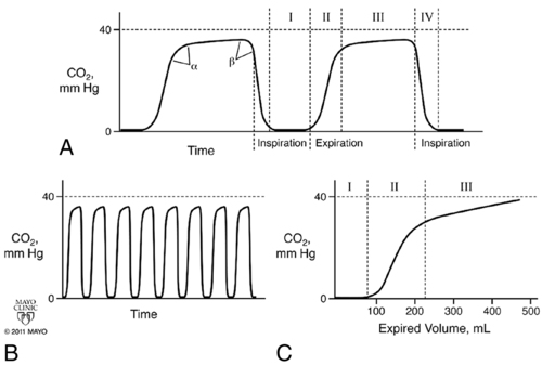
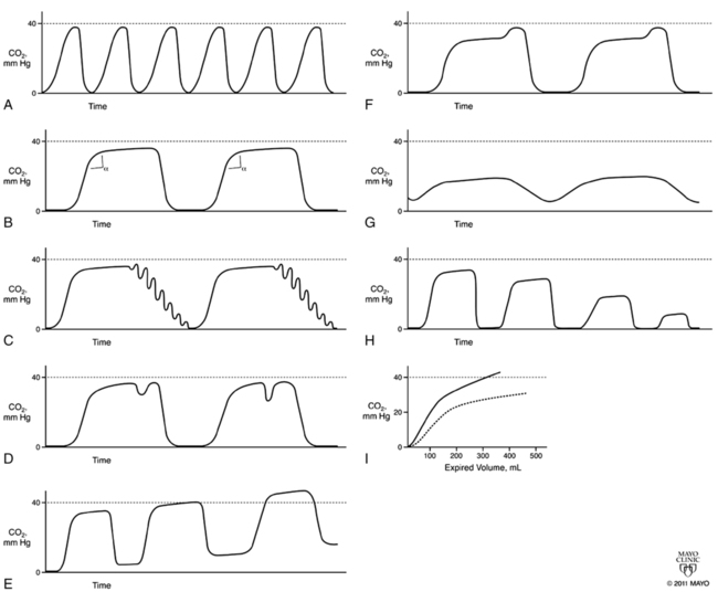
Increased dead space, whether mechanical or physiologic, can increase rebreathing and CO2 retention if the dead space is particularly large, especially in smaller patients. A heat and moisture exchanger in the breathing circuit may also be a source of dead space, with the larger the volume of the heat and moisture exchanger, the larger the dead space.

In the closed-circle systems used in modern anesthesia machines, minimal rebreathing of CO2, if any, should occur; however, malfunction of either of the unidirectional valves may lead to CO2 rebreathing. If an inspiratory valve is stuck open, rebreathing can occur because, during expiration, alveolar gas can backfill the inspiratory limb of the circle. A malfunctioning expiratory valve can lead to CO2 rebreathing in a spontaneously breathing patient because, during inspiration, the negative pressure generated by the patient can entrain alveolar gas from the expiratory limb of the circuit.

Other causes of inadvertent CO2 rebreathing usually involve the CO2 absorber. If the absorbent color indicator malfunctions—and, therefore, is not reflecting the true level of CO2 in the system—rebreathing can occur without the anesthesia provider being aware of the problem. In older anesthesia machines, the CO2 absorber could be bypassed. Older absorbent canisters had a rebreathing valve on them that, if engaged, would lead to CO2 rebreathing. Channeling of gas through the canister without contacting any active absorbent can also lead to CO2 rebreathing. Independent of the cause, absorbent malfunction is best corrected immediately by increasing the fresh gas flow and then troubleshooting the underlying cause.

Capnography

During induction of and emergence from anesthesia, rebreathing of CO2 will lengthen each process because of alterations in alveolar tensions associated with rebreathing of exhaled alveolar anesthetic gases. Capnography can help troubleshoot malfunctioning equipment, such as the problems noted above. The classic rebreathing pattern on the capnogram will show an elevation of the waveform baseline that does not return to 0, as well as a higher end-tidal CO2 (PETCO2) reading, although the PaCO2 value may be normal, depending on the degree of alveolar ventilation.

Sidestream versus mainstream sampling

The CO2 may be measured from a mainstream or sidestream device. Mainstream sampling uses a device that is placed close to the tracheal tube, with all the inhaled and exhaled gas flowing through the device. One benefit to the use of mainstream sampling is that the response time is faster, so no uncertainty exists regarding the rate of gas sampling. Drawbacks include the bulkiness of the device and the need for it to be heated to 40° C, therefore increasing the risk of burning the patient’s skin.

Sidestream sampling is the method most commonly used in today’s operating rooms. Sampling flow rate is an important aspect of this system and is usually in the range of 150 to 250 mL/min, with a mean of approximately 200 mL/min. If the sampling flow is less than 150 mL/min, the sampling time is too slow; if the flow is greater than 250 mL/min, the likelihood increases of fresh-gas contamination occurring. Water condensation can also be a problem as expired air condenses within the sampling tube walls. Various water-trap systems have been devised but may fail. Changing or flushing of the CO2 sampling tube with an air-filled syringe may alleviate some moisture and the sampling error, but a new filter is often required.

Co2 measurement methods

Infrared spectrometry is the most commonly used method of measuring gas concentration. Other methods include mass spectrometry, Raman scattering, and chemical colorimetric analysis.

Infrared spectrometry systems work by analyzing infrared light absorption from gas samples and comparing them with a known reference to determine the type and concentration of that particular gas. The advantages associated with the infrared system include the ability of the system to accurately analyze multiple gases—from CO2 to N2O and all the potent inhalation agents—as well as the fact that the unit is relatively small, lightweight, and inexpensive. The greatest disadvantage is that water vapor can interfere with measurements, resulting in falsely elevated readings of CO2 and inhalation agents.

Mass spectrometry can measure nearly every gas pertinent to anesthesia by separating gases and vapors according to differences in their mass-to-charge ratios, including O2 and N2, which cannot be measured by infrared. Mass spectrometry also has a relatively fast response time. The disadvantages to this system include the relatively large size of the unit, the need for warm-up time, and the cost.

Raman spectroscopy relies on the inelastic, or Raman, scattering of monochromatic light (e.g., a laser) by different gases, thereby providing information about the phonon modes (an excitation state, a quantum mechanical description of a special type of vibrational motion of molecules) in a system. The advantages to the use of Raman spectroscopy include the fact that multiple gases can be analyzed simultaneously, the system is very accurate, and the response time is rapid. However, this system is relatively large and expensive.

Colorimetric detection consists of a pH-sensitive paper within a chamber placed between the tracheal tube and ventilation device. The color change is reversible and can change from breath to breath. Several brands are marketed, but most use a color scale that is similar (i.e., purple: ETCO2 <4 mm Hg [<0.5% CO2]; tan: ETCO2 4-15 mm Hg [0.5% to 2% CO2]; yellow: ETCO2 >15 mm Hg [>2% CO2]). Advantages are portability, low cost, and no need for other equipment. The use of colorimetric detection is most applicable outside of the operating room to confirm tracheal-tube placement. The disadvantages are that the results are only semiquantitative.

Capnograms

Capnograms rely on time or volume to assess CO2 concentrations. Time capnograms are further divided into slow and fast tracings. All have their advantages, but the time capnogram is the most commonly used system (fast speed for trends and slow speed for detailed waveform analysis). Volume capnograms are unique in that a breath-by-breath measurement of CO2 concentration can be made, dead space can be divided into components, and significant changes in the morphology of the expired waveform can be detected as they relate to ventilation and perfusion. Normal CO2 waveforms and several abnormal capnograms are shown in Figures 9-1 and 9-2. PaCO2 and PETCO2 trends of metabolism, circulation, ventilation, and various equipment failures are summarized in Table 9-1. Changes in PETCO2 and the capnogram may indicate one of a number of potential changes in the patient’s condition, requiring the clinician to act appropriately.

Table 9-1

Causes of Altered End-Tidal CO2 during Anesthesia*

Cause PETCO2 PETCO2-to-PaCO2 Gradient
CO2 insufflation Increased Normal
Increased CO2 production Increased Normal
Right-to-left shunt Increased Widened
Increased physiologic or anatomic dead space, or both Decreased Widened
Increased apparatus dead space Increased Normal
Hyperventilation Decreased Normal
Hypoventilation Increased Normal
Leak in sampling line Decreased Widened
Poor seal around tracheal tube Decreased Widened
High sampling rate Decreased Widened
Low sampling rate Decreased Widened
Rebreathing due to malfunctioning breathing valve Increased Decreased
Rebreathing with low fresh gas in the Mapleson system Increased Decreased
Rebreathing with circle system (absorbent problem) Increased Normal

*Normal pressure of end-tidal CO2 (PETCO2) is 38 mm Hg (5%). The PaCO2-to-PETCO2 gradient is normally <5 mm Hg.

From hyperthermia, malignant hyperthermia, convulsions, pain, or bicarbonate administration.

Adapted, with permission, from Dorsch JA, Dorsch SE. Gas monitoring. In: Dorsch JA, Dorsch SE, eds. Understanding Anesthesia Equipment, 5th ed. Philadelphia: Lippincott, Williams & Wilkins; 2007:706-707.